. 2009 Jul;24(4):309-16.
doi: 10.1093/mutage/gep011. Epub 2009 Apr 16.

The Xpc gene markedly affects cell survival in mouse bone marrow

Affiliations

The Xpc gene markedly affects cell survival in mouse bone marrow

Joshua L Fischer et al. Mutagenesis. 2009 Jul.

Abstract

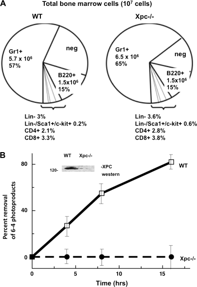
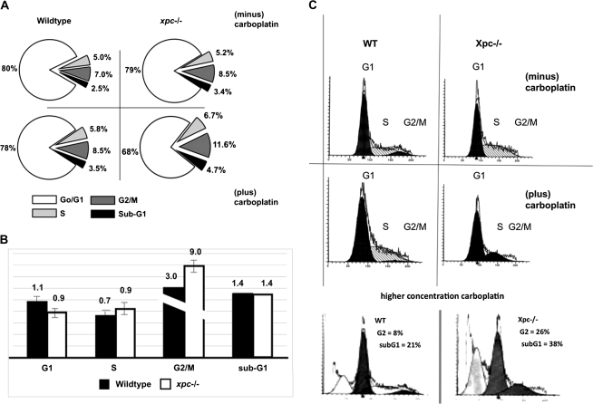
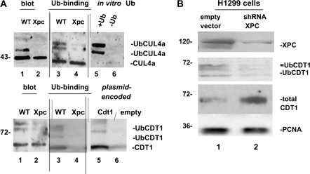
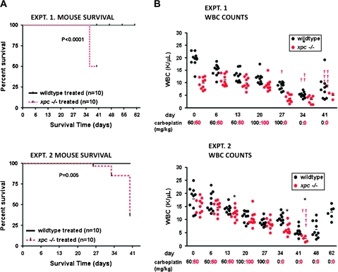
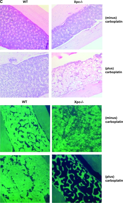
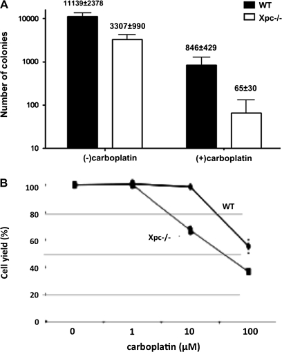
The XPC protein (encoded by the xeroderma pigmentosum Xpc gene) is a key DNA damage recognition factor that is required for global genomic nucleotide excision repair (G-NER). In contrast to transcription-coupled nucleotide excision repair (TC-NER), XPC and G-NER have been reported to contribute only modestly to cell survival after DNA damage. Previous studies were conducted using fibroblasts of human or mouse origin. Since the advent of Xpc-/- mice, no study has focused on the bone marrow of these mice. We used carboplatin to induce DNA damage in Xpc-/- and strain-matched wild-type mice. Using several independent methods, Xpc-/- bone marrow was approximately 10-fold more sensitive to carboplatin than the wild type. Importantly, 12/20 Xpc-/- mice died while 0/20 wild-type mice died. We conclude that G-NER, and XPC specifically, can contribute substantially to cell survival. The data are important in the context of cancer chemotherapy, where Xpc gene status and G-NER may be determinants of response to DNA-damaging agents including carboplatin. Additionally, altered cell cycles and altered DNA damage signalling may contribute to the cell survival end point.


Figures

Fig. 1
Fig. 1
G-NER defect in cultured Xpc−/− bone marrow. (A) For cell culture experiments (Figures 1–3) bone marrow was harvested from untreated wild-type or Xpc−/− mice. Cell yields were lower in Xpc−/− compared to wild type; therefore, cell numbers were normalized to 107 viable cells per genotype. Flow cytometry was conducted using six different lineage markers. No significant differences were observed in the lineage markers in comparing 107 cells of each genotype. As shown in the pie graph, lineage markers tested were Gr1+, B220+, Lin−, CD4+, CD8+ and the small subpopulation Lin−/Sca1+/c-kit+. The pie segment labelled ‘neg’ was unreactive for any of the lineage markers tested. (B) XPC protein was undetectable in Xpc−/− bone marrow (inset). Removal of DNA lesions was markedly slow in Xpc−/− bone marrow, consistent with the known rate-limiting role of XPC in G-NER. Pooled bone marrow of three or more untreated 10-week-old mice of each genotype was used. Cells were cultured in cytokine-containing medium for 15 h, irradiated with 20 J/m2 254 nm UV radiation in phosphate-buffered saline and returned to tissue culture. Time points were taken at 0, 4, 8 and 16 h and assayed using an antibody to 6-4 photoproducts (P < 0.02 by t-test).
Fig. 2
Fig. 2
Evidence of G1 and G2 cell cycle checkpoint alterations in cultured Xpc−/− bone marrow. (A) Bone marrow harvested from untreated 10-week-old mice was cultured in cytokine-containing medium for 15 h and then treated with 10-μM carboplatin for an additional 24 h. Cells were fixed in 70% ethanol and analysed by PI staining. By PI staining alone, cell cycle differences were not significant and 68–80% of cells were in G1. (B) Bone marrow harvested from untreated 10-week-old mice was stimulated with cytokines and cultured for 48 h in the presence of 10-μM BrdU to label proliferating cells, then treated with 10-μM carboplatin for 15 h and then fixed and stained with PI. A fluorescein isothiocyanate-conjugated antibody to BrdU was used to gate the BrdU-labelled population, which were then assayed for PI content. Values for carboplatin-treated bone marrow were divided by values for untreated bone marrow conducted side by side. The data represent three pooled mice of each genotype. Two separate experiments yielded similar results. A modest but significant decrease in G1 population was observed in Xpc−/− mice compared to wild-type mice (P < 0.05 by t-test). A significant increase was observed in the G2 population in Xpc−/− mice compared to wild-type mice (P < 0.02 by t-test). The plot shows relative cell cycle distribution after carboplatin treatment. (C) Raw flow cytometric data corresponding to the bar graph shown in panel (B). Cell number is plotted on the y-axis, at least 15 000 events per sample; PI staining is shown on the x-axis. Use of a higher concentration of carboplatin (40 μM) revealed a sub-G1 apoptotic population which was more pronounced in the Xpc−/− mutant bone marrow. The G2 population was also more pronounced in the mutant at the higher carboplatin concentration.
Fig. 3
Fig. 3
(A) CUL4A and CDT1 cell cycle checkpoint proteins in cultured wild-type and Xpc−/− bone marrow. We wanted to determine if the presence or absence of XPC would alter DNA damage signalling. Immunoblots were conducted using 50 μg of total cell lysates (lanes 1 and 2, upper and lower panels). Equal amounts of cellular proteins, 5 mg, were then affinity purified on a ubiquitin-binding resin and immunoblotting of bound proteins was conducted. The higher molecular weight ubiquitinated forms of CUL4A (upper panel) and CDT1 (lower panel) clearly differ between the two genotypes (lanes 3 and 4 of upper and lower panels). The data suggest a defect in CUL4A and CDT1 ubiquitin modification in Xpc−/− mice. To help identify ubiquitinated CUL4A and CDT1, we used in vitro ubiquitin-conjugated proteins. Omission of ubiquitin from the reaction shows that mainly ubiquitinated CUL4A is detected by the CUL4A antibody (lanes 5 and 6, upper panel). Plasmid-encoded CDT1 protein was also used as a marker (lanes 5 and 6, lower panel). (B) Knocking down XPC in H1299 cells alters the ubiquitin modification of CDT1. We used transiently transfected H1299 cells expressing an shRNA to XPC to test if the results shown in panel (A) were due directly to loss of XPC. Although Xpc was not completely silenced, it was clearly decreased by the shRNA. Higher molecular weight ubiquitinated forms of CDT1 were largely absent where XPC was knocked down, consistent with a role for XPC in DNA damage signalling to the CDT1 cell cycle checkpoint protein.
Fig. 4
Fig. 4
Effect of carboplatin administration in vivo in bone marrow of wild-type and Xpc−/− mice. (A) Kaplan–Meier plots of mouse survival. Twenty mice of each genotype received carboplatin or saline only as controls. Saline-only did not affect mouse survival nor alter WBC counts; saline-only control groups are not shown in order to simplify the figure. Mice received carboplatin at weekly intervals up until day 27. By day 41, 12/20 Xpc−/− mice died, while 0/10 wild-type mice died. (B) WBC counts were monitored during the course of the experiments. WBC counts were not significantly different prior to day 27, but were significantly different after day 27 (P < 0.05, lower panel). Each circle represents an individual mouse, black, wild type; red, Xpc−/−. The horizontal bars represent the mean of each group. By day 41, the few surviving Xpc−/− mice had significantly lower WBC counts (P < 0.05 by t-test). The carboplatin dosing schedule is shown in black, wild type; red, Xpc−/−. Note that wild-type mice received an additional dose on day 27 that was not administered to Xpc−/− mice. (C) Histological evaluation of wild-type and xpc−/− mice in carboplatin-treated and saline-control groups, day 41. Haematoxylin–eosin staining of formalin-fixed femur sections. Marked hypocellularity was observed in Xpc−/− mice receiving carboplatin (upper panel); 4′,6-diamidino-2-phenylindole staining (lower panel). The data are representative of at least three mice of each treatment group and genotype.
Fig. 4
Fig. 4
Effect of carboplatin administration in vivo in bone marrow of wild-type and Xpc−/− mice. (A) Kaplan–Meier plots of mouse survival. Twenty mice of each genotype received carboplatin or saline only as controls. Saline-only did not affect mouse survival nor alter WBC counts; saline-only control groups are not shown in order to simplify the figure. Mice received carboplatin at weekly intervals up until day 27. By day 41, 12/20 Xpc−/− mice died, while 0/10 wild-type mice died. (B) WBC counts were monitored during the course of the experiments. WBC counts were not significantly different prior to day 27, but were significantly different after day 27 (P < 0.05, lower panel). Each circle represents an individual mouse, black, wild type; red, Xpc−/−. The horizontal bars represent the mean of each group. By day 41, the few surviving Xpc−/− mice had significantly lower WBC counts (P < 0.05 by t-test). The carboplatin dosing schedule is shown in black, wild type; red, Xpc−/−. Note that wild-type mice received an additional dose on day 27 that was not administered to Xpc−/− mice. (C) Histological evaluation of wild-type and xpc−/− mice in carboplatin-treated and saline-control groups, day 41. Haematoxylin–eosin staining of formalin-fixed femur sections. Marked hypocellularity was observed in Xpc−/− mice receiving carboplatin (upper panel); 4′,6-diamidino-2-phenylindole staining (lower panel). The data are representative of at least three mice of each treatment group and genotype.
Fig. 5
Fig. 5
Quantification of bone marrow hypocellularity in Xpc−/− mice compared to wild type. (A) Colony-forming assays of bone marrow harvested from the respective genotypes and carboplatin treatment groups shown in Figure 4. Bone marrow was harvested and grown in complete methylcellulose medium containing interleukin-6 and stem cell factor for 10 days. Total colonies per femur are shown. Xpc−/− bone marrow in the carboplatin-treated group was decreased 10- to 12-fold compared to wild type (P < 0.008 by t-test). Xpc−/− bone marrow in the untreated group was decreased 3-fold compared to wild type (P < 0.02 by t-test). Each set of experiments utilized bone marrow from three or more mice of each genotype. (B) Assay of cell yield in bone marrow cells treated with carboplatin in vitro. Bone marrow of untreated 10-week-old wild-type and Xpc−/− mice was cultured for 24 h and then treated with indicated concentrations of carboplatin for 2 h. Cell yield after 72 h in culture is shown (P < 0.006 by t-test). The data shown were averaged from three experiments. The dose modification factor for 50% cell survival in the presence of carboplatin is ∼10-fold, consistent with the other data.

Similar articles

Cited by

References

    1. Friedberg EC, Aguilera AM, Gellert M, et al. DNA repair, from molecular mechanism to human disease. DNA Repair (Amst.) 2006;5:986–996. - PubMed
    1. Sands AT, Abuin A, Sanchez A, Conti CJ, Bradley A. High susceptibility to ultraviolet-induced carcinogenesis in mice lacking XPC. Nature. 1995;377:162–165. - PubMed
    1. Berg RJ, Ruven HJ, Sands AT, deGruiji FR, Mullenders LH. Defective global genome repair in XPC mice is associated with skin cancer susceptibility but not with sensitivity to UVB induced erythema and edema. J. Invest. Dermatol. 1998;110:405–409. - PubMed
    1. Hollander MC, Philburn RT, Patterson AD, Velasco-Miguel S, Friedberg EC, Linnoila RI, Fornace AJ., Jr Deletion of XPC leads to lung tumors in mice and is associated with early events in human lung carcinogenesis. Proc. Natl Acad. Sci USA. 2006;102:13200–13205. - PMC - PubMed
    1. Wolf T, Densmore JJ. Pegfilgrastim use during chemotherapy: current and future applications. Curr. Hematol. Rep. 2004;3:419–423. - PubMed

Publication types