. 2008 Dec;1(1):53-68.
doi: 10.1007/s12307-008-0006-3. Epub 2008 Mar 19.

A dialogue between the hypoxia-inducible factor and the tumor microenvironment

Affiliations

A dialogue between the hypoxia-inducible factor and the tumor microenvironment

Frédéric Dayan et al. Cancer Microenviron. 2008 Dec.

Abstract

The hypoxia-inducible factor is the key protein responsible for the cellular adaptation to low oxygen tension. This transcription factor becomes activated as a result of a drop in the partial pressure of oxygen, to hypoxic levels below 5% oxygen, and targets a panel of genes involved in maintenance of oxygen homeostasis. Hypoxia is a common characteristic of the microenvironment of solid tumors and, through activation of the hypoxia-inducible factor, is at the center of the growth dynamics of tumor cells. Not only does the microenvironment impact on the hypoxia-inducible factor but this factor impacts on microenvironmental features, such as pH, nutrient availability, metabolism and the extracellular matrix. In this review we discuss the influence the tumor environment has on the hypoxia-inducible factor and outline the role of this factor as a modulator of the microenvironment and as a powerful actor in tumor remodeling. From a fundamental research point of view the hypoxia-inducible factor is at the center of a signaling pathway that must be deciphered to fully understand the dynamics of the tumor microenvironment. From a translational and pharmacological research point of view the hypoxia-inducible factor and its induced downstream gene products may provide information on patient prognosis and offer promising targets that open perspectives for novel "anti-microenvironment" directed therapies.


Figures

Fig. 1
Fig. 1
Schematic of the structure of the three HIFα and the two HIFβ isoforms. NLS, nuclear localization signal; bHLH, basic helix-loop helix-domain; PAS, per arnt sim domain subdivided into PAS A and PAS B; ODD, oxygen-dependent degradation domain; TAD, transactivation domain. HIF-1α and HIF-2α have two distinct TAD, in the C- (C-TAD) and N- (N-TAD) terminal domains. The PAS and bHLH domains are dedicated to dimerization and recognition of target DNA sequences
Fig. 2
Fig. 2
Proline hydroxylation drives HIFα stability and asparagine hydroxylation drives HIF activity. Left panel: Under normoxic conditions, the interaction between two hydroxy-prolyls (Pro 402 and 564 for human HIF-1α on the schematic) and the VHL protein leads to the degradation of HIFα by the proteasome. Under hypoxic conditions, because of the lack of the oxygen substrate, the HIF-prolyl hydroxylase domain (PHD) proteins do not hydroxylate these two prolyl residues, leading to stabilization of HIF-1α. Right panel: In normoxia, the hydroxylation of an asparagine residue (Asn 803 for HIF-1α on the schematic) impairs interaction between the HIFα C-terminal transactivation domain (C-TAD) and its co-activator p300/CBP. Under hypoxic conditions, because of the lack of the substrate oxygen, HIF-asparagine hydroxylase (FIH) does not hydroxylate this asparagine residue, leading to increased activity of the HIFα C-TAD. Interestingly the N-TAD is not affected by asparaginyl hydroxylation. Symbols: formula image, stimulation; formula image, inhibition; formula image, interaction
Fig. 3
Fig. 3
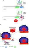
Model that questions the molecular significance of the two HIF-1α TAD and the impact of the oxygen sensor FIH on the intra-tumor location of gene expression as a function of the oxygen gradient. aUpper panel: If the two HIF-1α TAD are functionally different they would target different genes. In green are represented the potential N-TAD-only target genes while in blue are represented the potential C-TAD sensitive genes. Lower panel: By targeting specifically the C-TAD, FIH would inhibit only a subset of HIF-dependent genes. Consequently, FIH would not be a pure inhibitor but rather a switch between two categories of HIF spectrum genes. b In accordance with our working model, overexpression of FIH should delocalize C-TAD sensitive genes (blue dotted line) to highly hypoxic areas. In contrast FIH inhibition by siRNA should delocalize C-TAD sensitive genes to moderately hypoxic areas in the vicinity of blood vessels (lower panel). In parallel, N-TAD only genes (green dotted line) should not be sensitive to modulation of FIH activity
Fig. 4
Fig. 4
From oxygen to HIF and from HIF to oxygen. a A drop in the partial pressure of oxygen results in the inactivation of PHDs and FIH. Consequently the HIF pathway is activated, due respectively to inhibition of proteasomal degradation and a release of the C-TAD activity. b HIF in turn impacts on oxygen homeostasis by a double mechanism touching metabolism (green) and oxygen distribution/angiogenesis (red). HIF increases the glycolytic flux and represses the entrance of pyruvate into the Krebs cycle. The consequence is a decrease in oxygen consumption by mitochondrial respiration. In parallel HIF promotes angiogenesis via stimulation of vascular endothelial growth factor (VEGF), interleukin-A (IL-8) and angiopoitin-2 (Ang-2) leading to endothelial cell stimulation and blood vessel destabilization. In combination, HIF simultaneously increases tissue perfusion and decreases local oxygen consumption, thus promoting oxygen diffusion through hypoxic areas
Fig. 5
Fig. 5
From nutrients to HIF and from HIF to metabolism. a Two different pathways involved in the response to nutrients impact on HIF. First a decrease in the amount of glucose can inhibit the activity of FIH and PHD through decreased production of the co-substrate 2-oxoglutarate by the Krebs cycle. However, it is not sure that it is limiting in vivo. The question as to whether FIH and PHD might be nutritional sensors is still open to discussion. The second pathway responds to nutrient depletion reflecting a decrease in the ATP/AMP ratio. This nutritional stress activates the LKB1/AMPK/TSC1/TSC2 pathway resulting in an inhibition of mammalian target of rapamycin (mTOR). The action of mTOR on HIF is still a subject of debate. Essentially two options have been proposed: inhibition of HIF translation and/or inhibition of proteasomal degradation. Both converge in the activation of HIF under nutrient depletion conditions. b HIF can in turn impact on metabolism and cellular energy due to induction of a variety of target genes (here in green). It promotes glucose import via glucose transporters (GLUT1, 3) and it increases the rate of glucose consumption by inducing expression of glycolytic enzymes. HIF also promotes a shift to anaerobic metabolism by: favoring conversion of pyruvate to lactate through enhanced expression of the enzyme LDH-A and by increased expression of PDK-1 that counteracts the entrance of pyruvate into the Krebs cycle. Finally, through REDD-1, HIF stimulates the TSC1/2 complex, creating a negative feedback loop on mTOR. In summary, HIF allows cancer cells to shift to highly glycolytic anaerobic metabolism and to save energy by downregulating translation in a mTOR-dependent fashion
Fig. 6
Fig. 6
From pH to HIF and from HIF to pH. a It has been proposed that acidosis could protect HIFα from proteasomal degradation by sequestering the component of the VHL E3 ubiquitin ligase complex in nucleoli. b HIF acts both directly and indirectly on the pH. On the one hand, by stimulating glycolysis (green) it favors the production of lactate and protons that could potentially acidify the intracellular medium. On the other hand HIF promotes intracellular alkalinization (yellow) by activating monocarboxylate transporter (MCT1, 4) and Na+/H+ exchanger (NHE1, 6) that are able to evacuate lactate and protons, respectively. Simultaneously, the membrane carbonic anhydrases (CA) CA IX and CA XII isoforms acidify the extracellular matrix by converting CO2 and H2O into HCO3- and H+. Our group hypothesizes that while protons acidify the extracellular medium, bicarbonate could be recaptured in order to alkalinize the cell. In summary HIF compensates for the intracellular production of lactic acid associated with anaerobic metabolism by a process of intracellular alkalinization that is linked to extracellular acidification
Fig. 7
Fig. 7
From the extracellular matrix to HIF and from HIF to the extracellular matrix. a The extracellular matrix (ECM) is an essential component of the physicochemical environment of the cell, and includes fibrous proteins, glycoconjugates, growth factors and hormones. The chemical composition of the ECM can impact on HIF through growth factor stimulation. It has been shown that growth factors, by activating phosphatidylinositol-3-kinase (PI3K), can target mTOR and consequently HIF. As discussed above, the action of mTOR on HIF is a subject of debate. Essentially two options have been proposed: inhibition of HIF translation and/or inhibition of proteasomal degradation. Both converge to activate HIF. Growth factors could also activate HIF through the extracellular-regulated kinase (ERK) pathway. It has been proposed that phosphorylation of HIF by ERK could indirectly increase HIF activity by inhibiting the export of this transcription factor from the nucleus. As a consequence the activation of HIF target genes would be more efficient when HIF is phosphorylated. Moreover the vasoactive hormone angiotensin II has been shown to promote HIF-1α transcription through a protein kinase B (PKB) dependent mechanism. Finally, the physical density of the ECM could also indirectly influence the HIF pathway by modulating the oxygen diffusion length (not shown here). b A variety of HIF-induced genes have been shown to directly play a role on cell motility, invasion and extracellular matrix modulation (blue). A hypothetical mechanism for the loss of cell junctions under hypoxia could implicate the HIF-dependent activation of lysyl oxidase-like 2 (LOXL2) leading to stimulation of Snail. This transcriptional inhibitor could then downregulate E-cadherin (E-cad) and promote invasion. However, the subject of the ECM is also linked to other HIF-dependent actions, for instance, extracellular acidification could participate in mechanisms of invasion. Finally, the growth of neo-vessels, under the control of HIF, is also a crucial element in the penetration of cancer cells into the circulation. Autocrine motility factor (AMF); chemokine receptor CXCR4; receptor tyrosine kinase c-Met; lysyl oxidase (LOX); matrix metalloproteases (MMP)
Fig. 8
Fig. 8
Cyclic dynamics of tumor growth and putative anti-cancer approaches. Massive tumor cell proliferation leads to the formation of hypoxic zones at the periphery of blood vessels. The hypoxic signal stimulates a series of adaptation genes controlled by both the N-TAD and C-TAD of HIF-1α. Consequently, a set of adaptative changes (for instance involved in angiogenesis) make the microenvironment permissive for cell proliferation (for instance by reoxygenating hypoxic areas). From a therapeutic point of view, there are different ways to break this cycle. Classical chemotherapy inhibits cell proliferation but results in severe patient side-effects. Anti-angiogenics aim at blocking one feature of this adaptation phenomenon: the growth of new blood vessels. An alternative strategy consists in abolishing the overall adaptation mechanism by disorganizing the HIF-target genes. In addition, FIH inhibitors would represent a new class of anti-cancer drugs. Such candidate molecular targets may lead to innovative anti-microenvironmental adaptation approaches

Similar articles

Cited by

References

    1. {'text': '', 'ref_index': 1, 'ids': [{'type': 'PubMed', 'value': '11181773', 'is_inner': True, 'url': 'https://pubmed.ncbi.nlm.nih.gov/11181773/'}]}
    2. Hockel M, Vaupel P (2001) Tumor hypoxia: definitions and current clinical, biologic, and molecular aspects. J Natl Cancer Inst 93:266–276 - PubMed
    1. {'text': '', 'ref_index': 1, 'ids': [{'type': 'PMC', 'value': 'PMC1820761', 'is_inner': False, 'url': 'https://pmc.ncbi.nlm.nih.gov/articles/PMC1820761/'}, {'type': 'PubMed', 'value': '17043737', 'is_inner': True, 'url': 'https://pubmed.ncbi.nlm.nih.gov/17043737/'}]}
    2. Padhani AR, Krohn KA, Lewis JS et al (2007) Imaging oxygenation of human tumours. Eur Radiol 17:861–872 - PMC - PubMed
    1. {'text': '', 'ref_index': 1, 'ids': [{'type': 'PubMed', 'value': '10919633', 'is_inner': True, 'url': 'https://pubmed.ncbi.nlm.nih.gov/10919633/'}]}
    2. Baish JW, Jain RK (2000) Fractals and cancer. Cancer Res 60:3683–3688 - PubMed
    1. {'text': '', 'ref_index': 1, 'ids': [{'type': 'PMC', 'value': 'PMC549205', 'is_inner': False, 'url': 'https://pmc.ncbi.nlm.nih.gov/articles/PMC549205/'}, {'type': 'PubMed', 'value': '15701176', 'is_inner': True, 'url': 'https://pubmed.ncbi.nlm.nih.gov/15701176/'}]}
    2. Grizzi F, Russo C, Colombo P et al (2005) Quantitative evaluation and modeling of two-dimensional neovascular network complexity: the surface fractal dimension. BMC Cancer 5:14 - PMC - PubMed
    1. {'text': '', 'ref_index': 1, 'ids': [{'type': 'PubMed', 'value': '13130303', 'is_inner': True, 'url': 'https://pubmed.ncbi.nlm.nih.gov/13130303/'}]}
    2. Semenza GL (2003) Targeting HIF-1 for cancer therapy. Nat Rev Cancer 3:721–732 - PubMed

LinkOut - more resources