Vascular disease in mice with a dysfunctional circadian clock
- PMID: 19273720
- PMCID: PMC2761686
- DOI: 10.1161/CIRCULATIONAHA.108.827477
Vascular disease in mice with a dysfunctional circadian clock
Abstract
Background: Cardiovascular disease is the leading cause of death for both men and women in the United States and the world. A profound pattern exists in the time of day at which the death occurs; it is in the morning, when the endothelium is most vulnerable and blood pressure surges, that stroke and heart attack most frequently happen. Although the molecular components of circadian rhythms rhythmically oscillate in blood vessels, evidence of a direct function for the "circadian clock" in the progression to vascular disease is lacking.
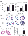
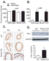
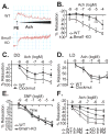
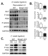
Methods and results: In the present study, we found increased pathological remodeling and vascular injury in mice with aberrant circadian rhythms, Bmal1-knockout and Clock mutant. In addition, naive aortas from Bmal1-knockout and Clock mutant mice exhibit endothelial dysfunction. Akt and subsequent nitric oxide signaling, a pathway critical to vascular function, was significantly attenuated in arteries from Bmal1-knockout mice.
Conclusions: Our data reveal a new role for the circadian clock during chronic vascular responses that may be of significance in the progression of vascular disease.
Figures


Comment in
-
Vascular rhythms and adaptation: do your arteries know what time it is?Circulation. 2009 Mar 24;119(11):1463-6. doi: 10.1161/CIRCULATIONAHA.108.847798. Epub 2009 Mar 9. Circulation. 2009. PMID: 19273717 Free PMC article. No abstract available.
Similar articles
-
Differential Regulation of BMAL1, CLOCK, and Endothelial Signaling in the Aortic Arch and Ligated Common Carotid Artery.J Vasc Res. 2016;53(5-6):269-278. doi: 10.1159/000452410. Epub 2016 Dec 7. J Vasc Res. 2016. PMID: 27923220 Free PMC article.
-
Genetic components of the circadian clock regulate thrombogenesis in vivo.Circulation. 2008 Apr 22;117(16):2087-95. doi: 10.1161/CIRCULATIONAHA.107.739227. Epub 2008 Apr 14. Circulation. 2008. PMID: 18413500
-
Dual role of the CLOCK/BMAL1 circadian complex in transcriptional regulation.FASEB J. 2006 Mar;20(3):530-2. doi: 10.1096/fj.05-5321fje. Epub 2006 Jan 25. FASEB J. 2006. PMID: 16507766
-
[Synchronization and genetic redundancy in circadian clocks].Med Sci (Paris). 2008 Mar;24(3):270-6. doi: 10.1051/medsci/2008243270. Med Sci (Paris). 2008. PMID: 18334175 Review. French.
-
[Biology and genetics of circadian rhythm].Encephale. 2009 Jan;35 Suppl 2:S53-7. doi: 10.1016/S0013-7006(09)75534-4. Encephale. 2009. PMID: 19268171 Review. French.
Cited by
-
Interorgan rhythmicity as a feature of healthful metabolism.Cell Metab. 2024 Apr 2;36(4):655-669. doi: 10.1016/j.cmet.2024.01.009. Epub 2024 Feb 8. Cell Metab. 2024. PMID: 38335957 Review.
-
Circadian rhythm disorder: a potential inducer of vascular calcification?J Physiol Biochem. 2020 Nov;76(4):513-524. doi: 10.1007/s13105-020-00767-9. Epub 2020 Sep 18. J Physiol Biochem. 2020. PMID: 32945991 Review.
-
Small molecule modifiers of circadian clocks.Cell Mol Life Sci. 2013 Aug;70(16):2985-98. doi: 10.1007/s00018-012-1207-y. Epub 2012 Nov 16. Cell Mol Life Sci. 2013. PMID: 23161063 Free PMC article. Review.
-
From Cultured Vascular Cells to Vessels: The Cellular and Molecular Basis of Vascular Dysfunction in Space.Front Bioeng Biotechnol. 2022 Apr 5;10:862059. doi: 10.3389/fbioe.2022.862059. eCollection 2022. Front Bioeng Biotechnol. 2022. PMID: 35480977 Free PMC article. Review.
-
Disrupting the circadian clock: gene-specific effects on aging, cancer, and other phenotypes.Aging (Albany NY). 2011 May;3(5):479-93. doi: 10.18632/aging.100323. Aging (Albany NY). 2011. PMID: 21566258 Free PMC article. Review.
References
-
- Hossmann V, Fitzgerald GA, Dollery CT. Circadian rhythm of baroreflex reactivity and adrenergic vascular response. Cardiovasc Res. 1980;14:125–129. - PubMed
-
- Millar-Craig MW, Bishop CN, Raftery EB. Circadian variation of blood-pressure. Lancet. 1978;1:795–797. - PubMed
-
- Panza JA, Epstein SE, Quyyumi AA. Circadian variation in vascular tone and its relation to alpha-sympathetic vasoconstrictor activity. N Engl J Med. 1991;325:986–990. - PubMed
-
- Boggild H, Knutsson A. Shift work, risk factors and cardiovascular disease. Scand J Work Environ Health. 1999;25:85–99. - PubMed
-
- Pierdomenico SD, Lapenna D, Guglielmi MD, Costantini F, Romano F, Schiavone C, Cuccurullo F, Mezzetti A. Arterial disease in dipper and nondipper hypertensive patients. Am J Hypertens. 1997;10:511–518. - PubMed
Publication types
MeSH terms
Substances
Grants and funding
LinkOut - more resources
Full Text Sources
Other Literature Sources
Molecular Biology Databases

