. 2009 May;29(10):2704-15.
doi: 10.1128/MCB.01811-08. Epub 2009 Mar 9.

The WD40 repeat protein WDR-23 functions with the CUL4/DDB1 ubiquitin ligase to regulate nuclear abundance and activity of SKN-1 in Caenorhabditis elegans

Affiliations

The WD40 repeat protein WDR-23 functions with the CUL4/DDB1 ubiquitin ligase to regulate nuclear abundance and activity of SKN-1 in Caenorhabditis elegans

Keith P Choe et al. Mol Cell Biol. 2009 May.

Abstract

The transcription factor SKN-1 protects Caenorhabditis elegans from stress and promotes longevity. SKN-1 is regulated by diverse signals that control metabolism, development, and stress responses, but the mechanisms of regulation and signal integration are unknown. We screened the C. elegans genome for regulators of cytoprotective gene expression and identified a new SKN-1 regulatory pathway. SKN-1 protein levels, nuclear accumulation, and activity are repressed by the WD40 repeat protein WDR-23, which interacts with the CUL-4/DDB-1 ubiquitin ligase to presumably target the transcription factor for proteasomal degradation. WDR-23 regulates SKN-1 target genes downstream from p38 mitogen-activated protein kinase, glycogen synthase kinase 3, and insulin-like receptor pathways, suggesting that phosphorylation of SKN-1 may function to modify its interaction with WDR-23 and/or CUL-4/DDB-1. These findings define the mechanism of SKN-1 accumulation in the cell nucleus and provide a new mechanistic framework for understanding how phosphorylation signals are integrated to regulate stress resistance and longevity.


Figures
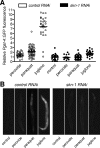
FIG. 1.
FIG. 1.
gst-4 transcription is regulated by SKN-1-dependent pathways that are activated by stress. Shown are relative Pgst-4::GFP fluorescence (A) and representative fluorescence micrographs (B) of worms fed control or skn-1 dsRNA and exposed to peroxide (5 mM for 20 min), paraquat (35 mM for 1 h), or juglone (38 μM for 1 h). Fluorescence was measured 6 to 8 h after exposure. Individual data points are plotted to illustrate variability. Means are denoted by lines (n = 48 to 152 worms). All three stressors induced significant (P < 0.001) Pgst-4::GFP fluorescence in control(RNAi) worms. RNAi knockdown of skn-1 significantly (P < 0.01) suppressed Pgst-4::GFP induction by all three stressors.
FIG. 2.
FIG. 2.
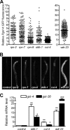
gst-4 transcription is repressed by the proteasome, DDB-1, CUL-4, and WDR-23. Shown are relative Pgst-4::GFP fluorescence (A) and representative fluorescence micrographs (B) of worms fed bacteria producing dsRNA to proteasome components (rpn-2, rpn-7, and rpn-8), ddb-1, cul-4, or wdr-23. RNAi or juglone exposure (Fig. 1) induced Pgst-4::GFP expression in multiple tissues. The strongest induction was observed in the intestine. (A) Individual data points are plotted to illustrate variability. Means are denoted by lines (n = 206 to 649 worms) and were all significantly (P < 0.001) different from a normalized value of 1.0 for control dsRNA-fed worms. (C) Relative gst-4 and gst-30 mRNA levels in worms fed bacteria producing dsRNA to ddb-1, cul-4, or wdr-23. The values are means plus standard errors (n = 4 populations of 20 to 30 worms). *, P < 0.05; **, P < 0.01; and ***, P < 0.001 compared to a normalized value of 1.0 for control dsRNA-fed worms.
FIG. 3.
FIG. 3.
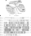
WDR-23 is evolutionarily conserved. (A) A phylogenetic tree was constructed using the neighbor-joining method with Poisson correction. BLAST searches were used to find the three closest homologues of WDR-23 in H. sapiens (human), D. melanogaster (fly), C. elegans (worm), A. thaliana (plant), and N. crassa (fungus). Human and C. elegans proteins are listed by their names; all other proteins are listed by their GenBank accession numbers. The Arabidopsis protein NP_176316 was the most divergent protein and was used to root the tree. G protein β polypeptide 3, one of the best characterized WD40-repeat proteins, is included for reference. The scale bar represents the fraction of amino acids replaced per site. (B) ClustalW alignment of C. elegans (Ce) full-length WDR-23a and WDR-23b with human (h) WDR23.1 and WDR23.2. Shading indicates conserved amino acids. Seven WD40 repeats are highlighted with solid boxes. The DWD box is highlighted with a dashed box. Asterisks indicate cysteine adjacent to a basic amino acid residue.
FIG. 4.
FIG. 4.
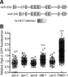
CUL-4, DDB-1, and the proteasome function with WDR-23. (A) The wdr-23 gene is predicted to encode two splice variants that differ at their amino termini (58). The tm1817 allele is a 635-bp deletion that completely removes exon 6 of the longer splice variant, WDR-23a, which is equivalent to exon 5 of the shorter splice variant, WDR-23b. This deletion is predicted to cause a frameshift and to encode a nonfunctional protein. (B) Effect of RNAi of rpn-2, rpn-7, rpn-8, ddb-1, cul-4, or F28D1.1 on Pgst-4::GFP fluorescence in wdr-23(tm1817) deletion mutants. Fluorescence is reported relative to that of deletion mutants fed control dsRNA bacteria. Individual data points are plotted to illustrate variability. Means are denoted by lines (n = 94 to 598 worms). ***, P < 0.001 greater than a normalized value of 1.0 for control dsRNA-fed wdr-23(tm1817) worms.
FIG. 5.
FIG. 5.
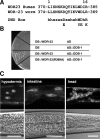
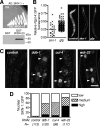
WDR-23 is expressed in cell nuclei and interacts with DDB-1. (A) Alignment of DWD boxes in human WDR23 and worm WDR-23 with the DWD box motif (h, hydrophobic; s, small; x, any residue). (B) Photograph of a histidine dropout- and 40 mM 3-amino-1,2,4-triazole-containing yeast plate streaked with yeast colonies (strain MaV203) coexpressing the GAL4 DNA binding domain (DB) fused to WDR-23 and the GAL4 activation domain (AD) alone; DB alone and AD fused to DDB-1; DB fused to WDR-23 and AD fused to DDB-1; or DB fused to a WDR-23 DWD box mutant (R389A) and AD fused to DDB-1. (C) Paired fluorescence (top) and differential interference contrast (bottom) micrographs of worms expressing the complete full-length WDR-23 fused to GFP. The WDR-23::GFP reporter is expressed in the nuclei of cells in the hypodermis, intestine, and head. Scale bars, 20 μm.
FIG. 6.
FIG. 6.
WDR-23 interacts with and regulates nuclear accumulation of SKN-1. (A) (Top) Photograph of a histidine dropout- and 40 mM 3-amino-1,2,4-triazole-containing plate streaked with yeast colonies (strain MaV203) coexpressing the complete reading frame of SKN-1c fused to the GAL4 activation domain (AD) and either the GAL4 DNA binding domain alone (DB) or the GAL4 DNA binding domain fused to full-length WDR-23 (DB::WDR-23). SKN-1- and WDR-23-coexpressing yeast cells also tested positive for uracil dropout, LacZ, and 5-fluoorotic acid phenotypes (not shown). (Bottom) Anti-V5 Western blot of lysates from CHO cells coexpressing V5-tagged SKN-1c and either GST alone or GST fused to full-length WDR-23. (B) Relative Pgst-4::GFP fluorescence (left) and representative fluorescence micrographs (right) of wdr-23(tm1817) deletion worms fed skn-1 or GFP dsRNA-producing bacteria. Individual data points are plotted to illustrate variability. Means are denoted by lines (n = 37 to 248 worms) and were significantly (P < 0.001) different from a normalized value of 1.0 for control dsRNA-fed worms. (C) Representative fluorescence micrographs of SKN-1::GFP-expressing worms fed control, ddb-1, cul4, or wdr-23 dsRNA-producing bacteria. The arrowheads mark the locations of nuclear SKN-1::GFP. Scale bar, 20 μm. (D) Quantification of nuclear SKN-1::GFP. Low, no visible nuclear GFP; medium, nuclear GFP visible only in anterior and/or posterior intestinal cells; high, nuclear GFP visible throughout the intestine.
FIG. 7.
FIG. 7.
WDR-23 regulates SKN-1 protein levels. (A) skn-1 mRNA levels in worms fed control, ddb-1, cul4, or wdr-23 dsRNA-producing bacteria. The values are means plus standard errors (n = 4 populations of 20 to 30 worms). (B) Ponceau S total-protein stain (left) and anti-SKN-1 immunoblot (right) of lysates from worms fed bacteria producing control or skn-1 dsRNA. (C) Ponceau S total-protein stain (left) and anti-SKN-1 immunoblot of lysates from N2 and wdr-23(tm1817) worms. Quantification of total protein and SKN-1 levels is shown on the far right. The values are means plus standard errors (n = 4 populations of ∼1,000 worms). **, P < 0.01 compared to N2.
FIG. 8.
FIG. 8.
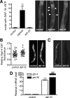
WDR-23 does not regulate DAF-16. (A) Numbers of nuclei with visible nuclear DAF-16::GFP expression (left) and representative fluorescence micrographs (right) of DAF-16::GFP-expressing worms treated with juglone (38 μM for 1 h) or fed bacteria producing wdr-23 dsRNA. Nuclear DAF-16::GFP was assessed in cells located between the pharynx and vulva. The values are means plus standard errors (n = 5 worms). **, P < 0.01 compared to control worms. (B) Relative Pgst-4::GFP fluorescence (left) and representative fluorescence micrographs (right) of wdr-23(tm1817) deletion mutants fed daf-16 dsRNA-producing bacteria. Individual data points are plotted to illustrate variability. Means are denoted by lines (n = 80 to 106 worms). (C) Representative fluorescence micrographs of DAF-16::GFP-expressing worms treated with control or daf-16 RNAi. Knockdown of DAF-16::GFP verified the efficiency of daf-16 RNAi. (D) Relative gst-4 and gst-30 mRNA levels in daf-16(mu86) worms fed bacteria producing wdr-23 dsRNA. The values are means plus standard errors (n = 4 populations of 20 to 30 worms). ***, P < 0.001.
FIG. 9.
FIG. 9.
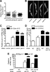
Stress, GSK-3, DAF-2, and SEK-1 may function upstream from WDR-23. Shown are relative Pgst-4::GFP fluorescence (A) and representative fluorescence micrographs (B) of wdr-23(tm1817) deletion mutants exposed to peroxide (5 mM for 20 min), paraquat (35 mM for 1 h), or juglone (38 μM for 1 h). Fluorescence was measured 6 to 8 h after exposure. Individual data points are plotted to illustrate variability. Means are denoted by lines (n = 137 to 269 worms). (C) Relative gst-4 mRNA levels in N2 and wdr-23(tm1817) worms fed bacteria producing control or gsk-3 dsRNA. (D) Relative gst-4 mRNA levels in N2 and daf-16(mgDf47);daf-2(e1370) worms fed bacteria producing control or wdr-23 dsRNA. (E) Relative gst-4 and gst-30 mRNA levels in N2 and sek-1(km4) worms fed bacteria producing control or wdr-23 dsRNA. The values in panels C, D, and E are means plus standard errors (n = 3 or 4 populations of 20 to 30 worms).
FIG. 10.
FIG. 10.
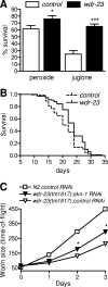
WDR-23 regulates stress resistance and longevity. (A) Percent survival of control and wdr-23 dsRNA-fed worms exposed to peroxide (5 mM for 30 min) or juglone (100 μM for 1 h). Survival was measured 24 h after exposure. The values are means plus standard errors (n = 8 populations of 22 to 143 worms). *, P < 0.05, and ***, P < 0.001 compared to control worms. (B) Longevity curves for control and wdr-23(RNAi) worms. Median longevities were 20 and 24 days for control and wdr-23 dsRNA-fed worms, respectively, and were significantly (P < 0.0001) different (n = 251 to 261 total worms from two experiments). A total of 26 control and 11 wdr-23(RNAi) worms were censored because of intestinal protrusion through the vulva. Day zero corresponds to worms at the L1 stage. Experiments were performed in RNAi-hypersensitive eri-1 mutant worms. (C) Loss of WDR-23 slows growth in a SKN-1-dependent manner. Relative worm length (time of flight) was measured with a COPAS Biosort beginning with eggs at day 0 and synchronized L1 larvae at day 1. The values are means plus standard errors (n = 99 to 803 worms). *, P < 0.001.
FIG. 11.
FIG. 11.
Working model of WDR-23 regulation of SKN-1. (A) We postulate that SKN-1 constitutively enters the nucleus in unstressed cells but does not accumulate or activate gene transcription because WDR-23 recruits it to CUL-4/DDB-1 ubiquitin ligase complexes, where it is presumably targeted for degradation by the proteasome. GSK-3- and the DAF-2-regulated kinases may inhibit SKN-1 activity upstream from WDR-23 (Fig. 9). (B) In cells exposed to oxidants or xenobiotics, SKN-1 bypasses WDR-23-mediated inhibition and thereby accumulates in the nucleus and activates the transcription of target genes (e.g., gst-4 and gst-30). SEK-1 functions upstream from WDR-23 (Fig. 9), suggesting that phosphorylation of SKN-1 by the p38 MAPK pathway may inhibit its binding with WDR-23 and/or ubiquitinylation. It is also possible that oxidants or xenobiotics may inhibit WDR-23 by other mechanisms (see Discussion).

Similar articles

Cited by

References

    1. An, J. H., and T. K. Blackwell. 2003. SKN-1 links C. elegans mesendodermal specification to a conserved oxidative stress response. Genes Dev. 171882-1893. - PMC - PubMed
    1. An, J. H., K. Vranas, M. Lucke, H. Inoue, N. Hisamoto, K. Matsumoto, and T. K. Blackwell. 2005. Regulation of the Caenorhabditis elegans oxidative stress defense protein SKN-1 by glycogen synthase kinase-3. Proc. Natl. Acad. Sci. USA 10216275-16280. - PMC - PubMed
    1. Angers, S., T. Li, X. Yi, M. J. MacCoss, R. T. Moon, and N. Zheng. 2006. Molecular architecture and assembly of the DDB1-CUL4A ubiquitin ligase machinery. Nature 443590-593. - PubMed
    1. Bernhardt, A., E. Lechner, P. Hano, V. Schade, M. Dieterle, M. Anders, M. J. Dubin, G. Benvenuto, C. Bowler, P. Genschik, and H. Hellmann. 2006. CUL4 associates with DDB1 and DET1 and its downregulation affects diverse aspects of development in Arabidopsis thaliana. Plant J. 47591-603. - PubMed
    1. Bishop, N. A., and L. Guarente. 2007. Two neurons mediate diet-restriction-induced longevity in C. elegans. Nature 447545-549. - PubMed

Publication types

MeSH terms

LinkOut - more resources