Tumor-promoting phenotype of CD90hi prostate cancer-associated fibroblasts
- PMID: 19267366
- PMCID: PMC2736596
- DOI: 10.1002/pros.20946
Tumor-promoting phenotype of CD90hi prostate cancer-associated fibroblasts
Abstract
Background: Cancer-associated stroma contributes to the malignant behavior of adenocarcinomas of the prostate and other organs. CD90 is a marker of mesenchymal stem cells (MSCs) and its expression is higher in prostate cancer stroma compared to normal tissue. Cultured prostate cancer-associated fibroblasts (CAFs) expressing high versus low levels of CD90 were analyzed for an MSC-like or tumor-promoting phenotype.
Methods: CD90(hi) and CD90(lo) cells were collected by fluorescence-activated cell sorting (FACS). Expression of genes associated with MSCs and/or tumor-promoting activities was measured by quantitative polymerase chain reaction (qPCR). Effects of stromal cell co-culture or conditioned media were tested on BPH-1 epithelial cells.
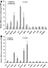
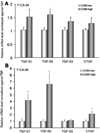
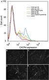
Results: The pattern of gene expression did not support the hypothesis that CD90(hi) cells were MSCs. However, CD90(hi) cells expressed higher levels of many genes associated with tumor promotion, including cytokines, angiogenic factors, hedgehog signaling components, and transforming growth factor (TGF)-beta. Co-culture or conditioned medium from CD90(hi) cells increased CXCR4 expression in BPH-1 cells, at least in part due to TGF-beta, and protected BPH-1 cells from apoptosis.
Conclusions: Our results suggest that the elevated expression of CD90 previously observed in the cancer-associated stroma of the human prostate is biologically significant. Although our results do not support the idea that CD90(hi) cells cultured from the cancer stroma are MSCs, our findings suggest that the phenotype of these cells is more tumor-promoting than that of cells expressing low CD90.
Figures


Similar articles
-
A bioengineered microenvironment to quantitatively measure the tumorigenic properties of cancer-associated fibroblasts in human prostate cancer.Biomaterials. 2013 Jul;34(20):4777-85. doi: 10.1016/j.biomaterials.2013.03.005. Epub 2013 Apr 2. Biomaterials. 2013. PMID: 23562048
-
Aberrant Transforming Growth Factor-β Activation Recruits Mesenchymal Stem Cells During Prostatic Hyperplasia.Stem Cells Transl Med. 2017 Feb;6(2):394-404. doi: 10.5966/sctm.2015-0411. Epub 2016 Sep 7. Stem Cells Transl Med. 2017. PMID: 28191756 Free PMC article.
-
Cathepsin D acts as an essential mediator to promote malignancy of benign prostatic epithelium.Prostate. 2013 Apr;73(5):476-88. doi: 10.1002/pros.22589. Epub 2012 Sep 19. Prostate. 2013. PMID: 22996917 Free PMC article.
-
Deciphering the role of stroma in pancreatic cancer.Curr Opin Gastroenterol. 2013 Sep;29(5):537-43. doi: 10.1097/MOG.0b013e328363affe. Curr Opin Gastroenterol. 2013. PMID: 23892539 Free PMC article. Review.
-
A historical perspective on the role of stroma in the pathogenesis of benign prostatic hyperplasia.Differentiation. 2011 Nov-Dec;82(4-5):168-72. doi: 10.1016/j.diff.2011.04.002. Epub 2011 Jun 30. Differentiation. 2011. PMID: 21723032 Free PMC article. Review.
Cited by
-
Novel preconditioning strategies for enhancing the migratory ability of mesenchymal stem cells in acute kidney injury.Stem Cell Res Ther. 2018 Aug 23;9(1):225. doi: 10.1186/s13287-018-0973-3. Stem Cell Res Ther. 2018. PMID: 30139368 Free PMC article. Review.
-
Therapeutic targeting of the prostate cancer microenvironment.Nat Rev Urol. 2010 Sep;7(9):494-509. doi: 10.1038/nrurol.2010.134. Nat Rev Urol. 2010. PMID: 20818327 Review.
-
Loss of stromal androgen receptor leads to suppressed prostate tumourigenesis via modulation of pro-inflammatory cytokines/chemokines.EMBO Mol Med. 2012 Aug;4(8):791-807. doi: 10.1002/emmm.201101140. Epub 2012 Jun 29. EMBO Mol Med. 2012. PMID: 22745041 Free PMC article.
-
Gene expression down-regulation in CD90+ prostate tumor-associated stromal cells involves potential organ-specific genes.BMC Cancer. 2009 Sep 8;9:317. doi: 10.1186/1471-2407-9-317. BMC Cancer. 2009. PMID: 19737398 Free PMC article.
-
Cancer Associated Fibroblasts: Naughty Neighbors That Drive Ovarian Cancer Progression.Cancers (Basel). 2018 Oct 29;10(11):406. doi: 10.3390/cancers10110406. Cancers (Basel). 2018. PMID: 30380628 Free PMC article. Review.
References
-
- Tuxhorn JA, Ayala GE, Rowley DR. Reactive stroma in prostate cancer progression. J Urol. 2001;166(6):2472–2483. - PubMed
-
- Cunha GR, Hayward SW, Wang YZ, Ricke WA. Role of the stromal microenvironment in carcinogenesis of the prostate. Int J Cancer. 2003;107(1):1–10. - PubMed
-
- Berry PA, Maitland NJ, Collins AT. Androgen receptor signalling in prostate: Effects of stromal factors on normal and cancer stem cells. Mol Cell Endocrinol. 2008;288(l–2):30–37. - PubMed
-
- Larue L, Bellacosa A. Epithelial-mesenchymal transition in development and cancer: Role of phosphatidylinositol 3′ kinase/AKT pathways. Oncogene. 2005;24(50):7443–7454. - PubMed
-
- Hugo H, Ackland ML, Blick T, Lawrence MG, Clements JA, Williams ED, Thompson EW. Epithelial-mesenchymal and mesenchymal-epithelial transitions in carcinoma progression. J Cell Physiol. 2007;213(2):374–383. - PubMed
Publication types
MeSH terms
Substances
Grants and funding
LinkOut - more resources
Full Text Sources
Other Literature Sources
Medical

