. 2009 Mar 9;184(5):677-90.
doi: 10.1083/jcb.200810091. Epub 2009 Mar 2.

Requirements for NuMA in maintenance and establishment of mammalian spindle poles

Affiliations

Requirements for NuMA in maintenance and establishment of mammalian spindle poles

Alain D Silk et al. J Cell Biol. .

Abstract

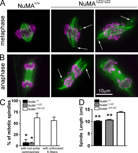
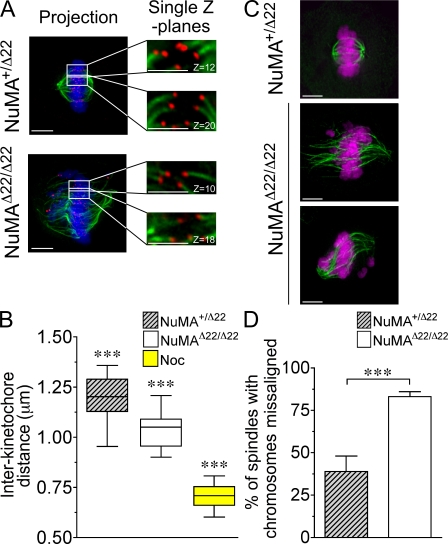
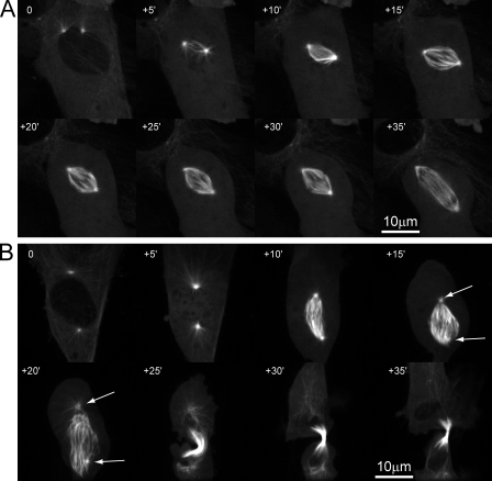
Microtubules of the mitotic spindle in mammalian somatic cells are focused at spindle poles, a process thought to include direct capture by astral microtubules of kinetochores and/or noncentrosomally nucleated microtubule bundles. By construction and analysis of a conditional loss of mitotic function allele of the nuclear mitotic apparatus (NuMA) protein in mice and cultured primary cells, we demonstrate that NuMA is an essential mitotic component with distinct contributions to the establishment and maintenance of focused spindle poles. When mitotic NuMA function is disrupted, centrosomes provide initial focusing activity, but continued centrosome attachment to spindle fibers under tension is defective, and the maintenance of focused kinetochore fibers at spindle poles throughout mitosis is prevented. Without centrosomes and NuMA, initial establishment of spindle microtubule focusing completely fails. Thus, NuMA is a defining feature of the mammalian spindle pole and functions as an essential tether linking bulk microtubules of the spindle to centrosomes.


Figures

Figure 1.
Figure 1.
Creation of conditional and disrupted NuMA alleles. (A) Schematic representations of (i) a portion of the mouse NuMA gene including exons 16–25 and EcoRI restriction sites, (ii) the exon 22 targeting vector showing the neomycin resistance (Neo) and diphtheria toxin (DT) cassettes and placement of loxP and FRT sites, (iii) the structure of the correctly targeted allele with the introduced SacII restriction site and locations of genotyping primers, (iv) the conditional allele (flox) produced by Flp-enhanced recombinase–mediated recombination of FRT sites flanking Neo, and (v) the deletion allele (Δ) produced by Cre recombination of loxP sites surrounding exon 22. Red bars indicate exon 22. (B) Genomic DNA blotting from neomycin-resistant ES clones after digestion with either EcoRI alone or EcoRI and SacII in combination and hybridization with the 5′ probe shown in A. (C) Predicted PCR fragment sizes for wild-type, Neo, flox, and Δ22 alleles of NuMA using primer sets shown in A. (D) PCR products from neomycin-resistant ES clones using primers i, ii, and iv. (E) PCR products from mouse tail DNA using primers i, ii, and iii. (F) Four independently targeted ES clones and a dilution series blotted with antibodies to NuMA and tubulin. NR, nonrecombined; HR, homologously recombined.
Figure 2.
Figure 2.
Tamoxifen-induced disruption of NuMA inhibits proliferation of primary embryo fibroblasts. (A) Timeline showing experimental design in which confluent primary cells are treated with 0.1 µM 4-OHT in 2% serum for 48 h. Cells were washed and maintained in 2% serum for 48 h before trypsinization and dilution into media containing 15% serum for subsequent analysis. (B) Conversion of NuMAflox to NuMAΔ22 in two independent primary NuMAflox/flox cell lines carrying the Cre-ERTM transgene. Recombination was monitored by PCR 48 h after treatment with 4-OHT. (C) qPCR using primers within the floxed region of NuMA was used to measure the efficiency of Cre-mediated recombination in the genomic DNA of fibroblasts. (D) Immunoblotting of NuMA and tubulin in NuMAflox/flox,Cre-ERTM and a dilution series of NuMA+/+,Cre-ERTM fibroblasts at day 4 of the experimental timeline. (E) Growth curves of primary fibroblasts after 4-OHT–mediated NuMA deletion; n = 3–4 experiments per cell line. Time in days follows timeline shown in A. (F) Mitotic index of primary MEFs treated with 4-OHT. For each genotype, >2,000 cells were counted in two separate cell lines. (G) Duration of mitosis in wild-type (+/+, Cre) and NuMA-disrupted (flox/flox, Cre) immortalized embryo fibroblasts. Results represent the mean of two independent experiments. Error bars represent SEM.
Figure 3.
Figure 3.
Spindle defects in primary NuMAΔ22/Δ22 fibroblasts. Primary fibroblasts were processed for immunofluorescence on experimental day 5, as shown in Fig. 2 A. (A) Example of metaphase in a control cell (NuMA+/+) and two exon 22–deleted (NuMAΔ22/Δ22) primary fibroblasts. Arrows indicate centrosomes. (B) Anaphase in wild-type and two NuMAΔ22/Δ22 primary cells. Each image represents a maximum intensity projection of a deconvolved series of z sections spanning the entire cell in 0.2-µm intervals. Arrows indicate centrosomes. Tubulin is shown in green, and phosphorylated histone H3 is shown in purple. (C) Frequencies of spindle–centrosome dissociation and pole splaying defects seen in control and NuMA-depleted metaphase cells. Cells were scored as phenotypic if at least one centrosome was nonpolar or at least one pole displayed an obvious lack of microtubule focusing. Two independent cell lines were examined per genotype, with >50 cells counted for NuMA+/+ and >130 cells for each of NuMA+/Δ22 and NuMAΔ22/Δ22 fibroblasts. (D) Spindle length in MG132-arrested primary fibroblasts measured as the linear distance between spindle poles or the approximate position of most spindle microtubule ends in defocused poles. At least 20 spindles per genotype were examined. *, P < 0.01 using Bonferroni's multiple comparison test and compared with NuMAΔ22/Δ22; **, P < 0.001. Error bars indicate SEM.
Figure 4.
Figure 4.
Reduced spindle tension and efficiency of chromosome alignment in the absence of NuMA. (A) Distance between sister kinetochores in cells arrested in metaphase with MG132 and incubated on ice for 10 min to selectively depolymerize nonkinetochore fiber microtubules. Images represent maximum intensity projections of a deconvolved series of z sections spanning the entire cell in 0.2-µm intervals (projection) or single deconvolved z sections. Kinetochore pairs were identified in single z sections by the relative positioning of kinetochores and orientation of associated kinetochore fibers. Blue, DNA; green, microtubules; red, kinetochores. Bars: (left) 5 µm; and (right) 2.5 µm. (B) Interkinetochore distances of paired sister chromatids in NuMA+/Δ22, NuMAΔ22/Δ22, and nocodazole (Noc)-treated control cells. The boxes represent the interquartile (middle 50%), and the whiskers represent the full range. Horizontal lines represent the median value. (C) Examples of NuMA+/Δ22 cells with fully aligned chromosomes and NuMAΔ22/Δ22 fibroblasts with chromosome alignment defects. Cells were treated as in A and processed for immunofluorescence to visualize DNA (purple) and tubulin (green). Bars, 5 µm. (D) Percentage of spindles showing chromosome alignments defects. ***, P < 0.0001. Error bars indicate SEM.
Figure 5.
Figure 5.
Bipolar spindle formation precedes centrosome detachment in the absence of mitotic NuMA function. (A and B) Selected images from videos of primary embryo fibroblasts transduced with retrovirus encoding tubulin-YFP, either heterozygous (A; Video 1, available at http://www.jcb.org/cgi/content/full/jcb.200810091/DC1) or homozygous (B; Video 2) for NuMAΔ22, undergoing the first mitosis after 4-OHT treatment and release from growth arrest. Arrows indicate centrosomes, and each time point shows a maximum intensity projection of five confocal fluorescence z sections acquired in 2-µm intervals.
Figure 6.
Figure 6.
Prometaphase centrosome separation and centrosome-independent spindle pole focusing require NuMA. (A) Primary MEFs were arrested in mitosis by treatment with STLC and processed for immunofluorescence. Green, α-tubulin; blue, DNA; red, γ-tubulin. (B) Frequencies of monopolar spindles as shown in A. Two independent cell lines per genotype were used, and >200 mitoses per genotype were counted. (C) Anaphase in NuMA+/Δ22 control and NuMAΔ22/Δ22 cells 1 h after washout of STLC. In merged images, DNA is shown in purple and γ-tubulin in green. Arrows indicate centrosomes. (D) Stills from videos of NuMA+/Δ22 (Video 3, available at http://www.jcb.org/cgi/content/full/jcb.200810091/DC1) and NuMAΔ22/Δ22 (Video 4) primary embryo fibroblasts transduced with retrovirus encoding tubulin-YFP. Cells were arrested with STLC for 3–4 h and filmed after washout of the drug. Scoring for centrosome separation was performed blinded to genotype. Each time point shows a maximum intensity projection of 12 confocal fluorescence z sections acquired in 1-µm intervals. Error bars indicate SEM.
Figure 7.
Figure 7.
Kinetochore–microtubule attachments are required for centrosome separation in prometaphase. (A) Immunoblot of an extract of HeLa cells 48 h after transfection with siRNA oligos against glyceraldehyde 3-phosphate dehydrogenase (GAPDH) or Nuf2. (B) Still images from videos of HeLa cells coexpressing H2B–monomeric RFP and tubulin-YFP. Cells were transfected with siRNA oligos against GAPDH or Nuf2 and 48 h later were arrested with STLC. Time is given in minutes relative to STLC washout. Images are maximum intensity projections of five confocal z sections spaced 2 µm apart. Green, tubulin; purple, histone H2B. (C) Frequency of spindles that remained monopolar 90 min after release from STLC in cells treated with siRNA to GAPDH or Nuf2. Error bars represent the mean and SEM of three separate experiments; n = 192 cells GAPDH siRNA; n = 228 cells Nuf2 siRNA; ***, P < 0.0001 by Fisher's exact test.

Similar articles

Cited by

References

    1. Chang W., Dynek J.N., Smith S. 2005. NuMA is a major acceptor of poly(ADP-ribosyl)ation by tankyrase 1 in mitosis.Biochem. J. 391:177–184 - PMC - PubMed
    1. Chatzimeletiou K., Morrison E.E., Prapas N., Prapas Y., Handyside A.H. 2008. The centrosome and early embryogenesis: clinical insights.Reprod. Biomed. Online. 16:485–491 - PubMed
    1. Cheeseman I.M., Desai A. 2005. A combined approach for the localization and tandem affinity purification of protein complexes from metazoans.Sci. STKE. - PubMed
    1. Compton D.A. 1998. Focusing on spindle poles.J. Cell Sci. 111(Pt 11)1477–1481 - PubMed
    1. Dasso M. 2001. Running on Ran: nuclear transport and the mitotic spindle.Cell. 104:321–324 - PubMed

Publication types

MeSH terms