

Membrane type 1 matrix metalloproteinase is a crucial promoter of synovial invasion in human rheumatoid arthritis
- PMID: 19248098
- PMCID: PMC2819053
- DOI: 10.1002/art.24331
Membrane type 1 matrix metalloproteinase is a crucial promoter of synovial invasion in human rheumatoid arthritis
Abstract
Objective: A hallmark of rheumatoid arthritis (RA) is invasion of the synovial pannus into cartilage, and this process requires degradation of the collagen matrix. The aim of this study was to explore the role of one of the collagen-degrading matrix metalloproteinases (MMPs), membrane type 1 MMP (MT1-MMP), in synovial pannus invasiveness.
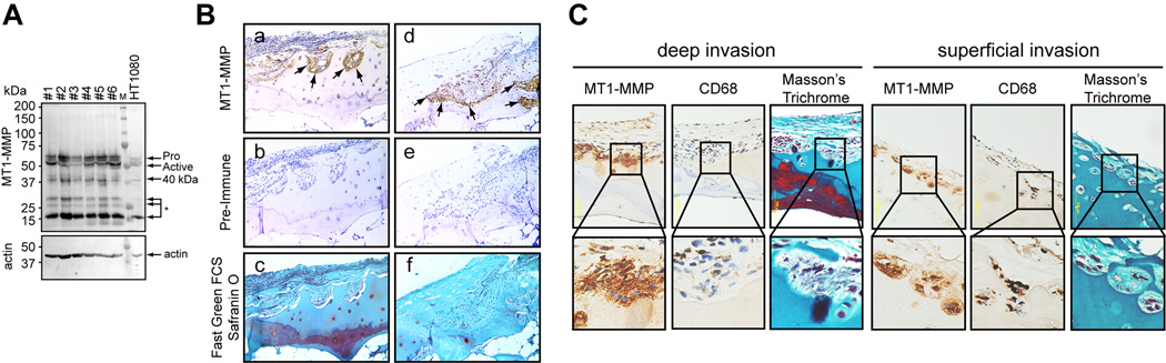
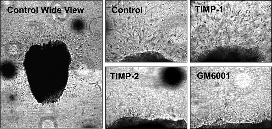
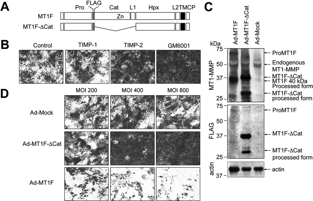
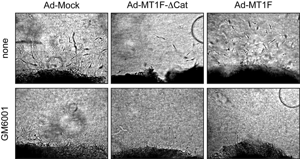
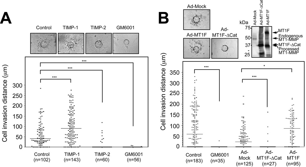
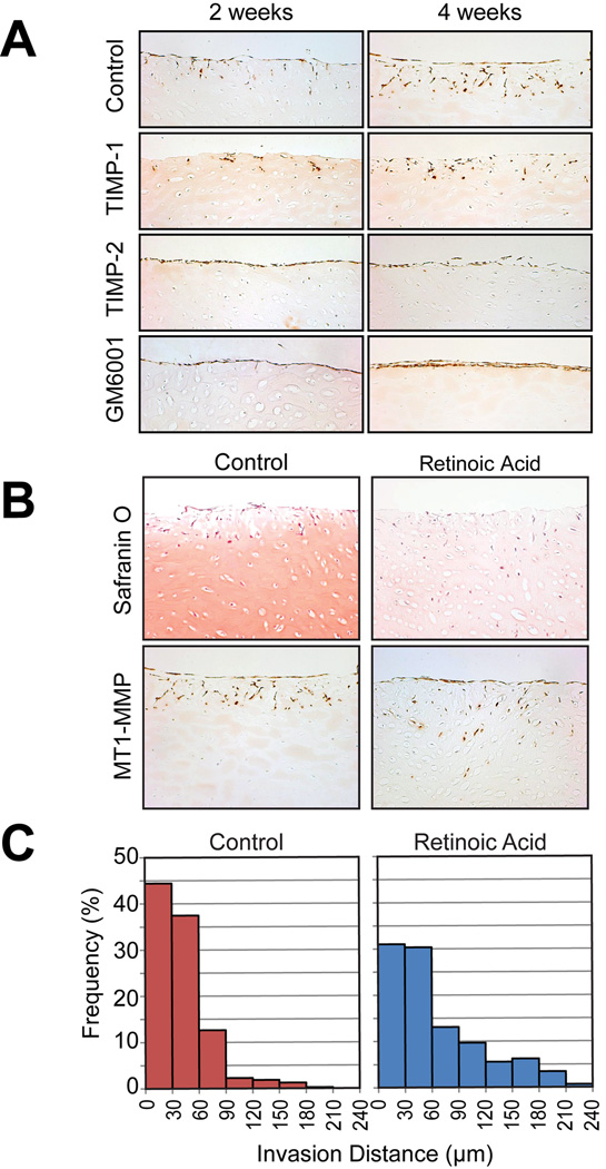
Methods: The expression and localization of MT1-MMP in human RA pannus were investigated by Western blot analysis of primary synovial cells and immunohistochemical analysis of RA joint specimens. The functional role of MT1-MMP was analyzed by 3-dimensional (3-D) collagen invasion assays and a cartilage invasion assay in the presence or absence of tissue inhibitor of metalloproteinases 1 (TIMP-1), TIMP-2, or GM6001. The effect of adenoviral expression of a dominant-negative MT1-MMP construct lacking a catalytic domain was also examined.
Results: MT1-MMP was highly expressed at the pannus-cartilage junction in RA joints. Freshly isolated rheumatoid synovial tissue and isolated RA synovial fibroblasts invaded into a 3-D collagen matrix in an MT1-MMP-dependent manner. Invasion was blocked by TIMP-2 and GM6001 but not by TIMP-1. Invasion was also inhibited by the overexpression of a dominant-negative MT1-MMP, which inhibits collagenolytic activity and proMMP-2 activation by MT1-MMP on the cell surface. Synovial fibroblasts also invaded into cartilage in an MT1-MMP-dependent manner. This process was further enhanced by removing aggrecan from the cartilage matrix.
Conclusion: MT1-MMP serves as an essential collagen-degrading proteinase during pannus invasion in human RA. Specific inhibition of MT1-MMP-dependent invasion may represent a novel therapeutic strategy for RA.
Figures
Similar articles
-
Invasive potential of human rheumatoid tenosynovial cells is in part MT1-MMP dependent.J Hand Surg Am. 2009 Sep;34(7):1282-90. doi: 10.1016/j.jhsa.2009.04.015. Epub 2009 Jul 12. J Hand Surg Am. 2009. PMID: 19596176
-
New collagenolytic enzymes/cascade identified at the pannus-hard tissue junction in rheumatoid arthritis: destruction from above.Matrix Biol. 1998 Dec;17(8-9):585-601. doi: 10.1016/s0945-053x(98)90110-x. Matrix Biol. 1998. PMID: 9923652
-
The arthritis severity locus Cia5d is a novel genetic regulator of the invasive properties of synovial fibroblasts.Arthritis Rheum. 2008 Aug;58(8):2296-306. doi: 10.1002/art.23610. Arthritis Rheum. 2008. PMID: 18668563 Free PMC article.
-
Coordinate action of membrane-type matrix metalloproteinase-1 (MT1-MMP) and MMP-2 enhances pericellular proteolysis and invasion.Cancer Sci. 2010 Apr;101(4):843-7. doi: 10.1111/j.1349-7006.2010.01498.x. Epub 2010 Jan 18. Cancer Sci. 2010. PMID: 20148894 Free PMC article. Review.
-
MT1-MMP as a Key Regulator of Metastasis.Cells. 2023 Aug 31;12(17):2187. doi: 10.3390/cells12172187. Cells. 2023. PMID: 37681919 Free PMC article. Review.
Cited by
-
Research Progress of Therapeutic Enzymes and Their Derivatives: Based on Herbal Medicinal Products in Rheumatoid Arthritis.Front Pharmacol. 2021 Mar 16;12:626342. doi: 10.3389/fphar.2021.626342. eCollection 2021. Front Pharmacol. 2021. PMID: 33796022 Free PMC article. Review.
-
Selective Inhibition of Membrane Type 1 Matrix Metalloproteinase Abrogates Progression of Experimental Inflammatory Arthritis: Synergy With Tumor Necrosis Factor Blockade.Arthritis Rheumatol. 2016 Feb;68(2):521-31. doi: 10.1002/art.39414. Arthritis Rheumatol. 2016. PMID: 26315469 Free PMC article.
-
The Use of Human Mesenchymal Stem Cells as Therapeutic Agents for the in vivo Treatment of Immune-Related Diseases: A Systematic Review.Front Immunol. 2018 Sep 11;9:2056. doi: 10.3389/fimmu.2018.02056. eCollection 2018. Front Immunol. 2018. PMID: 30254638 Free PMC article.
-
Proteases involved in cartilage matrix degradation in osteoarthritis.Biochim Biophys Acta. 2012 Jan;1824(1):133-45. doi: 10.1016/j.bbapap.2011.06.020. Epub 2011 Jul 8. Biochim Biophys Acta. 2012. PMID: 21777704 Free PMC article. Review.
-
Transient collagen triple helix binding to a key metalloproteinase in invasion and development.Structure. 2015 Feb 3;23(2):257-69. doi: 10.1016/j.str.2014.11.021. Structure. 2015. PMID: 25651059 Free PMC article.
References
-
- Murphy G, Nagase H. Reappraising metalloproteinases in rheumatoid arthritis and osteoarthritis: destruction or repair? Nat Clin Pract Rheumatol. 2008;4(3):128–135. - PubMed
-
- Sledge C, Reddi A, Walsh D, Blake D. Biology of the Normal Joint. In: S R, Harris EJ, CB S, editors. Kelly's Textbook of Rheumatology. 6 ed. Philadelphia: W.B: Saunders Company; 2001. pp. 1–26.
-
- Heinegård D, Lorenzo P, Saxne T. Matrix Glycoproteins, Proteoglycans, and Cartilage. In: S R, Harris EJ, CB S, editors. Kelly's Textbook of Rheumatology. 6 ed. Philadelphia: W.B: Saunders Company; 2001. pp. 41–53.
-
- Visse R, Nagase H. Matrix metalloproteinases and tissue inhibitors of metalloproteinases: structure, function, and biochemistry. Circ Res. 2003;92(8):827–839. - PubMed
Publication types
MeSH terms
Substances
Grants and funding
LinkOut - more resources
Full Text Sources
Medical
Research Materials
Miscellaneous
