

Invited article: an MRI-based approach to the diagnosis of white matter disorders
- PMID: 19237705
- PMCID: PMC2677542
- DOI: 10.1212/01.wnl.0000343049.00540.c8
Invited article: an MRI-based approach to the diagnosis of white matter disorders
Abstract
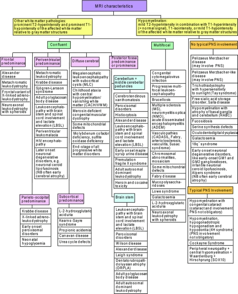
Background: There are many different white matter disorders, both inherited and acquired, and consequently the diagnostic process is difficult. Establishing a specific diagnosis is often delayed at great emotional and financial costs. The pattern of brain structures involved, as visualized by MRI, has proven to often have a high diagnostic specificity.
Methods: We developed a comprehensive practical algorithm that relies mainly on the characteristics of brain MRI.
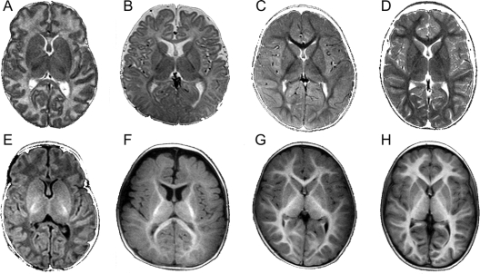
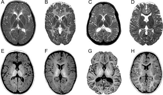
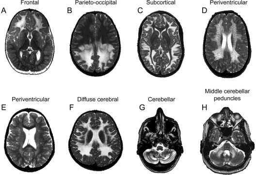
Results: The initial decision point defines a hypomyelination pattern, in which the cerebral white matter is hyperintense (normal), isointense, or slightly hypointense relative to the cortex on T1-weighted images, vs other pathologies with more prominent hypointensity of the cerebral white matter on T1-weighted images. In all types of pathology, the affected white matter is hyperintense on T2-weighted images, but, as a rule, the T2 hyperintensity is less marked in hypomyelination than in other pathologies. Some hypomyelinating disorders are typically associated with peripheral nerve involvement, while others are not. Lesions in patients with pathologies other than hypomyelination can be either confluent or isolated and multifocal. Among the diseases with confluent lesions, the distribution of the abnormalities is of high diagnostic value. Additional MRI features, such as white matter rarefaction, the presence of cysts, contrast enhancement, and the presence of calcifications, further narrow the diagnostic possibilities.
Conclusion: Application of a systematic decision tree in MRI of white matter disorders facilitates the diagnosis of specific etiologic entities.
Figures
Similar articles
-
Delayed encephalopathy of acute carbon monoxide intoxication: diffusivity of cerebral white matter lesions.AJNR Am J Neuroradiol. 2003 Sep;24(8):1592-7. AJNR Am J Neuroradiol. 2003. PMID: 13679276 Free PMC article.
-
Characterization of multiple sclerosis plaques with T1-weighted MR and quantitative magnetization transfer.AJNR Am J Neuroradiol. 1995 Aug;16(7):1473-9. AJNR Am J Neuroradiol. 1995. PMID: 7484636 Free PMC article.
-
Differential diagnostic approach to MR imaging of white matter diseases.Top Magn Reson Imaging. 2006 Aug;17(4):243-63. doi: 10.1097/01.rmr.0000248666.91834.af. Top Magn Reson Imaging. 2006. PMID: 17414999 Review.
-
[Cerebral white matter lesions: differential diagnosis on magnetic resonance imaging].Brain Nerve. 2015 Apr;67(4):467-74. doi: 10.11477/mf.1416200163. Brain Nerve. 2015. PMID: 25846595 Review. Japanese.
-
Inversion-recovery echo-planar MR in adult brain neoplasia.AJNR Am J Neuroradiol. 1998 Feb;19(2):267-73. AJNR Am J Neuroradiol. 1998. PMID: 9504476 Free PMC article.
Cited by
-
DARS-associated leukoencephalopathy can mimic a steroid-responsive neuroinflammatory disorder.Neurology. 2015 Jan 20;84(3):226-30. doi: 10.1212/WNL.0000000000001157. Epub 2014 Dec 19. Neurology. 2015. PMID: 25527264 Free PMC article.
-
LEUDEN Syndrome: A Novel Hypomyelinating Leukoencephalopathy in a 1-Year-Old Girl.Ann Indian Acad Neurol. 2024 Jul 1;27(4):467-469. doi: 10.4103/aian.aian_257_24. Epub 2024 Aug 16. Ann Indian Acad Neurol. 2024. PMID: 39150464 Free PMC article. No abstract available.
-
A Positive Association between the Atherogenic Index of Plasma and White Matter Hyperintensity.Korean J Fam Med. 2022 May;43(3):193-198. doi: 10.4082/kjfm.21.0129. Epub 2022 May 20. Korean J Fam Med. 2022. PMID: 35610965 Free PMC article.
-
AARS2 leukoencephalopathy: A new variant of mitochondrial encephalomyopathy.Mol Genet Genomic Med. 2019 Apr;7(4):e00582. doi: 10.1002/mgg3.582. Epub 2019 Jan 31. Mol Genet Genomic Med. 2019. PMID: 30706699 Free PMC article.
-
AIMP1/p43 mutation and PMLD.Am J Hum Genet. 2011 Mar 11;88(3):391; author reply 393-5. doi: 10.1016/j.ajhg.2011.02.003. Am J Hum Genet. 2011. PMID: 21397066 Free PMC article. No abstract available.
References
-
- van der Voorn JP, Pouwels PJ, Hart AA, et al. Childhood white matter disorders: quantitative MR imaging and spectroscopy. Radiology 2006;241:510–517. - PubMed
-
- Van der Knaap MS, Valk J. Magnetic Resonance of Myelination and Myelin Disorders, 3rd ed. Berlin: Springer; 2005.
-
- van der Knaap MS, Breiter SN, Naidu S, Hart AA, Valk J. Defining and categorizing leukoencephalopathies of unknown origin: MR imaging approach. Radiology 1999;213:121–133. - PubMed
-
- van der Knaap MS, Valk J, de Neeling N, Nauta JJ. Pattern recognition in magnetic resonance imaging of white matter disorders in children and young adults. Neuroradiol 1991;33:478–493. - PubMed
-
- Schiffmann R, Moller JR, Trapp BD, et al. Childhood ataxia with diffuse central nervous system hypomyelination. Ann Neurol 1994;35:331–340. - PubMed
Publication types
MeSH terms
Grants and funding
LinkOut - more resources
Full Text Sources
Other Literature Sources
Medical