. 2009 Mar 27;33(6):717-26.
doi: 10.1016/j.molcel.2009.01.026. Epub 2009 Feb 12.

An architectural role for a nuclear noncoding RNA: NEAT1 RNA is essential for the structure of paraspeckles

Affiliations

An architectural role for a nuclear noncoding RNA: NEAT1 RNA is essential for the structure of paraspeckles

Christine M Clemson et al. Mol Cell. .

Abstract

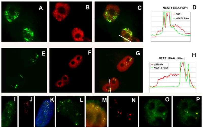
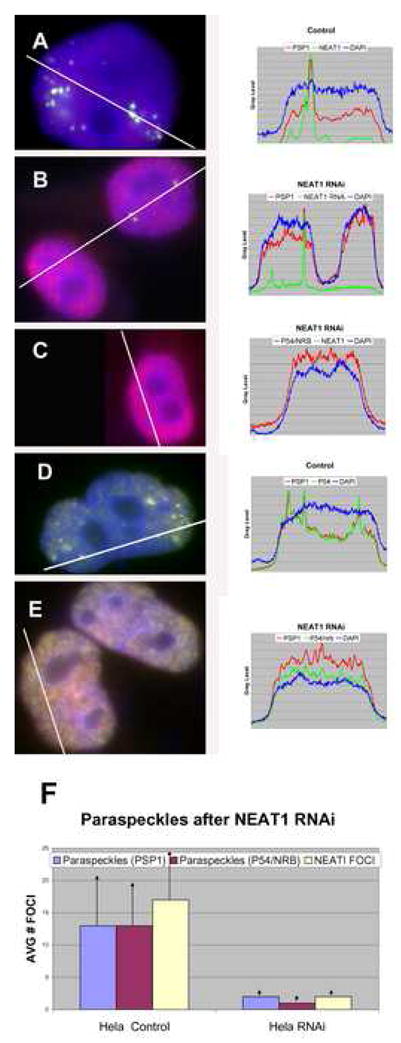
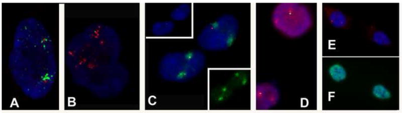
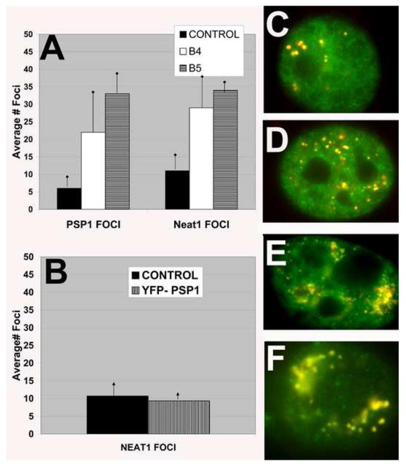
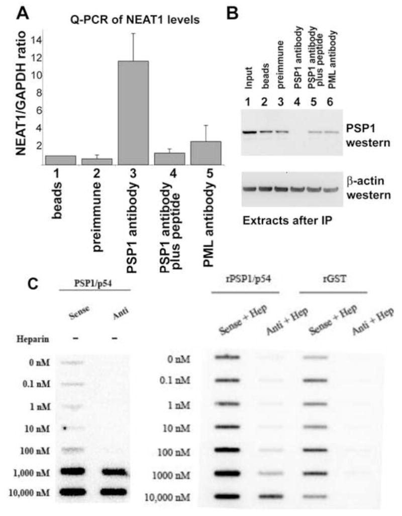
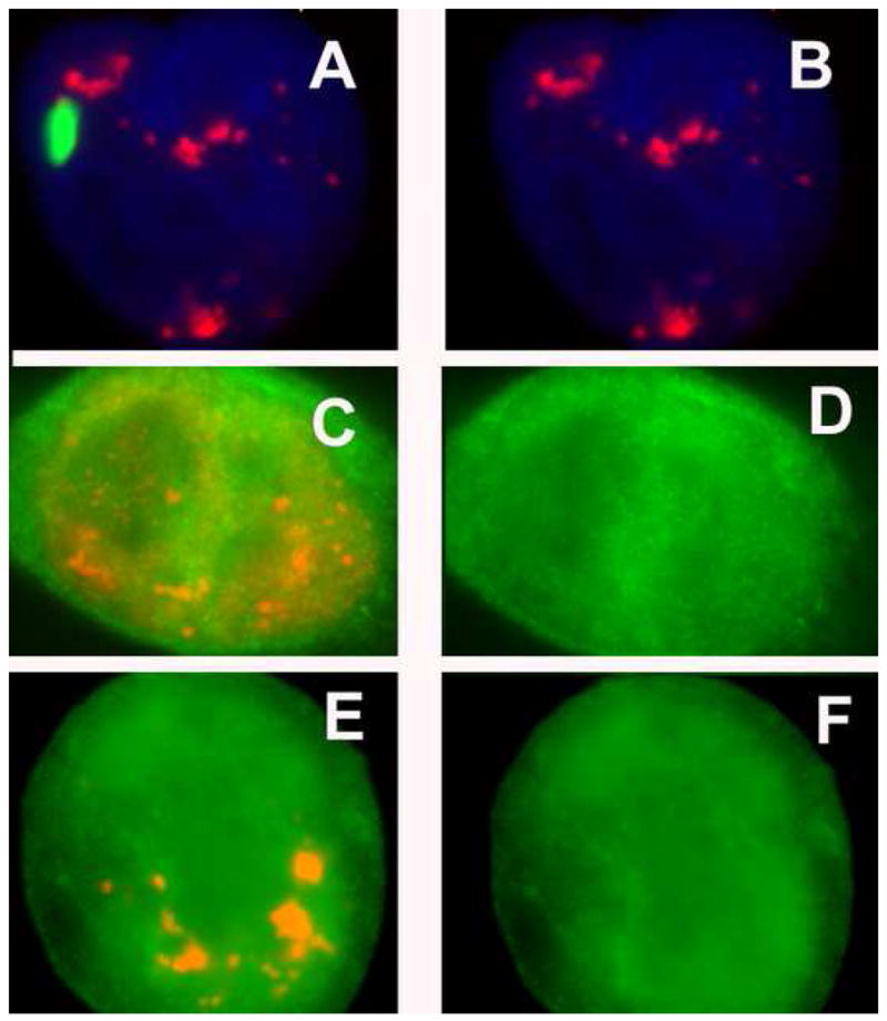
NEAT1 RNA, a highly abundant 4 kb ncRNA, is retained in nuclei in approximately 10 to 20 large foci that we show are completely coincident with paraspeckles, nuclear domains implicated in mRNA nuclear retention. Depletion of NEAT1 RNA via RNAi eradicates paraspeckles, suggesting that it controls sequestration of the paraspeckle proteins PSP1 and p54, factors linked to A-I editing. Unlike overexpression of PSP1, NEAT1 overexpression increases paraspeckle number, and paraspeckles emanate exclusively from the NEAT1 transcription site. The PSP-1 RNA binding domain is required for its colocalization with NEAT1 RNA in paraspeckles, and biochemical analyses support that NEAT1 RNA binds with paraspeckle proteins. Unlike other nuclear-retained RNAs, NEAT1 RNA is not A-I edited, consistent with a structural role in paraspeckles. Collectively, results demonstrate that NEAT1 functions as an essential structural determinant of paraspeckles, providing a precedent for a ncRNA as the foundation of a nuclear domain.


Figures

Figure 1
Figure 1
NEAT1 RNA Overlaps with Paraspeckles in Multiple Human and Mouse Cells. Colocalization of NEAT1 RNA with paraspeckle proteins. A–D) NEAT1 RNA (green) localizes as bright discrete foci with little nucleoplasmic signal in HeLa cells. B) PSP1, a marker for paraspeckles (red), gives a broad nucleoplasmic signal coupled with clusters of more intense foci C) Overlap (yellow) of the numerous NEAT1 RNA foci (green) colocalize precisely with the accumulations of PSP1 antibody (red) that demark paraspeckles. E–H) NEAT1 RNA foci (E: green) also precisely align with accumulations of p54 antibody (F: red) that demark paraspeckles (G: overlap) in HeLa cells. D, H) Linescan analysis demonstrates that NEAT1 RNA (green) is primarily in paraspeckle foci, with little nucleoplasmic signal, while paraspeckle proteins (red) are found throughout the nucleoplasm and concentrate in paraspeckles. NEAT1 RNA strictly and consistently overlaps with PSP1 antibody in all human cells examined including: I–K (same cell): Tig1. I) NEAT1 RNA (green), J) PSP1 (red), K) merged: Nucleus is stained with DAPI (blue); L) HT-1080 (NEAT1 RNA - green, PSP1 antibody, red); and M) 293 (NEAT1 RNA – green, PSP1 antibody -red). N–P) Mouse cells (MEFs) display fewer Neat1 foci (panel N red) which overlap and correspond with fewer PSP1 antibody foci (panel O green – merged images panel P).
Figure 2
Figure 2
Depletion of NEAT1 RNA by RNA inhibition causes Paraspeckles to Disappear NEAT1 RNA (green) and PSP1 (red) in cells transfected twice over 48 hours with control siRNA (A) and NEAT1 RNA siRNA at a concentration of 100nM using Lipofectamine RNAimax (Invitrogen). (B). A) NEAT1 RNA is found as multiple foci throughout the nucleus unlike markers for paraspeckles (PSP1 – red) which are also found throughout the nucleoplasm (linescan). B) This representative example shows that while RNAi is successful in reducing most NEAT1 RNA (green) signal (bottom cell), occasionally a few clusters of NEAT1 signal remain (top cell), typically associated with sites of new NEAT1 transcription (Figure S2F). PSP1 enrichment in paraspeckles occurs where the NEAT1 clusters remain, while the nucleoplasmic PSP1 (red) is elevated relative to control (linescan). C) NEAT1 RNA (green) and p54 (red) in cells transfected with NEAT1 siRNA. No foci of p54 are detected in NEAT1 RNA depleted cells, but the nucleoplasmic levels of p54 is elevated relative to the control (linescan). PSP1 (red) and p54 (green) in cells transfected with control siRNA (D) and NEAT1 RNA siRNA (E): D) PSP1 (red) and p54 (green), while found throughout the nucleoplasm, concentrate in paraspeckles. E) PSP1 and p54 no longer accumulate in paraspeckles in NEAT1 knockdowns, but nucleoplasmic levels of both proteins increase above the DAPI signal relative to control (linescan). F) Blind computer analysis and quantitation (see suppl. Methods) was used to count the number of paraspeckles using NEAT1 RNA, PSP1 or p54 antibody staining (bars represent standard error). All 3 paraspeckle markers show concomitant reduction in paraspeckle number after NEAT1 knockdown.
Figure 3
Figure 3
NEAT2/MALAT-1 RNAi Does not affect SC-35 Domains A) SC-35 domains (red) in HeLa cells overlap with B) NEAT2 RNA (green) in cells treated with control siRNA duplex. C) In cells inhibited for NEAT2 RNA (green), SC-35 domains (red) remain intact. Note that, similar to NEAT1 RNAi, while most of the NEAT2 RNA is effectively eliminated, occasionally some RNA associated with the site of transcription remains. D) Paraspeckles (PSP1 antibody -green) are unaffected by inhibition of NEAT2 RNA (red).
Figure 4
Figure 4
Paraspeckles Form as NEAT1 Foci Spread from the Site of Transcription. Dual NEAT1 RNA or PSP1 antibody and chr 11q13 detection. A) In interphase nuclei NEAT1 RNA foci (green) cluster near and spread from the site of transcription at 11q 13 (marked by the UHG locus–red). B) Similarly, paraspeckles (marked by PSP1 antibody – red) are often seen as a similar bipolar pattern that cluster and spread from 11q (UHG locus – green). C) In re-forming G1 daughter nuclei two large foci of NEAT1 RNA (red and top inset) are detected next to chromosome 11 (green and bottom inset). D) As G1 progresses, the earliest paraspeckles (PSP1 -red) are detected near 11q13 (UHG – green). E and F – (same cell) Later G1 daughter cells that have begun transcribing NEAT1 RNA (E: red) show bright nucleoplasmic PSP1 (F: green) signal and paraspeckle formation coincident with a single NEAT1 locus.
Figure 5
Figure 5
Overexpression of Neat1, but not PSP1, increases the number of Paraspeckles 3T3 or HeLa cells were transfected with full length mNeat1 or hYFP-PSP1 respectively (or an empty vector control). qPCR demonstrated that cell line B4 and B5 expressed Neat1 RNA at a level 20% and 50% above the control, respectively. Immunoblotting showed that the YFP-PSP1 is expressed at a level 4X higher than endogenous PSP1 (Fox et al., 2002). Quantitation of Neat1 foci and paraspeckles was performed using blind computer scoring (see suppl Methods.). A) Increasing levels of mNeat1 expression causes a concommitant increase in paraspeckle number (as detected with anti-PSP1). B) HeLa cells overexpressing YFP-PSP1 show no change in paraspeckle number. Both NEAT1 RNA and PSP1 antibody, which detects both endogenous and transfected full length YFP-PSP1 (Fox et al., 2002), were used to mark paraspeckles with similar results (not shown). Bars represent standard error. C–E) Colocalization of Neat1 RNA (red) with PSP1 antibody (green) in: C) control; D) B4 cells and E) B5 cells. F) Colocalization of NEAT1 RNA (red) with YFP-PSP1 (green) in HeLa cells.
Figure 6
Figure 6
NEAT1 binds with paraspeckle proteins in vivo and in vitro. (A) NEAT1 and GAPDH RT-PCR analyzed by quantitative PCR (qPCR). Lane 1: beads alone, lane 2: rabbit preimmune serum, lane 3 anti PSP1 serum, lane 4: anti PSP1 serum pre-bound with the cognate peptide the antibody was raised against, lane 5: anti PML. The NEAT1 q-PCR data is based on amplification of a region at the 5′ end of NEAT1 (see Exptl. Proc.). The q-PCR data shown is derived from two individual IPRTPCR experiments, in which each sample was analyzed in duplicate. To control for non-specific binding, values were first normalized against the q-PCR GAPDH levels of the same samples and then adjusted such that the NEAT1 levels of the beads alone control had a value of 1. (B) Western blotting shows that only the PSP1 IP depletes PSP1 from a nuclear extract. Equal amounts of extract before (lane 1) and after the IP (lanes 2–6) were transferred to a membrane and probed for the presence of PSP1 (upper panel) and beta-actin (lower panel). (C) Nitrocellulose filter binding assays show that in vitro transcribed NEAT1 RNA binds with increasing amounts of recombinant PSP1/p54 (left panel). Heparin, which abolishes non-specific RNA-protein interactions (Wang and Gegenheimer, 1990), does not affect the binding of the sense strand of NEAT1 RNA to the recombinant paraspeckle proteins, but does significantly diminish the binding by the NEAT1 RNA antisense strand. (Replicate experiments and RNA binding curves are shown in Figure S6).
Figure 7
Figure 7
Deletion of functional domains abrogates the association of PSP1 with NEAT1 RNA in Paraspeckles A: YFP PSP1-236-523 (green) which is missing the RNA recognition motif does not localize with NEAT1 RNA (red - panel A&B) in paraspeckles. C–F: YFP PSP1 1-236 (green panel C–F) which is lacking the coiled coil domain (previously shown to be important for its interaction with p54 -Fox et al, 2005) is found diffusely throughout the nucleoplasm and does not localize to paraspeckles demarked both by PSP1 antibody (red panel C) and NEAT1 RNA (red panel E). Note: for the RRM mutant, paraspeckles are demarked by NEAT1 RNA only as the PSP1 antibody cross reacts to the PSP1-236-523 mutant protein; whereas both PSP1 antibody and NEAT1 RNA were used to demark paraspeckles with the PSP1-236-523 mutant protein as it is not detected by the PSP1 antibody (Fox et al, 2002).

Similar articles

Cited by

References

    1. Arya R, Mallik M, Lakhotia SC. Heat shock genes-integrating cell survival and death. J Biosci. 2007;32:595–610. - PubMed
    1. Auboeuf D, Dowhan DH, Li X, Larkin K, Ko L, Berget SM, O’Malley BW. CoAA, a nuclear receptor coactivator protein at the interface of transcriptional coactivation and RNA splicing. Mol Cell Biol. 2004;24:442–453. - PMC - PubMed
    1. Bass BL. RNA editing by adenosine deaminases that act on RNA. Annu Rev Biochem. 2002;71:817–846. - PMC - PubMed
    1. Brockdorff N, Duthie SM. X chromosome inactivation and the Xist gene. Cellular and Molecular Life Sciences. 1998;54:104–112. - PMC - PubMed
    1. Brown SDM. XIST and the mapping of the X chromosome inactivation centre. BioEssays. 1991;13(11):607–612. - PubMed

Publication types