The macrophage colony-stimulating factor 1 response signature in breast carcinoma
- PMID: 19188147
- PMCID: PMC2987696
- DOI: 10.1158/1078-0432.CCR-08-1283
The macrophage colony-stimulating factor 1 response signature in breast carcinoma
Abstract
Purpose: Macrophages play an important role in breast carcinogenesis. The pathways that mediate the macrophage contribution to breast cancer and the heterogeneity that exists within macrophages are incompletely understood. Macrophage colony-stimulating factor 1 (CSF1) is the primary regulator of tissue macrophages. The purpose of this study was to define a novel CSF1 response signature and to evaluate its clinical and biological significance in breast cancer.
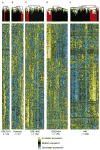
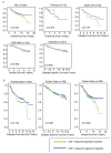
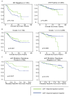
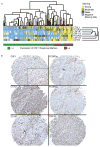
Experimental design: We defined the CSF1 response signature by identifying genes overexpressed in tenosynovial giant cell tumor and pigmented villonodular synovitis (tumors composed predominantly of macrophages recruited in response to the overexpression of CSF1) compared with desmoid-type fibromatosis and solitary fibrous tumor. To characterize the CSF1 response signature in breast cancer, we analyzed the expression of CSF1 response signature genes in eight published breast cancer gene expression data sets (n = 982) and did immunohistochemistry and in situ hybridization for CSF1 response genes on a breast cancer tissue microarray (n = 283).
Results: In both the gene microarray and tissue microarray analyses, a consistent subset (17-25%) of breast cancers shows the CSF1 response signature. The signature is associated with higher tumor grade, decreased expression of estrogen receptor, decreased expression of progesterone receptor, and increased TP53 mutations (P < 0.001).
Conclusions: Our data show that the CSF1 response signature is consistently seen in a subset of breast carcinomas and correlates with biological features of the tumor. Our findings provide insight into macrophage biology and may facilitate the development of personalized therapy for patients most likely to benefit from CSF1-targeted treatments.
Conflict of interest statement
Conflict of Interest: There are no conflicts of interest.
Figures


Similar articles
-
Translocation and expression of CSF1 in pigmented villonodular synovitis, tenosynovial giant cell tumor, rheumatoid arthritis and other reactive synovitides.Am J Surg Pathol. 2007 Jun;31(6):970-6. doi: 10.1097/PAS.0b013e31802b86f8. Am J Surg Pathol. 2007. PMID: 17527089
-
Stromal signatures in endometrioid endometrial carcinomas.Mod Pathol. 2014 Apr;27(4):631-9. doi: 10.1038/modpathol.2013.131. Epub 2013 Nov 22. Mod Pathol. 2014. PMID: 24263966
-
Endogenous versus tumor-specific host response to breast carcinoma: a study of stromal response in synchronous breast primaries and biopsy site changes.Clin Cancer Res. 2011 Feb 1;17(3):437-46. doi: 10.1158/1078-0432.CCR-10-1709. Epub 2010 Nov 22. Clin Cancer Res. 2011. PMID: 21098336 Free PMC article.
-
Molecular pathways involved in synovial cell inflammation and tumoral proliferation in diffuse pigmented villonodular synovitis.Autoimmun Rev. 2010 Sep;9(11):780-4. doi: 10.1016/j.autrev.2010.07.001. Epub 2010 Jul 8. Autoimmun Rev. 2010. PMID: 20620241 Review.
-
Current Systemic Treatment Options for Tenosynovial Giant Cell Tumor/Pigmented Villonodular Synovitis: Targeting the CSF1/CSF1R Axis.Curr Treat Options Oncol. 2016 Feb;17(2):10. doi: 10.1007/s11864-015-0385-x. Curr Treat Options Oncol. 2016. PMID: 26820289 Review.
Cited by
-
Tissues and Tumor Microenvironment (TME) in 3D: Models to Shed Light on Immunosuppression in Cancer.Cells. 2021 Apr 7;10(4):831. doi: 10.3390/cells10040831. Cells. 2021. PMID: 33917037 Free PMC article. Review.
-
Gene expression in extratumoral microenvironment predicts clinical outcome in breast cancer patients.Breast Cancer Res. 2012 Mar 19;14(2):R51. doi: 10.1186/bcr3152. Breast Cancer Res. 2012. PMID: 22429463 Free PMC article.
-
Phase Ib study of anti-CSF-1R antibody emactuzumab in combination with CD40 agonist selicrelumab in advanced solid tumor patients.J Immunother Cancer. 2020 Oct;8(2):e001153. doi: 10.1136/jitc-2020-001153. J Immunother Cancer. 2020. PMID: 33097612 Free PMC article. Clinical Trial.
-
Classification, molecular characterization, and the significance of pten alteration in leiomyosarcoma.Sarcoma. 2012;2012:380896. doi: 10.1155/2012/380896. Epub 2012 Feb 15. Sarcoma. 2012. PMID: 22448121 Free PMC article.
-
Co-dependencies in the tumor immune microenvironment.Oncogene. 2022 Jul;41(31):3821-3829. doi: 10.1038/s41388-022-02406-7. Epub 2022 Jul 11. Oncogene. 2022. PMID: 35817840 Free PMC article. Review.
References
-
- Condeelis J, Pollard JW. Macrophages: obligate partners for tumor cell migration, invasion, and metastasis. Cell. 2006;124:263–6. - PubMed
-
- Pollard JW. Tumour-educated macrophages promote tumour progression and metastasis. Nat Rev Cancer. 2004;4:71–8. - PubMed
-
- Sapi E. The role of CSF-1 in normal physiology of mammary gland and breast cancer: an update. Exp Biol Med (Maywood) 2004;229:1–11. - PubMed
-
- Scholl SM, Pallud C, Beuvon F, et al. Anti-colony-stimulating factor-1 antibody staining in primary breast adenocarcinomas correlates with marked inflammatory cell infiltrates and prognosis. J Natl Cancer Inst. 1994;86:120–6. - PubMed
-
- Maher MG, Sapi E, Turner B, et al. Prognostic significance of colony-stimulating factor receptor expression in ipsilateral breast cancer recurrence. Clin Cancer Res. 1998;4:1851–6. - PubMed
Publication types
MeSH terms
Substances
Grants and funding
LinkOut - more resources
Full Text Sources
Other Literature Sources
Medical
Research Materials
Miscellaneous

