

Gene expression profiling of the tumor microenvironment during breast cancer progression
- PMID: 19187537
- PMCID: PMC2687710
- DOI: 10.1186/bcr2222
Gene expression profiling of the tumor microenvironment during breast cancer progression
Abstract
Introduction: The importance of the tumor microenvironment in breast cancer has been increasingly recognized. Critical molecular changes in the tumor stroma accompanying cancer progression, however, remain largely unknown. We conducted a comparative analysis of global gene expression changes in the stromal and epithelial compartments during breast cancer progression from normal to preinvasive to invasive ductal carcinoma.
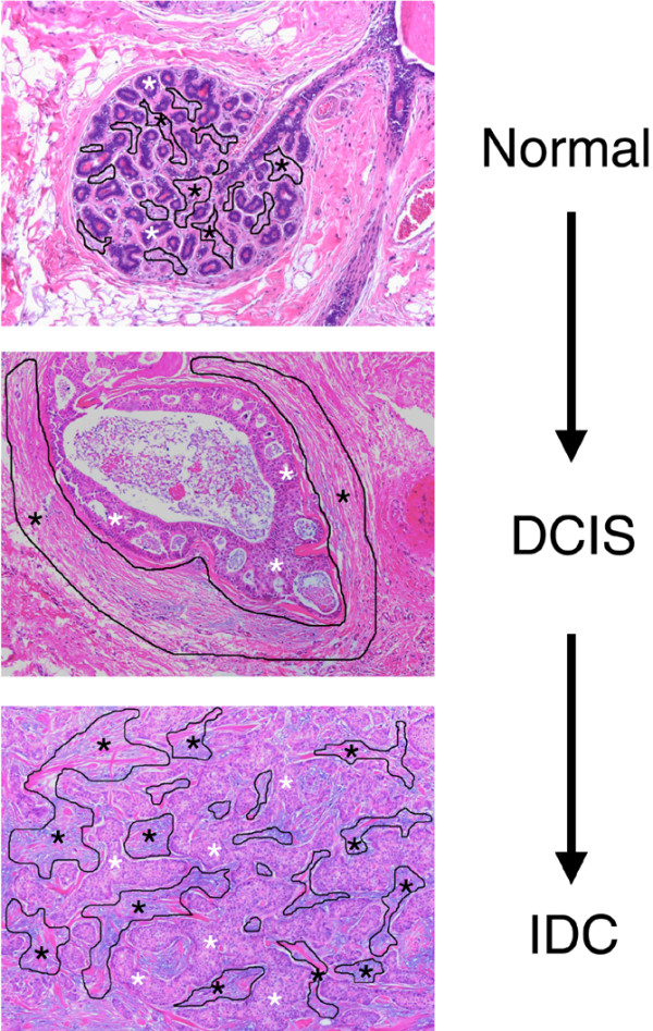
Methods: We combined laser capture microdissection and gene expression microarrays to analyze 14 patient-matched normal epithelium, normal stroma, tumor epithelium and tumor-associated stroma specimens. Differential gene expression and gene ontology analyses were performed.
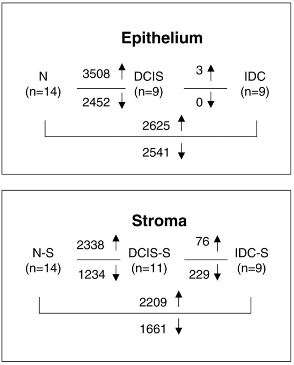
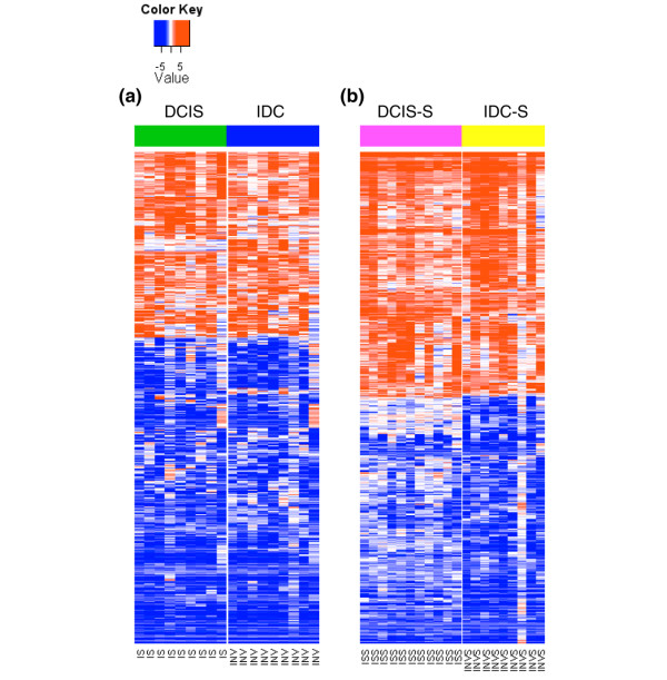
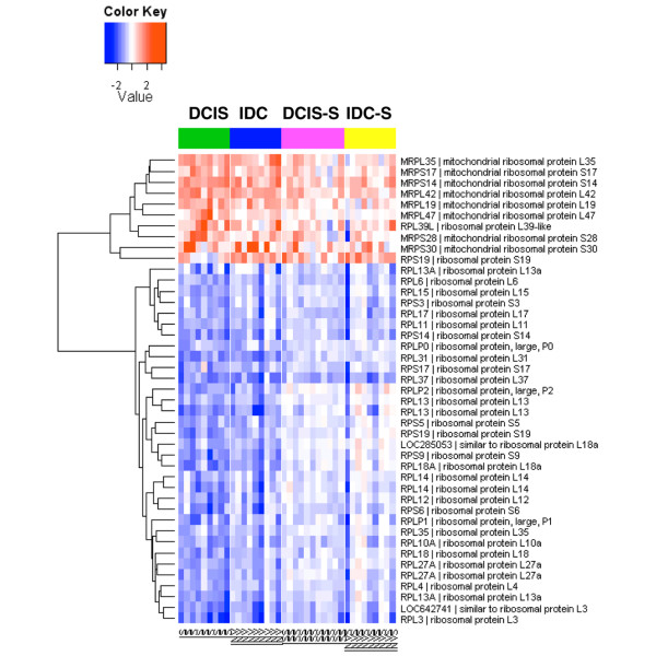
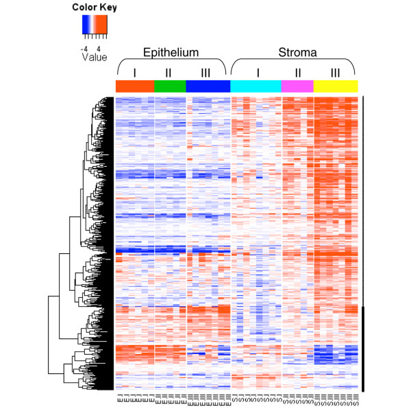
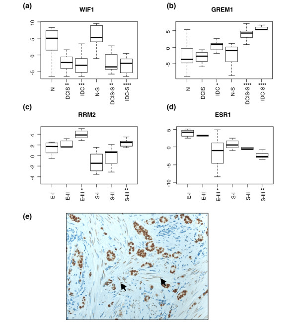
Results: Tumor-associated stroma undergoes extensive gene expression changes during cancer progression, to a similar extent as that seen in the malignant epithelium. Highly upregulated genes in the tumor-associated stroma include constituents of the extracellular matrix and matrix metalloproteases, and cell-cycle-related genes. Decreased expression of cytoplasmic ribosomal proteins and increased expression of mitochondrial ribosomal proteins were observed in both the tumor epithelium and the stroma. The transition from preinvasive to invasive growth was accompanied by increased expression of several matrix metalloproteases (MMP2, MMP11 and MMP14). Furthermore, as observed in malignant epithelium, a gene expression signature of histological tumor grade also exists in the stroma, with high-grade tumors associated with increased expression of genes involved in immune response.
Conclusions: Our results suggest that the tumor microenvironment participates in tumorigenesis even before tumor cells invade into stroma, and that it may play important roles in the transition from preinvasive to invasive growth. The immune cells in the tumor stroma may be exploited by the malignant epithelial cells in high-grade tumors for aggressive invasive growth.
Figures
Comment in
-
Breaking down barriers: the importance of the stromal microenvironment in acquiring invasiveness in young women's breast cancer.Breast Cancer Res. 2009;11(2):102. doi: 10.1186/bcr2235. Epub 2009 Mar 26. Breast Cancer Res. 2009. PMID: 19344495 Free PMC article.
Similar articles
-
Gene expression profiling of tumour epithelial and stromal compartments during breast cancer progression.Breast Cancer Res Treat. 2012 Aug;135(1):153-65. doi: 10.1007/s10549-012-2123-4. Epub 2012 Jun 21. Breast Cancer Res Treat. 2012. PMID: 22718308
-
Breaking down barriers: the importance of the stromal microenvironment in acquiring invasiveness in young women's breast cancer.Breast Cancer Res. 2009;11(2):102. doi: 10.1186/bcr2235. Epub 2009 Mar 26. Breast Cancer Res. 2009. PMID: 19344495 Free PMC article.
-
The urokinase-system in tumor tissue stroma of the breast and breast cancer cell invasion.Int J Oncol. 2009 Jan;34(1):15-23. Int J Oncol. 2009. PMID: 19082473
-
Tumor-extracellular matrix interactions: Identification of tools associated with breast cancer progression.Semin Cancer Biol. 2015 Dec;35:3-10. doi: 10.1016/j.semcancer.2015.09.012. Epub 2015 Sep 28. Semin Cancer Biol. 2015. PMID: 26416466 Review.
-
Identifying progression predictors of breast ductal carcinoma in situ.J Clin Pathol. 2017 Feb;70(2):102-108. doi: 10.1136/jclinpath-2016-204154. Epub 2016 Nov 18. J Clin Pathol. 2017. PMID: 27864452 Review.
Cited by
-
RORα suppresses breast tumor invasion by inducing SEMA3F expression.Cancer Res. 2012 Apr 1;72(7):1728-39. doi: 10.1158/0008-5472.CAN-11-2762. Epub 2012 Feb 20. Cancer Res. 2012. PMID: 22350413 Free PMC article.
-
Int6 reduction activates stromal fibroblasts to enhance transforming activity in breast epithelial cells.Cell Biosci. 2015 Mar 8;5:10. doi: 10.1186/s13578-015-0001-6. eCollection 2015. Cell Biosci. 2015. PMID: 25774287 Free PMC article.
-
Preinvasive breast cancer.Annu Rev Pathol. 2010;5:193-221. doi: 10.1146/annurev.pathol.4.110807.092306. Annu Rev Pathol. 2010. PMID: 19824828 Free PMC article. Review.
-
Breast tumour stroma is a prognostic indicator and target for therapy.Breast Cancer Res. 2009;11 Suppl 3(Suppl 3):S16. doi: 10.1186/bcr2435. Epub 2009 Dec 18. Breast Cancer Res. 2009. PMID: 20030867 Free PMC article. No abstract available.
-
A comprehensive overview of genomic imprinting in breast and its deregulation in cancer.Nat Commun. 2018 Oct 8;9(1):4120. doi: 10.1038/s41467-018-06566-7. Nat Commun. 2018. PMID: 30297886 Free PMC article. Review.
References
-
- de Visser KE, Coussens LM. The inflammatory tumor microenvironment and its impact on cancer development. Contrib Microbiol. 2006;13:118–137. - PubMed
-
- Liotta LA, Kohn EC. The microenvironment of the tumour-host interface. Nature. 2001;411:375–379. - PubMed
-
- Muller A, Homey B, Soto H, Ge N, Catron D, Buchanan ME, McClanahan T, Murphy E, Yuan W, Wagner SN, Barrera JL, Mohar A, Verastegui E, Zlotnik A. Involvement of chemokine receptors in breast cancer metastasis. Nature. 2001;410:50–56. - PubMed
Publication types
MeSH terms
Substances
Grants and funding
LinkOut - more resources
Full Text Sources
Other Literature Sources
Medical
Molecular Biology Databases
Miscellaneous
