. 2009 Apr;296(4):G899-909.
doi: 10.1152/ajpgi.90507.2008. Epub 2009 Jan 22.

Spermidine/spermine-N1-acetyltransferase ablation protects against liver and kidney ischemia-reperfusion injury in mice

Affiliations

Spermidine/spermine-N1-acetyltransferase ablation protects against liver and kidney ischemia-reperfusion injury in mice

Kamyar Zahedi et al. Am J Physiol Gastrointest Liver Physiol. 2009 Apr.

Abstract

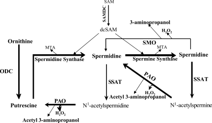
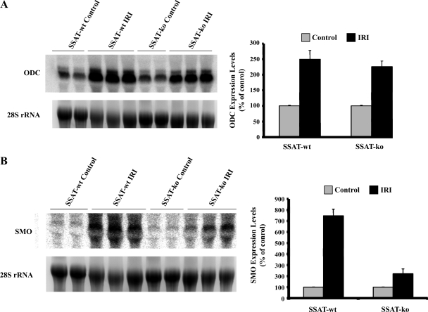
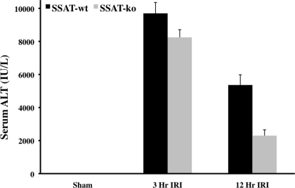
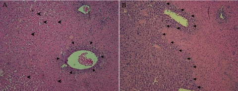
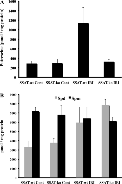
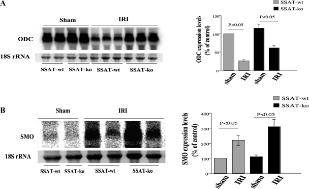
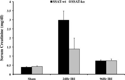
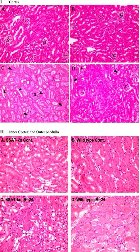
Expression of spermine/spermidine-N1-acetyltransferase (SSAT), the rate-limiting enzyme of polyamine backconversion cascade, increases after ischemia-reperfusion injuries (IRI). We hypothesized that SSAT plays an important role in the mediation of IRI. To test our hypothesis, wild-type (SSAT-wt) and SSAT-deficient (SSAT-ko) mice were subjected to liver or kidney IRI by ligation of hepatic or renal arteries. The liver and kidney content of putrescine (Put), a downstream by-product of SSAT activity, increased in SSAT-wt animals but not in SSAT-ko animals after IRI, indicating that polyamine backconversion is not functional in SSAT-deficient mice. When subjected to hepatic IRI, SSAT-ko mice were significantly protected against liver damage compared with SSAT-wt mice. Similarly, SSAT-ko animals subjected to renal IRI showed significantly greater protection against damage to kidney tubules than SSAT-wt mice. These studies indicate that SSAT-deficient animals are protected against IRI and suggest that SSAT is an important mediator of the tissue damage in IRI.


Figures

Fig. 1.
Fig. 1.
Schematic diagram depicting the polyamine metabolic pathway. Spermidine (Spd) and spermine (Spm) are synthesized by the sequential addition of aminopropyl groups derived from decarboxylated S-adenosylmethionine (dcSAM) onto putrescine (Put) and Spd to form Spd and Spm, respectively. Catabolism of polyamines proceeds via the backconversion pathway or via the 1-step Spm oxidation by spermine oxidase (SMO). In the spermidine/spermine N-1-acetyltransferase (SSAT)/polyamine oxidase (PAO)-mediated polyamine backconversion pathway, Spm and Spd are first acetylated by SSAT, the rate-limiting enzyme of the pathway, and then oxidized by PAO, leading to Put and cytotoxic by-products (i.e., H2O2, acetyl-3-aminopropionaldehyde). SAM, S-adenosylmethionine; SAMDC, S-adenosylmethionine decarboxylase; MTA, 5-methylthioadenosine; ODC, ornithine decarboxylase.
Fig. 2.
Fig. 2.
Genotyping of wild-type (SSAT-wt) and SSAT-deficient (SSAT-ko) mice. Genomic DNA was isolated and subjected to PCR amplification by using specific primers to identify the SSAT-wt and SSAT-ko mice. All animals depicted except the heterozygous animal were males. The amplified DNA was size fractionated on a 1% agarose-Tris/acetate/EDTA gel and analyzed for the presence of 408 bp (SSAT-wt) and 680 bp (SSAT-ko) PCR amplification products. Lane marked as Std contains DNA size standard.
Fig. 3.
Fig. 3.
Comparison of polyamine levels in SSAT-wt and SSAT ko mice after liver ischemia-reperfusion injury (IRI). A: liver Put levels. Liver Put levels were measured in injured and sham-operated (Cont) SSAT-wt and SSAT-ko animals. The Put content in the livers of both SSAT-wt and -ko mice were similar in sham-operated animals. However, in IRI animals, Put levels increased by ∼6-fold in the livers of wild-type animals compared with sham-operated animals of the same genotype. The hepatic Put levels in the IRI SSAT-ko animals were similar to those of the SSAT-ko sham-operated mice. B: liver Spd and Spm levels. The concentration of Spm and Spd in the livers of uninjured SSAT-wt and SSAT-ko animals were similar. However, the concentrations of Spm and Spd in the livers of SSAT-wt mice were significantly lower than those of sham-operated and SSAT-ko mice.
Fig. 4.
Fig. 4.
Northern blot analysis of ODC (A) and SMO (B) mRNA levels in livers of SSAT-wt and SSAT-ko animals after IRI. The expression of ODC increased (A, left) whereas the expression of SMO was only weakly induced (B, left) after IRI in both SSAT-wt and SSAT-ko animals. Densitometric analysis of the Northern blot results and normalization of the signal values against 28s rRNA were used to evaluate the induction profiles of each transcript. The semiquantitative values of the induction profiles of ODC and SMO are presented on the right of A and B, respectively.
Fig. 5.
Fig. 5.
Assessment of liver damage in SSAT-wt and SSAT-ko mice after IRI. The loss of liver function associated with liver IRI was compared in SSAT-wt and SSAT-ko mice (n = 4 per group) by examining serum alanine aminotransferase (ALT) levels. The serum ALT levels of sham-operated SSAT-wt and -ko mice were similar. The liver ALT of both genotypes were similarly elevated as early as 3 h after the onset of reperfusion; however, at 12 h after onset of reperfusion the SSAT-ko animals had significantly (P < 0.05) lower serum ALT levels than their SSAT-wt counterparts.
Fig. 6.
Fig. 6.
Comparison of the histopathology of liver after partial hepatic IRI in SSAT-wt and SSAT-ko mice. The histology of livers of SSAT-wt (A) and -ko (B) animals subjected to 90 min of ischemia and 12 h of reperfusion were examined (n = 4/group). Histological comparison of uninjured SSAT-wt and -ko liver did not reveal any structural differences (data not shown). Histological assessment of livers from SSAT-wt and SSAT-ko (A and B, respectively) mice after induction of liver injury showed the typical extensive hemorrhagic necrosis and inflammatory infiltrates seen after IRI. However, in livers from SSAT-ko mice (B), there was far less congestion and the extent of necrosis was substantially reduced (small arrows delineate the injured vs. uninjured areas; large arrows indicate the areas of congestion).
Fig. 7.
Fig. 7.
Comparison of polyamine levels in SSAT-wt and SSAT ko mice after kidney IRI. A: kidney Put levels. Kidney Put levels were measured in injured and sham-operated SSAT-wt and -ko animals. The Put content in the kidneys of SSAT-wt were similar in sham-operated animals. After IRI, Put levels increased by ∼4-fold in the kidneys of SSAT-wt animals compared with sham-operated animals of the same genotype. The kidney Put levels in the injured SSAT-ko animals were not different from those of the SSAT-ko and SSAT-wt sham-operated mice. B: kidney Spd and Spm levels. The kidney Spd levels increased in SSAT-wt and SSAT-ko mice by ∼2-fold after IRI whereas the Spm levels were only marginally reduced in the injured kidneys of either SSAT-wt or SSAT-ko animals.
Fig. 8.
Fig. 8.
Northern blot analysis of ODC (A) and SMO (B) mRNA expression in kidneys of SSAT-wt and SSAT-ko animals after sham operation or IRI. The expression of ODC decreased (A, left) whereas the expression of SMO increased (B, left) after IRI in both SSAT-wt and SSAT-ko animals. Densitometric scanning of the gels and normalization of the values against 28s rRNA was used to evaluate the signals. The semi-quantitative values of the induction profiles of ODC and SMO are presented on the right of A and B, respectively.
Fig. 9.
Fig. 9.
Assessment of kidney damage in SSAT-wt and SSAT ko mice after IRI. The loss of renal function caused by bilateral IRI was compared in SSAT-wt and -ko mice (n = 6 per group) by comparing serum creatinine levels. Whereas serum creatinine levels of sham-operated SSAT-ko and -wt mice were similar, the SSAT-ko animals subjected to renal IRI had significantly (P < 0.05) lower serum creatinine levels than SSAT-wt animals subjected to renal IRI. The serum creatinine levels in both genotypes were significantly reduced and near normal levels by 96 h postreperfusion.
Fig. 10.
Fig. 10.
Comparison of the histopathology of kidneys after renal IRI in SSAT-wt and SSAT-ko mice. I: cortex. The histology of the kidney cortical regions of SSAT-ko and SSAT-wt sham-operated animals (A and B, respectively) and animals subjected to 30 min of ischemia and 24 h of reperfusion (C and D, respectively) were examined microscopically. Comparison of the kidneys of sham-operated animals did not reveal any differences between the SSAT-wt and SSAT-ko mice. Our results indicated that after IRI, the kidneys of SSAT-ko had reduced tubular dilatation (arrowheads) as well as edema, and cast formation (small arrows) relative to the kidneys of SSAT-wt animals. G, glomeruli. II: inner cortex and outer medulla. The histology of the corticomedullary region of kidneys of SSAT-ko and SSAT-wt sham-operated animals (A and B, respectively) and animals subjected to 30 min of ischemia and 24 h of reperfusion (C and D) were examined microscopically. Comparison of the kidneys of sham-operated animals from SSAT-wt and -ko genotypes (B vs. A) did not reveal any differences in the renal architecture. Kidneys from SSAT-ko animals (C) subjected to IRI display significant protection against structural damage in the corticomedullary (inner cortex/outer medulla) region relative to SSAT-wt animals (D).

Similar articles

Cited by

References

    1. Alhonen L, Karppinen A, Uusi-Oukari M, Vujcic S, Korhonen VP, Halmekyto M, Kramer DL, Hines R, Janne J, Porter CW. Correlation of polyamine and growth responses to N1,N11-diethylnorspermine in primary fetal fibroblasts derived from transgenic mice overexpressing spermidine/spermine N1-acetyltransferase. J Biol Chem 273: 1964–1969, 1998. - PubMed
    1. Alhonen L, Parkkinen JJ, Keinanen T, Sinervirta R, Herzig KH, Janne J. Activation of polyamine catabolism in transgenic rats induces acute pancreatitis. Proc Natl Acad Sci USA 97: 8290–8295, 2000. - PMC - PubMed
    1. Alhonen L, Rasanen TL, Sinervirta R, Parkkinen JJ, Korhonen VP, Pietila M, Janne J. Polyamines are required for the initiation of rat liver regeneration. Biochem J 362: 149–153, 2002. - PMC - PubMed
    1. Barone S, Okaya T, Rudich S, Petrovic S, Tehrani K, Wang Z, Zahedi K, Casero RA, Lentsch AB, Soleimani M. Distinct and sequential upregulation of genes regulating cell growth and cell cycle progression during hepatic ischemia/reperfusion injury. Am J Physiol Cell Physiol 289: C826–C835, 2005. - PubMed
    1. Baskaya MK, Rao AM, Dogan A, Donaldson D, Gellin G, Dempsey RJ. Regional brain polyamine levels in permanent focal cerebral ischemia. Brain Res 744: 302–308, 1997. - PubMed

Publication types