. 2009 Mar 27;284(13):8557-66.
doi: 10.1074/jbc.M806702200. Epub 2009 Jan 21.

Ubiquitin Ligase RLIM Modulates Telomere Length Homeostasis through a Proteolysis of TRF1

Affiliations

Ubiquitin Ligase RLIM Modulates Telomere Length Homeostasis through a Proteolysis of TRF1

Yoon Ra Her et al. J Biol Chem. .

Abstract

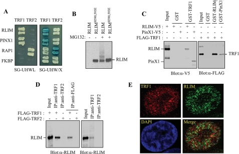
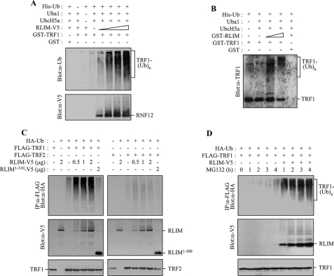
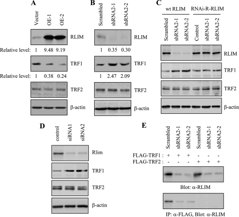
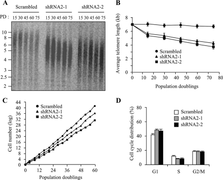
The telomeric protein TRF1 negatively regulates telomere length by inhibiting telomerase access at the telomere termini, suggesting that the protein level of TRF1 at telomeres is tightly regulated. Regulation of TRF1 protein abundance is essential for proper telomere function and occurs primarily through post-translational modifications of TRF1. Here we describe RLIM, a RING H2 zinc finger protein with intrinsic ubiquitin ligase activity, as a TRF1-interacting protein. RLIM increases TRF1 turnover by targeting it for degradation by the proteasome in a ubiquitin-dependent manner, independently of Fbx4, which is known to interact with and negatively regulate TRF1. Whereas overexpression of RLIM decreases the level of TRF1 protein, depletion of endogenous RLIM expression by small hairpin RNA increases the level of TRF1 and leads to telomere shortening, thereby impairing cell growth. These results demonstrate that RLIM is involved in the negative regulation of TRF1 function through physical interaction and ubiquitin-mediated proteolysis. Hence, RLIM represents a new pathway for telomere maintenance by modulating the level of TRF1 at telomeres.


Figures

FIGURE 1.
FIGURE 1.
Physical interaction between TRF1 and LRIM. A, analysis of the physical interaction between TRF1 and RLIM using the yeast two-hybrid assay. PinX1, RAP1, and unrelated FKBP52 were used as the TRF1-binding, TRF2-binding, and negative control, respectively. The growth on the SG-HWUL plate and the blue signal on the SG-HWU/X plate indicate activation of the reporter genes, LacZ and LEU2, respectively. S, synthetic; G, galactose; H, histidine (-); W, tryptophan (-); U, uracil, (-); L, leucine (-); X, 5-bromo-4-chloro-3-indolyl-β-d-galactopyranoside (X-gal). B, H1299 cells were transfected with V5-RLIM or V5-RLIMH590,593E and either untreated or treated with 10 μm MG132 for 4 h. Cell lysates were analyzed by immunoblotting with anti-V5 antibody probe. C, interaction between TRF1 and RLIM in vitro. Left, GST and GST-TRF1 were immobilized on glutathione-Sepharose and incubated with exogenously expressed RLIM-V5 and PinX1-V5. After washing and SDS-PAGE, bound RLIM and PinX1 were detected by immunoblotting with anti-V5 antibody. Right, GST, GST-RLIM, and GST-PinX1 were incubated with exogenously expressed FLAG-TRF1 followed by immunoblotting with anti-FLAG antibody. D, coimmunoprecipitation of TRF1 and RLIM. Left, H1299 cells were transfected with FLAG-TRF1 or FLAG-TRF2 and then subjected to immunoprecipitation as indicated followed by immunoblotting with anti-RLIM antibody. Right, H1299 cells were subjected to immunoprecipitation with either anti-TRF1 or -TRF2 antibodies followed by immunoblotting with anti-RLIM antibody. E, H1299 cells were treated with 10 μm MG132 for 4 h and analyzed by indirect immunofluorescence. Paraformaldehyde-fixed cells were stained with anti-TRF1 (red) and anti-RLIM antibodies (green). DNA was stained with 4,6-diamidino-2-phenylindole (blue).
FIGURE 2.
FIGURE 2.
Mapping the interaction domains on TRF1 and RLIM. A, schematic representation of the region of RLIM involved in binding to TRF1. B, the region of RLIM involved in binding to TRF1 was analyzed in a GST pulldown assay. H1299 cells were transfected with various RLIM-V5 mutants and subjected to immunoblotting analysis with anti-V5 antibody directly (left) or were first precipitated by GST or GST-TRF1 (right). C, H1299 cells transfected with FLAG-TRF1 were precipitated by GST or the various truncated GST-RLIM fusion proteins followed by immunoblotting with anti-FLAG antibody. D, schematic representation of TRF1 truncations fused to GST. E, GST or the various truncated GST-TRF1 fusion proteins were affinity-purified and incubated with H1299 cell extracts followed by detection of endogenous RLIM by immunoblotting with anti-RLIM.
FIGURE 3.
FIGURE 3.
RLIM serves as an E3 ubiquitin ligase for TRF1. A, RLIM-V5 was expressed in H1299 cells and affinity-purified from cells pretreated with 10 μm MG132 for 4 h using anti-V5 antibody-coupled with protein A-Sepharose beads. GST-TRF1 was incubated with increasing amounts of RLIM-V5 in the presence of E1, UbcH5a (E2), and His-ubiquitin (Ub) as indicated. After the ubiquitination reaction, the samples were analyzed by SDS-PAGE and immunoblotting with anti-ubiquitin antibody to reveal ubiquitinated products. B, GST-TRF1 was incubated with increasing amounts of GST-RLIM expressed in bacteria as indicated. Ubiquitinated products were detected by immunoblotting with anti-TRF1 antibody. C, H1299 cells were co-transfected with HA-ubiquitin, FLAG-TRF1 (or FLAG-TRF2), and together with either increasing amounts of RLIM-V5 or RLIM-(1–310)-V5 as specified and treated with 10 μm MG132 for 4 h. Immunoprecipitation was performed with anti-FLAG antibody before probing with anti-HA antibody. D, H1299 cells were co-transfected with HA-ubiquitin, FLAG-TRF1, and together with or without RLIM-V5 as indicated and treated with 10 μm MG132 for the indicated times. Anti-FLAG immunoprecipitates and cell lysates were analyzed by immunoblotting with anti-HA antibody and anti-V5 antibody probes, respectively.
FIGURE 4.
FIGURE 4.
Overexpression of RLIM reduces the steady-state level of TRF1 protein. A, H1299 cells were co-transfected with FLAG-TRF1 (or FLAG-TRF2) and either increasing amounts of RLIM-V5 or RLIM-(1–310)-V5 as specified. The levels of ectopically expressed TRF1 (or TRF2) and RLIM were determined by immunoblotting with anti-FLAG antibody and anti-V5 antibody, respectively. An antibody against β-actin was used as a loading control. B, representative results of reverse transcription-PCR analysis for the expression of RLIM and TRF1 genes in H1299 cells transfected with RLIM. Reverse transcription-PCR products from each sample were normalized to glyceraldehyde-3-phosphate dehydrogenase (GAPDH) signal. C, H1299 cells were transfected with RLIM-V5, HA-Fbx4, or both and treated with 100 μg/ml cycloheximide together with, or without, 10 μm MG132 for the indicated times. The nuclear fractions were analyzed on immunoblots with anti-TRF1 antibody or anti-TATA-binding protein (TBP) antibody. D, graphical representation of relative TRF1 levels normalized against the TATA-binding protein loading control. The graph represents an average of three independent experiments.
FIGURE 5.
FIGURE 5.
RLIM regulates the levels of endogenous TRF1 protein. A, HT1080 clones stably expressing RLIM-V5 (OE-1 and OE-2) or the control empty vector were harvested at 75 population doublings and subjected to immunoblotting with anti-RLIM, anti-TRF1, anti-TRF2, or β-actin antibodies. The relative levels of RLIM and TRF1 were determined from three independent experiments using β-actin as a loading control. B, HT1080 clones stably expressing RLIM shRNA2 (shRNA2-1 and shRNA2-2) or scrambled shRNA were harvested at 75 population doublings and subjected to immunoblotting with anti-RLIM, anti-TRF1, anti-TRF2, or β-actin antibodies. The relative levels of RLIM and TRF1 were determined from three independent experiments using β-actin as a loading control. C, HT1080 cells stably expressing RLIM shRNA2 (shRNA2-1 and shRNA2-2) or scrambled shRNA were transfected with wild type RLIM (wt RLIM) or RNAi-R-RLIM and examined for expression of endogenous RLIM, TRF1, and TRF2 by immunoblotting. D, HT1080 cells were transiently transfected with siRNA duplexes, either nontargeting control or targeting RLIM (siRNA1 and siRNA2) for 3 days and examined for the expression of endogenous RLIM, TRF1, and TRF2 by immunoblotting. E, HT1080 cells stably expressing RLIM shRNA2 (shRNA2-1 and shRNA2-2) or scrambled shRNA were transfected with FLAG-TRF1 or FLAG-TRF2 and subjected to immunoprecipitation (IP) with anti-FLAG antibody, followed by immunoblotting with anti-RLIM antibody.
FIGURE 6.
FIGURE 6.
RLIM regulates the half-life of endogenous TRF1. A, stable clones expressing the control vector, RLIM-V5 (OE-1), or shRNA (shRNA2-1) were treated with 100 μg/ml cycloheximide together with, or without, 10 μm MG132 for the indicated times followed by immunoblotting with anti-TRF1 antibody or anti-actin antibodies. B, graphical representation of relative TRF1 levels normalized against the β-actin loading control. The graph represents an average of three independent experiments.
FIGURE 7.
FIGURE 7.
Depletion of RLIM leads to progressive telomere shortening and impaired cell growth. A, genomic blot of telomere restriction fragments in stable HT1080 clones expressing RLIM shRNA2 (shRNA2-1 or shRNA2-2) or the control scrambled shRNA. Genomic DNA was isolated at the indicated population doublings (PD), digested with RsaI and HinfI, and analyzed by Southern blotting using a telomere repeat probe. B, graphical representation of average terminal restriction fragment length versus population doubling number from two independent experiments. C, growth curves of stable HT1080 clones expressing RLIM shRNA2 (shRNA2-1 or shRNA2-2) or the control scrambled shRNA. Stable clones were replated every 3–4 days to maintain log-phase growth and to calculate the growth rate. D, cell cycle profiles of stable clones expressing RLIM shRNA2. Stable clones were harvested at 60 population doublings, and cell cycle profiles were determined by propidium iodide staining and flow cytometry. The results represent the average of three independent experiments.

Similar articles

Cited by

References

    1. Smogorzewska, A., and de Lange, T. (2004) Annu. Rev. Biochem. 73 177-208 - PubMed
    1. Blasco, M. A., Lee, H. W., Hande, M. P., Samper, E., Lansdorp, P. M., DePinho, R. A., and Greider, C. W. (1997) Cell 91 25-34 - PubMed
    1. Harrington, L. (2005) Chromosome Res. 13 493-504 - PubMed
    1. Lingner, J., Cooper, J. P., and Cech, T. R. (1995) Science 269 1533-1534 - PubMed
    1. Cerone, M. A., Autexier, C., Londoño-Vallejo, J. A., and Bacchetti, S. (2005) Oncogene 24 7893-7901 - PubMed

Publication types

MeSH terms

LinkOut - more resources