. 2009 Feb;11(2):172-82.
doi: 10.1038/ncb1831. Epub 2009 Jan 18.

CHD8 suppresses p53-mediated apoptosis through histone H1 recruitment during early embryogenesis

Affiliations

CHD8 suppresses p53-mediated apoptosis through histone H1 recruitment during early embryogenesis

Masaaki Nishiyama et al. Nat Cell Biol. 2009 Feb.

Abstract

The chromodomain helicase DNA-binding (CHD) family of enzymes is thought to regulate gene expression, but their role in the regulation of specific genes has been unclear. Here we show that CHD8 is expressed at a high level during early embryogenesis and prevents apoptosis mediated by the tumour suppressor protein p53. CHD8 was found to bind to p53 and to suppress its transactivation activity. CHD8 promoted the association of p53 and histone H1, forming a trimeric complex on chromatin that was required for inhibition of p53-dependent transactivation and apoptosis. Depletion of CHD8 or histone H1 resulted in p53 activation and apoptosis. Furthermore, Chd8(-/-) mice died early during embryogenesis, manifesting widespread apoptosis, whereas deletion of p53 ameliorated this developmental arrest. These observations reveal a mode of p53 regulation mediated by CHD8, which may set a threshold for induction of apoptosis during early embryogenesis by counteracting p53 function through recruitment of histone H1.


Figures
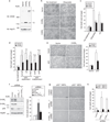
Figure 1
Figure 1
Anti-apoptotic activity of CHD8. (a–c) NIH 3T3 cells overexpressing CHD8S or CHD8L were subjected to immunoblot (IB, a) analysis with anti-CHD8 and exposed to genotoxic stress. Cells were examined by phase-contrast microscopy (b) and the percentage of dead cells was determined by trypan blue staining (c). Data in c are mean ± s.d., n = 3. (d) NIH 3T3 cells overexpressing CHD8S were exposed to etoposide (ETOP, 50 µM), cycloheximide (CHX, 100 µg ml−1), staurosporine (STR, 1 µM), UV radiation or c-Myc overexpression. Data in d are mean ± s.d., n = 3 (**P < 0.01; n.s., not significant; P > 0.05; Student’s t-test). (e) U2OS cells were infected with retroviral vectors for CHD8S or p53 and were stained with trypan blue (left panel). Data shown in the right panel are mean ± s.d., n = 3. (f) U2OS cells were infected with retroviral vectors encoding shRNAs specific for p53, both CHD8S and CHD8L (CHD8S+L-1) or EGFP (control), subjected to immunoblotting (left panel), stained with trypan blue and the percentage of dead cells determined (right panel). Data shown in the right panel are mean ± s.d., n = 3. (g, h) p53+/+ or p53−/− MEFs infected with retroviral vectors for CHD8S+L-1 or CHD8S+L-2 shRNAs were examined by phasecontrast microscopy (g) and the percentage of dead cells determined by trypan blue staining (h). Data in h are mean ± s.d., n = 3. Scale bars are 100 µm (b, e, g).
Figure 2
Figure 2
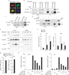
CHD8 interacts with and inhibits transactivation by p53. (a) HeLa cells expressing Myc–CHD8S were immunostained with anti-Myc or p53. Scale bar, 5 µm. (b) U2OS cells were incubated with etoposide and then subjected to immunoprecipitation (IP) with an anti-CHD8 antibody, and immunoblot analysis with an anti-p53 antibody. (c) Immunoprecipitation of U2OS lysates with anti-p53 and immunoblot analysis with an anti-CHD8 antibodies. (d) In vitro binding assay for recombinant CHD8S and p53. (e) U2OS cells overexpressing CHD8S were incubated with doxorubicin (DXR) and then subjected to immunoblot analysis with the indicated antibodies. (f) U2OS cells overexpressing CHD8S were treated with the genotoxic agents DXR (0.5 µM) and etoposide (ETOP, 20 µM) and then subjected to qRT–PCR. (g) U2OS cells infected with a retroviral vector encoding CHD8S+L-1 shRNA were subjected to qRT–PCR. (h) Luciferase assay using either wild-type or mutant CHD8SΔ53. Data are mean ± s.d., n = 3 (fh).
Figure 3
Figure 3
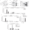
CHD8 binds to the promoters of p53 target genes. (a) U2OS cells infected with a retroviral vector encoding p53 shRNA were incubated with the genotoxic agents etoposide (ETOP, 20 µM) and doxorubicin (DXR, 0.5 µM). The cells were then subjected to ChIP with an anti-CHD8 antibody and the precipitated DNA was quantified by real-time PCR with primers specific for p53-responsive elements (p53REs) 1 or 2 or a control region (CR) of the p21 promoter or for the p27 promoter. (b) U2OS cells overexpressing CHD8S were treated with ETOP (20 µM) and DXR (0.5 µM) and subjected to ChIP with an anti-p53 antibody. Data are mean ± s.d., n = 3 (a, b). (c) U2OS cells overexpressing CHD8S were incubated with ETOP and then subjected to ChIP with an anti-CHD8 antibody. The immunoprecipitates were subjected to ChIP with an anti-p53 antibody.
Figure 4
Figure 4
CHD8 recruits histone H1 to the promoters of p53 target genes. (a) HEK293T cells expressing 3 × Flag–histone H1c were subjected to immunoprecipitation with anti-Flag and immunoblot analysis with anti-CHD8 antibodies. (b) Immunoprecipitation of HEK293T lysates with anti-CHD8 and immunoblot analysis with anti-histone H1 antibodies. (c) In vitro binding assay for recombinant CHD8S and histone H1c. (d) U2OS cells overexpressing CHD8S were treated with the genotoxic agents etoposide (ETOP, 20 µM) and doxorubicin (DXR, 0.5 µM) and subjected to ChIP with an anti-histone H1 antibody. (e) ChIP was performed for the BAX promoter (n.d., not detected). (f) U2OS cells infected with a retroviral vector for CHD8S+L-1 shRNA were subjected to ChIP. (g) U2OS cells overexpressing CHD8S were infected with a retroviral vector for p53 shRNA, incubated with ETOP and then subjected to ChIP. Data are mean ± s.d., n = 3 (dg).
Figure 5
Figure 5
Interaction of CHD8 with p53 and histone H1 is necessary for recruitment of histone H1 to the promoters of p53 target genes. (a) Schematic representation of CHD8S derivatives. (b–d) U2OS cells overexpressing CHD8S derivatives were exposed to genotoxic stress (etoposide, ETOP, 20 µM and doxorubicin, DXR, 0.5 µM) and then subjected to ChIP (b), qRT–PCR (c) and trypan blue staining (d). Data are mean ± s.d., n = 3.
Figure 6
Figure 6
Requirement for histone H1 in repression of p53-mediated transcription. (a, b) Wild-type (WT) or HH1c−/−HH1d−/−HH1e−/− triple-knockout (TKO) ES cells were incubated with etoposide (ETOP, µ2 M) and doxorubicin (DXR, 0.05 µM) and then subjected to qRT–PCR (a) and trypan blue staining (b). (c) U2OS cells were infected with a retroviral vector for histone H1 (HH1) shRNA and then subjected to immunoblot analysis. (d) The cells were incubated with ETOP (2 µM) and DXR (0.05 µM) and then subjected to qRT–PCR. Data are mean ± s.d., n = 3 (a, b, d).
Figure 7
Figure 7
Deletion of p53 rescues the phenotype of CHD8-deficient mice. (a) qRT–PCR for CHD8S and CHD8L mRNAs in mouse embryos at the indicated stages. Data are mean ± s.d., n = 3. (b) Immunoblot analysis of CHD8S in mouse embryos as well as in U2OS cells and those overexpressing (OE) CHD8S. (c) Histopathological examination of Chd8+/+p53+/+, Chd8−/−p53+/+, Chd8−/−p53+/− and Chd8−/−p53−/− embryos stained with haematoxylin and eosin. (d) Higher-magnification views of the boxed regions in c. Arrowheads indicate a layer of mesoderm. (e) In vitro culture of blastocysts of the indicated genotypes. Scale bars are 100 µm (c–e).
Figure 8
Figure 8
CHD8 sets a threshold for induction of apoptosis during early embryogenesis by counteracting p53 function. (a, b) Immunoprecipitation of ES cell lysates with an anti-CHD8 antibody, and immunoblot analysis with either anti-p53 (a) or anti-histone H1 (b) antibodies. (c–e) ES cells infected with retroviral vectors for CHD8S+L-1 or CHD8S+L-2 shRNAs were subjected to trypan blue staining (c, quantification shown in d) and qRT–PCR (e). (f) Chd8+/+ or Chd8−/− blastocysts were subjected to qRT–PCR for p21 and Noxa. Data are mean ± s.d., n = 3 (d–f). (g, h) Sections of Chd8+/+p53+/+, Chd8−/−p53+/+ and Chd8−/−p53−/− embryos were subjected to TUNEL assay (g) or to immunohistochemistry (h) with anti-Mdm2 antibody. Scale bars are 100 µm (c, g, h). (i) Model for the biological role of CHD8. When cells proliferate extensively during early embryogenesis, CHD8 is expressed at high levels and suppresses p53 function through histone H1 recruitment to prevent unwanted apoptosis. Once the level of CHD8 expression decreases, during mid to late embryogenesis, some cells undergo apoptosis for organogenesis.

Similar articles

Cited by

References

    1. Vousden KH, Lu X. Live or let die: the cell’s response to p53. Nature Rev. Cancer. 2002;2:594–604. - PubMed
    1. Harris SL, Levine AJ. The p53 pathway: positive and negative feedback loops. Oncogene. 2005;24:2899–2908. - PubMed
    1. Vogelstein B, Lane D, Levine AJ. Surfing the p53 network. Nature. 2000;408:307–310. - PubMed
    1. Laptenko O, Prives C. Transcriptional regulation by p53: one protein, many possibilities. Cell Death Differ. 2006;13:951–961. - PubMed
    1. Aylon Y, Oren M. Living with p53, dying of p53. Cell. 2007;130:597–600. - PubMed

Publication types