Enhanced replication and pathogenesis of Moloney murine leukemia virus in mice defective in the murine APOBEC3 gene
- PMID: 19150103
- PMCID: PMC3124558
- DOI: 10.1016/j.virol.2008.11.051
Enhanced replication and pathogenesis of Moloney murine leukemia virus in mice defective in the murine APOBEC3 gene
Abstract
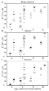
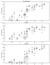
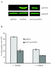
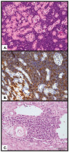
Human APOBEC3G (hA3G), a member of the AID/APOBEC family of deaminases, is a restriction factor for human immunodeficiency virus (HIV). In the absence of the viral Vif protein hA3G is packaged into virions and during reverse transcription in a recipient cell it deaminates cytosines, leading to G-->A hypermutation and inactivation of the viral DNA. Unlike humans, who carry seven APOBEC3 genes, mice only carry one, mA3. Thus the role of mA3 in restriction of retroviral infection could be studied in mA3 -/- knockout mice, where the gene is inactivated. M-MuLV-infected mA3 -/- mice showed substantially higher levels of infection at very early times compared to wild-type mice (ca. 2 logs at 0-10 days), particularly in the bone marrow and spleen. Restriction of M-MuLV infection was studied ex vivo in primary bone marrow-derived dendritic cells (BMDCs) that express or lack mA3, using an M-MuLV-based retroviral vector expressing enhanced jellyfish green fluorescent protein (EGFP). The results indicated that mA3 within the virions as well as mA3 in the recipient cell contribute to resistance to infection in BMDCs. Finally, M-MuLV-infected mA3 +/+ mice developed leukemia more slowly compared to animals lacking one or both copies of mA3 although the resulting disease was similar (T-lymphoma). These studies indicate that mA3 restricts replication and pathogenesis of M-MuLV in vivo.
Figures


Similar articles
-
Interactions of murine APOBEC3 and human APOBEC3G with murine leukemia viruses.J Virol. 2008 Jul;82(13):6566-75. doi: 10.1128/JVI.01357-07. Epub 2008 Apr 30. J Virol. 2008. PMID: 18448535 Free PMC article.
-
Murine Leukemia Virus P50 Protein Counteracts APOBEC3 by Blocking Its Packaging.J Virol. 2020 Aug 31;94(18):e00032-20. doi: 10.1128/JVI.00032-20. Print 2020 Aug 31. J Virol. 2020. PMID: 32641479 Free PMC article.
-
The incorporation of APOBEC3 proteins into murine leukemia viruses.Virology. 2008 Aug 15;378(1):69-78. doi: 10.1016/j.virol.2008.05.006. Epub 2008 Jun 24. Virology. 2008. PMID: 18572219
-
Biochemical and biological studies of mouse APOBEC3.J Virol. 2014 Apr;88(7):3850-60. doi: 10.1128/JVI.03456-13. Epub 2014 Jan 22. J Virol. 2014. PMID: 24453360 Free PMC article.
-
[Recent advances in the study of mechanism of APOBEC3G against virus].Yao Xue Xue Bao. 2014 Jan;49(1):30-6. Yao Xue Xue Bao. 2014. PMID: 24783502 Review. Chinese.
Cited by
-
Nucleic acid recognition orchestrates the anti-viral response to retroviruses.Cell Host Microbe. 2015 Apr 8;17(4):478-88. doi: 10.1016/j.chom.2015.02.021. Epub 2015 Mar 26. Cell Host Microbe. 2015. PMID: 25816774 Free PMC article.
-
ZASC1 knockout mice exhibit an early bone marrow-specific defect in murine leukemia virus replication.Virol J. 2013 Apr 24;10:130. doi: 10.1186/1743-422X-10-130. Virol J. 2013. PMID: 23617998 Free PMC article.
-
BST-2/tetherin-mediated restriction of chikungunya (CHIKV) VLP budding is counteracted by CHIKV non-structural protein 1 (nsP1).Virology. 2013 Mar 30;438(1):37-49. doi: 10.1016/j.virol.2013.01.010. Epub 2013 Feb 12. Virology. 2013. PMID: 23411007 Free PMC article.
-
Murine leukemia virus infection of non-dividing dendritic cells is dependent on nucleoporins.PLoS Pathog. 2024 Jan 12;20(1):e1011640. doi: 10.1371/journal.ppat.1011640. eCollection 2024 Jan. PLoS Pathog. 2024. PMID: 38215165 Free PMC article.
-
Noninfectious retrovirus particles drive the APOBEC3/Rfv3 dependent neutralizing antibody response.PLoS Pathog. 2011 Oct;7(10):e1002284. doi: 10.1371/journal.ppat.1002284. Epub 2011 Oct 6. PLoS Pathog. 2011. PMID: 21998583 Free PMC article.
References
-
- Abudu A, Takaori-Kondo A, Izumi T, Shirakawa K, Kobayashi M, Sasada A, Fukunaga K, Uchiyama T. Murine retrovirus escapes from murine APOBEC3 via two distinct novel mechanisms. Curr Biol. 2006;16(15):1565–70. - PubMed
-
- Bishop KN, Holmes RK, Sheehy AM, Davidson NO, Cho SJ, Malim MH. Cytidine deamination of retroviral DNA by diverse APOBEC proteins. Curr Biol. 2004;14(15):1392–6. - PubMed
Publication types
MeSH terms
Substances
Grants and funding
LinkOut - more resources
Full Text Sources
Other Literature Sources
Molecular Biology Databases

