. 2009 Apr;16(4):593-602.
doi: 10.1038/cdd.2008.183. Epub 2009 Jan 9.

TGF-beta induces apoptosis in human B cells by transcriptional regulation of BIK and BCL-XL

Affiliations

TGF-beta induces apoptosis in human B cells by transcriptional regulation of BIK and BCL-XL

L C Spender et al. Cell Death Differ. 2009 Apr.

Abstract

Transforming growth factor-beta (TGF-beta) potently induces apoptosis in Burkitt's lymphoma (BL) cell lines and in explanted primary human B lymphocytes. The physiological relevance and mechanism of TGF-beta-mediated apoptosis induction in these cells remains to be determined. Here we demonstrate the requirement for TGF-beta-mediated regulation of BIK and BCL-X(L) to activate an intrinsic apoptotic pathway in centroblastic BL cells. TGF-beta directly induced transcription of BIK and a consensus Smad-binding element identified in the BIK promoter recruits TGF-beta-activated Smad transcription factor complexes in vivo. TGF-beta also transcriptionally repressed expression of the apoptosis inhibitor BCL-X(L). Inhibition of BCL-X(L) sensitised BL cells to TGF-beta-induced apoptosis whereas overexpression of BCL-X(L) or suppression of BIK by shRNA, diminished TGF-beta-induced apoptosis. BIK and BCL-X(L) were also identified as TGF-beta target genes in purified normal human centroblast B cells and immunohistochemical analyses of tonsil tissue revealed widespread TGF-beta receptor-regulated Smad activation and a focal pattern of BIK expression. Furthermore, using a selective inhibitor of the TGF-beta receptor we provide evidence that autocrine TGF-beta signalling through ALK5 contributes to the default apoptotic programme in normal human centroblasts undergoing spontaneous apoptosis. Our data suggests that TGF-beta may act as a physiological mediator of human germinal centre homoeostasis by regulation of BIK and BCL-X(L).


Figures
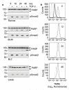
Figure 1
Figure 1
TGF-β activates an intrinsic apoptotic program in centroblastic BL cells. (a) Western blot analysis of apoptosis (cleavage of the caspase substrate PARP) and TGF-β signaling (pSmad2) in a panel of BL cell lines treated with 1ng/ml TGF-β for the times indicated. (b) Loss of mitochondrial inner membrane potential (ΔΨm) during TGF-β-induced apoptosis of Ramos cells shown by reduced mean fluorescence of cells labelled with the mitochondrial stain tetramethlyrhodamine ethyl ester (TMRE). Cells were untreated (dash), labelled with TMRE alone (solid) or labelled with TMRE and treated with TGF-β (dotted). Cells were also treated with cyanide-m-chlorophenylhydrazone (CCCP) (thick solid) to cause complete membrane depolarisation and serve as a positive control for ΔΨm. Cells were treated for the times indicated on each histogram and were analysed by flow cytometry.
Figure 2
Figure 2
TGF-β-induced apoptosis in BL cells is associated with induction of BIK and downregulation of BCL-XL. (a) Analysis of transcripts encoding BCL-2 family members by multiprobe RNAse protection assay (RPA) following TGF-β treatment of BL2 cells. Total RNA from untreated or TGF-β treated cells was analysed during a 6hr time course. Lanes corresponding to yeast negative control RNA (Y), undigested probe (P) and 32P-labelled pBR322 size markers (M) are shown. Sizes of the probe template set are also indicated. Confirmation of BIK induction (b) and BCL-XL downregulation (c) by Western blot analysis of RIPA lysates prepared from untreated and TGF-β (1ng/ml) treated BL cell lines. BIMEL expression was also measured and Smad2/3 levels are shown as a loading control. TGF-β signaling was analysed by Western blotting for phosphorylation of Smad3 (p-Smad3). Actin blots are shown as a loading control.
Figure 3
Figure 3
Regulation of BCL-XL and BIK are critical for optimal TGF-β-mediated apoptosis. (a) Flow cytometry analysis of active caspase-3 positive cells (mean ± s.d., n=3) induced by 24hr TGF-β treatment in L3055 BL cells and BCL-XL stable transfectants. (b) Western blot analysis of RIPA extracts from L3055 BL cells and BCL-XL stable transfectants after 48hrs TGF-β treatment for PARP cleavage, BCL-XL and phospho-Smad2 (p-Smad2). (c) Western blot analysis of 50μg RIPA extracts from untreated BL cells as indicated. Expression levels of BCL-XL and BCL-2 in each cell line are shown. A western blot for actin is included as a loading control. (d) BL2, Ramos and BL40 cells were seeded overnight at 5 × 10⁵/ml and then treated for 48 hours with the BCL-XL inhibitor BH3i-2′ (Calbiochem) as indicated. Apoptosis induction was determined by PI staining and flow cytometry (mean ± s.d., sub-G1 DNA content (n=3)). (e) Ramos cells were left untreated or treated for 48 hours with TGF-β in the presence or absence of low dose BH3i-2′ (5μM). Apoptosis induction was determined by PI staining and flow cytometry (mean ± s.d., sub-G1 DNA content (n=3)). (f) Independent stable cell lines (labelled 1 and 2) expressing non-silencing (NS) and shRNAs targeted against Bik (Bik#1 and Bik#2) were generated by retroviral infection of Ramos BL cells. Quantitative real time PCR (qRT-PCR) was used to demonstrate knockdown of BIK in stable Ramos BL cells. Mean relative amounts of BIK RNA are expressed after normalisation to 18s rRNA levels. (g) Determination of apoptosis (mean ± s.d., sub-G1 DNA content (n=3)) by PI staining and flow cytometry following TGF-β treatment of cell lines described in (f).
Figure 4
Figure 4
TGF-β directly regulates BIK transcription and induces recruitment of activated Smad complexes to the BIK promoter. (a) RPAs for BIK and BCL-XL, expression in cells treated with or without TGF-β and the protein synthesis inhibitors cycloheximide and anisomycin (C+A) as indicated. Time points -2 and 0 are samples harvested prior to and following inhibitor treatment respectively. TGF-β was added at 0 hours. (b) RPA for BIK and the loading control GAPDH following TGF-β treatment of BL cells pretreated for 1 hour with or without the transcription inhibitor actinomycin D (ActD). Time points -1 and 0 are samples harvested prior to and following inhibitor treatment respectively. TGF-β was added at 0 hours. M, P and Y are as described in Figure 2. (c) Real time qRT-PCR carried out on RNA isolated from BL cell lines treated with TGF-β in the presence or absence of the HDAC inhibitor TSA. Shown is the mean ± s.d. amount of RNA normalised to 18s levels and relative to TGF-β untreated samples. (d) Western blot analysis of 50μg RIPA extracts from BL cells treated with and without TGF-β in the presence or absence of TSA as indicated. TGF-β signaling was analysed by Western blotting for phosphorylation of Smads. A western blot for total Smad2/3 is included as a loading control. (e) Potential Smad binding regions (SBRs) within the human Bik promoter. Arrows indicate positions of PCR primers used in ChIP assays shown in (f). (f) ChIP assay for Smad recruitment to the endogenous Bik promoter in BL cells cultured with and without TGF-β for 1 hour. Input lanes are from 10% of samples used in the IPs performed with control IgG, Smad3 and Smad4 antibodies. (g) Sequence of ds oligos used in EMSAs shown in (h) containing the potential wildtype and mutant SBR2 (mSBR2). SBE sequences are shown in bold. (h) EMSA of CA46 nuclear extracts treated with and without TGF-β for 1 hour as indicated and incubated with wild type SBR1 and SBR2 and mSBR2. TGF-β inducible and antibody supershifted complexes are shown (arrowed).
Figure 5
Figure 5
Autocrine TGF-β signaling contributes to spontaneous centroblast apoptosis. Flow cytometry analysis of centroblasts in preparations of explanted normal human tonsil mononuclear cells (TMCs) showing the percent survival of CD77/CD38 +ve cells (a) and the percentage of anti-CD77-FITC stained cells remaining after 24 and 48 hours in vitro culture of TMCs (b) with either exogenous TGF-β or an inhibitor of TGF-β signaling (SB-431542, 10μM) or solvent control (DMSO). (c) Western blot analysis for PARP cleavage and actin of extracts from 1×10⁵ purified centroblasts incubated for 36 hours with TGF-β, SB-431542 or DMSO solvent control. Actin levels are shown as a loading control. (d) Percent of active caspase-3 negative cells (mean ± s.d., n=3) determined by PhiPhiLux (G1D2) fluorescent caspase substrate and flow cytometry in purified centroblasts treated with DMSO (-) or SB-431542 for the indicated times.
Figure 6
Figure 6
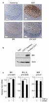
TGF-β-mediated regulation of BIK and BCL-XL in primary human centroblasts. (a) Sections of formalin fixed paraffin-embedded human tonsil tissue were stained with antibodies as indicated and counterstained with haematoxylin. Scale bars represent 200μM. (b) Western blot analysis of BIK expression in TMCs, TMCs depleted of centroblasts (CD77 depleted) and isolated centroblasts (purified CD77). A western blot for actin is included as a loading control. (c) Real time qRT-PCR carried out on RNA isolated from purified centroblasts treated with zVAD-fmk and -/+ TGF-β for 2-4h (BIK and BIM) or 16-24h (BCL-XL). The mean fold changes in relative RNA expression compared to untreated controls are shown. Statistical analysis was carried out using paired t-tests (n=3). Statistically significant (p) values of <0.005 (**) are indicated.
Figure 7
Figure 7. Model of centroblastic cell apoptosis pathways
The contribution of TGF-β signaling to regulation of BCL-2 family members is included alongside known apoptosis pathways functional in germinal centres. The three separate apoptosis pathways converge to regulate death of human centroblasts. FAS signaling activates the extrinsic caspase-8/caspase-3 cascade while TGF-β signaling via its receptor complex represses the prosurvival factor BCL-XL and induces BIK to activate the intrinsic pathway. Fas and TGF-β signaling contribute to the default apoptotic state of ‘death by neglect’ while B cell receptor signaling also activates the mitochondrial apoptosis pathway in cells lacking environmental survival cues. Loss of TGF-β signaling through inhibition of TGF-β receptor signaling by SB-431542 would reduce the amount of caspase-3 activation and raise the threshold for apoptosis induction. The model highlights the requirement for maintenance of cFLIP and BCL-XL levels for cells to survive during germinal centre reactions.

Similar articles

Cited by

References

    1. Shi Y, Massague J. Mechanisms of TGF-beta signaling from cell membrane to the nucleus. Cell. 2003;113:685–700. - PubMed
    1. Schuster N, Krieglstein K. Mechanisms of TGF-beta-mediated apoptosis. Cell Tissue Res. 2002;307:1–14. - PubMed
    1. Li MO, Wan YY, Sanjabi S, Robertson AK, Flavell RA. Transforming growth factor-beta regulation of immune responses. Annu Rev Immunol. 2006;24:99–146. - PubMed
    1. Dong M, Blobe GC. Role of transforming growth factor-beta in hematologic malignancies. Blood. 2006;107:4589–96. - PMC - PubMed
    1. Inman GJ, Allday MJ. Resistance to TGF-beta1 correlates with a reduction of TGF-beta type II receptor expression in Burkitt’s lymphoma and Epstein-Barr virus-transformed B lymphoblastoid cell lines. J Gen Virol. 2000;81:1567–78. - PubMed

MeSH terms