. 2009 Mar;83(6):2406-16.
doi: 10.1128/JVI.01972-08. Epub 2009 Jan 7.

E4orf1 limits the oncolytic potential of the E1B-55K deletion mutant adenovirus

Affiliations

E4orf1 limits the oncolytic potential of the E1B-55K deletion mutant adenovirus

Michael A Thomas et al. J Virol. 2009 Mar.

Abstract

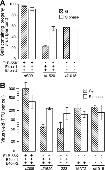
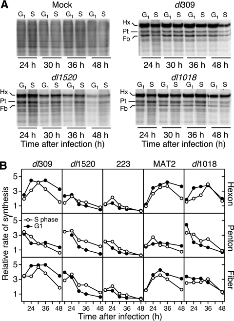
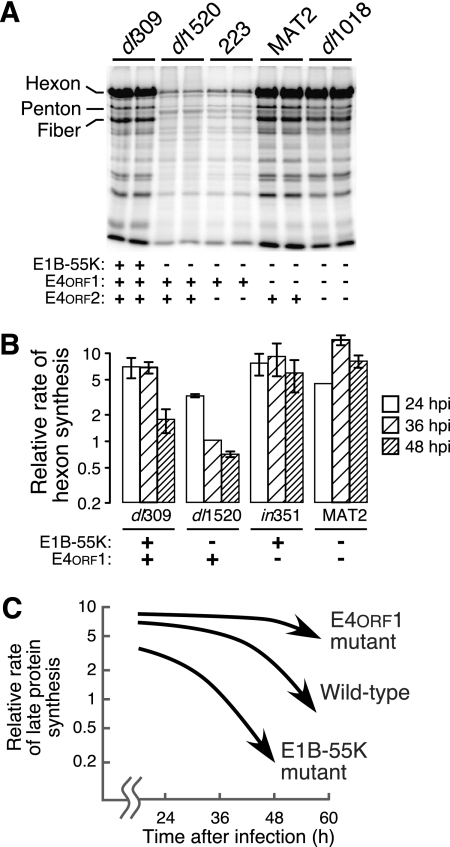
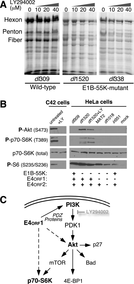
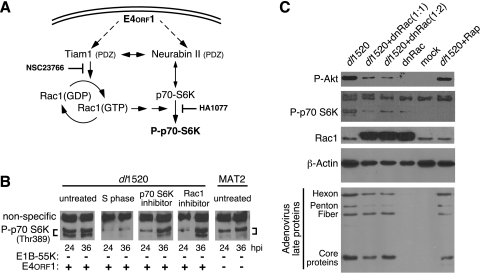
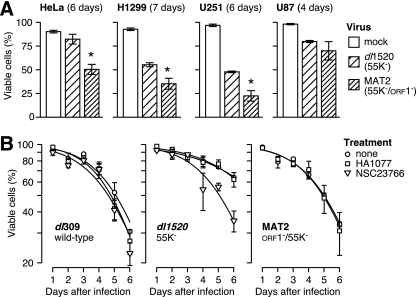
Clinical trials have shown oncolytic adenoviruses to be tumor selective with minimal toxicity toward normal tissue. The virus ONYX-015, in which the gene encoding the early region 1B 55-kDa (E1B-55K) protein is deleted, has been most effective when used in combination with either chemotherapy or radiation therapy. Therefore, improving the oncolytic nature of tumor-selective adenoviruses remains an important objective for improving this form of cancer therapy. Cells infected during the G(1) phase of the cell cycle with the E1B-55K deletion mutant virus exhibit a reduced rate of viral late protein synthesis, produce fewer viral progeny, and are less efficiently killed than cells infected during the S phase. Here we demonstrate that the G(1) restriction imposed on the E1B-55K deletion mutant virus is due to the viral oncogene encoded by open reading frame 1 of early region 4 (E4orf1). E4orf1 has been reported to signal through the phosphatidylinositol 3'-kinase pathway leading to the activation of Akt, mTOR, and p70 S6K. Evidence presented here shows that E4orf1 may also induce the phosphorylation of Akt and p70 S6K in a manner that depends on Rac1 and its guanine nucleotide exchange factor Tiam1. Accordingly, agents that have been reported to disrupt the Tiam1-Rac1 interaction or to prevent phosphorylation of the ribosomal S6 kinase partially alleviated the E4orf1 restriction to late viral protein synthesis and enhanced tumor cell killing by the E1B-55K mutant virus. These results demonstrate that E4orf1 limits the oncolytic nature of a conditionally replicating adenovirus such as ONYX-015. The therapeutic value of similar oncolytic adenoviruses may be improved by abrogating E4orf1 function.


Figures

FIG. 1.
FIG. 1.
E4orf1 imposes a G1 restriction on virus production directed by E1B-55K deletion mutant adenoviruses. Synchronously dividing cultures of HeLa cells were generated by mitotic detachment and HU treatment as described in Materials and Methods. Cells were infected at early G1 (hatched bars) or early S phase (light bars) with the indicated viruses. (A) Infected cells were harvested after 32 h, and at least 100 infected cells from multiple samples were evaluated by transmission electron microscopy for the presence of progeny viral particles. The percentages of cells containing progeny viral particles from four (dl309 and dl1520) and two (dl1018) independent experiments are shown with the standard deviation indicated by error bars. (B) Synchronously dividing HeLa cells were generated and infected as in panel A with the viruses indicated in panel B. After 72 h, the yield of progeny virus was determined by plaque assay and is presented as the infectious virus per infected cell. Results from three independent experiments are represented as the mean with standard error of the mean. The status of the relevant viral gene is indicated below each bar.
FIG. 2.
FIG. 2.
E4orf1 imposes a G1 restriction on viral late protein synthesis directed by E1B-55K deletion mutant adenoviruses. (A) Synchronously dividing cultures of HeLa cells were generated by mitotic detachment and HU treatment and mock infected or infected with the indicated viruses at early G1 or early S phase. Infected cells were pulse-labeled for 1 h with 35S-labeled amino acids at 24, 30, 36, and 48 hpi. Labeled proteins were separated by SDS-PAGE, and the newly synthesized protein was visualized and quantified by phosphorescence imaging. Representative gels are shown with the viral late structural proteins hexon, penton, and fiber indicated as Hx, Pt, and Fb, respectively. (B) The relative rates of synthesis of the viral hexon, penton, and fiber proteins were determined by pulse-labeling as indicated for panel A. The rate of synthesis of each protein was normalized to the rate measured for dl1520 at 36 hpi in cells infected in G1. G1 rates are shown with closed circles, and S-phase rates are shown with open circles.
FIG. 3.
FIG. 3.
E4orf1 restricts viral late protein synthesis in adenovirus-infected cells. (A) Duplicate cultures of asynchronously dividing HeLa cells were infected with the indicated viruses and pulse-labeled with 35S-labeled amino acids at 36 hpi. Labeled proteins were separated by electrophoresis and visualized by phosphorescence imaging. The late structural proteins hexon, penton, and fiber are indicated. The status of the relevant viral gene is indicated below each lane. (B) Asynchronously dividing HeLa cells were infected with the indicated viruses and pulse-labeled for 1 h with 35S-labeled amino acids at 24, 36, and 48 hpi. Radioactivity incorporated into the hexon protein was quantified by phosphorescence imaging. This value was normalized to the amount of radioactivity incorporated into hexon in dl1520 virus-infected cells at 36 hpi. The values shown are the mean of 9 to 23 independent infections with the standard error of the mean indicated. The status of the relevant viral gene is indicated below each virus. (C) The relative rate of late protein synthesis directed by representative viruses at late times after infection is schematically represented for an E4orf1 deletion mutant virus, an E1B-55K deletion mutant virus, and the wild-type virus. E4orf1 restricts viral late protein synthesis at all times of infection in the E1B-55K mutant background and at very late times in the context of a wild-type virus infection.
FIG. 4.
FIG. 4.
The PI3-kinase inhibitor LY294002 diminishes viral late protein synthesis and does not reduce Akt and p70 S6K phosphorylation in adenovirus-infected cells. (A) Asynchronously dividing HeLa cells were infected with either the wild-type virus dl309 or the E1B-55K deletion mutant virus dl1520 or dl338. Four hours after infection, LY294002 was added at the indicated concentration. At 36 hpi, the cells were pulse-labeled with 35S-labeled amino acids and the labeled proteins were visualized by SDS-PAGE and phosphorescence imaging. (B) Akt and p70 S6K are phosphorylated in dl1520 virus-infected cells. The C4-2 prostatic cancer cell line with constitutive activation of the PI3-kinase pathway was left untreated or exposed to 50 μM LY294002 (LY) for 36 h before cellular proteins were separated by SDS-PAGE, transferred to nitrocellulose, and sequentially analyzed by blotting with antibodies specific for phospho-Akt (Ser473), phospho-p70-S6K (Thr389), total p70-S6K, and phosphoribosomal protein S6 (serines 235 and 236). Asynchronously dividing HeLa cells were mock infected or infected with the indicated viruses and exposed to 50 μM LY294002 at 4 h after infection. At 36 hpi, cells were harvested and protein was separated by SDS-PAGE, transferred to nitrocellulose, and analyzed with the indicated antibodies. The status of the relevant viral gene is indicated below each lane.
FIG. 5.
FIG. 5.
E4orf1-induced phosphorylation of p70 S6K and Akt may involve Rac1. (A) Schematic representation of a potential signaling pathway initiated by E4orf1 through the PDZ domain-containing proteins Tiam1 and neurabin II. Solid arrows identify known pathways and interactions. (B) HeLa cells were blocked at the G1/S border by exposure to HU for 24 h, released from the block, and infected 1 h later (S phase) or infected as an asynchronously dividing culture with the E1B-55K single-mutant virus dl1520 or the E1B-55K/E4orf1 double-mutant virus MAT2. At 4 hpi, the growth medium was replaced with fresh medium or with medium containing the p70 S6K inhibitor HA1077 at 50 mM or the Tiam1-Rac1 inhibitor NSC23766 at 40 μM. At 24 and 36 hpi, cellular proteins were analyzed by immunoblotting for phosphorylation of Thr389 on p70 S6K. A nonspecific cross-reacting protein is indicated which served as a loading control. The status of the relevant viral gene is indicated below each lane. (C) Asynchronously dividing HeLa cells were mock infected or infected with a nonreplicating adenovirus vector expressing dnRac1 (dnRac) at a multiplicity of 20 (1:1) or 40 (1:2). After 24 h, the cells were again mock infected or infected with the E1B-55K deletion mutant virus dl1520 at a multiplicity of 20. After 1 h, rapamycin (Rap) was added to 50 nM to one culture. Cells were harvested 36 h after infection with dl1520. Infected cell proteins were separated by SDS-PAGE and analyzed by immunoblotting with monospecific antibodies (phospho-Akt, phospho-P70 S6K, Rac1, β-actin) or polyclonal serum specific for the adenovirus late proteins.
FIG. 6.
FIG. 6.
E4orf1 restricts the tumor cell-killing potential of E1B-55K mutant virus dl1520 (ONYX-015) in a Tiam1/Rac1-dependent manner. (A) Cell lines derived from human cervical carcinoma (HeLa), large-cell lung carcinoma (H1299), and malignant glioblastoma (U251, U87) were mock infected or infected with the indicated viruses at a multiplicity sufficient to infect all of the cells. The viability of the cells was determined by trypan blue dye exclusion on the indicated day after infection. Values represent the mean of three independent infections with the standard error of the mean. The asterisk identifies values for MAT2-infected cells that are significantly (P < 0.05) less than the viability of dl1520 virus-infected cells. (B) HeLa cells were infected with the wild-type virus (dl309), the E1B-55K mutant virus (dl1520), or the E1B-55K/E4orf1 double-mutant virus (MAT2) at a multiplicity of 20 and left untreated (circles) or were adjusted to 50 mM HA1077 (squares) or 40 μM NSC2366 (triangles) at 4 hpi. The fraction of viable cells was determined daily for 6 days after infection by trypan blue dye exclusion. The plotted values represent the mean of three independent infections with the standard error of the mean indicated by error bars. The lines represent best-fit curves determined by logistic regression with a quasibinomial model.

Similar articles

Cited by

References

    1. Araujo, F. D., T. H. Stracker, C. T. Carson, D. V. Lee, and M. D. Weitzman. 2005. Adenovirus type 5 E4orf3 protein targets the Mre11 complex to cytoplasmic aggresomes. J. Virol. 7911382-11391. - PMC - PubMed
    1. Babiss, L. E., and H. S. Ginsberg. 1984. Adenovirus type 5 early region 1b gene product is required for efficient shutoff of host protein synthesis. J. Virol. 50202-212. - PMC - PubMed
    1. Baker, A., K. J. Rohleder, L. A. Hanakahi, and G. Ketner. 2007. Adenovirus E4 34k and E1b 55k oncoproteins target host DNA ligase IV for proteasomal degradation. J. Virol. 817034-7040. - PMC - PubMed
    1. Barker, D. D., and A. J. Berk. 1987. Adenovirus proteins from both E1B reading frames are required for transformation of rodent cells by viral infection and DNA transfection. Virology 156107-121. - PubMed
    1. Barraud, P., J. C. Paillart, R. Marquet, and C. Tisne. 2008. Advances in the structural understanding of Vif proteins. Curr. HIV Res. 691-99. - PMC - PubMed

Publication types

MeSH terms

Substances