Comparative Study
. 2009 Jan 8:10:10.
doi: 10.1186/1471-2164-10-10.

A gene expression signature shared by human mature oocytes and embryonic stem cells

Affiliations
Comparative Study

A gene expression signature shared by human mature oocytes and embryonic stem cells

Said Assou et al. BMC Genomics. .

Abstract

Background: The first week of human pre-embryo development is characterized by the induction of totipotency and then pluripotency. The understanding of this delicate process will have far reaching implication for in vitro fertilization and regenerative medicine. Human mature MII oocytes and embryonic stem (ES) cells are both able to achieve the feat of cell reprogramming towards pluripotency, either by somatic cell nuclear transfer or by cell fusion, respectively. Comparison of the transcriptome of these two cell types may highlight genes that are involved in pluripotency initiation.

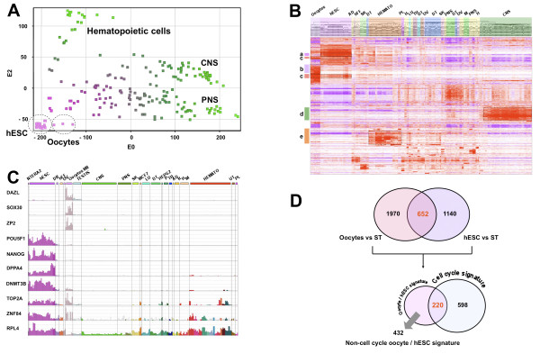
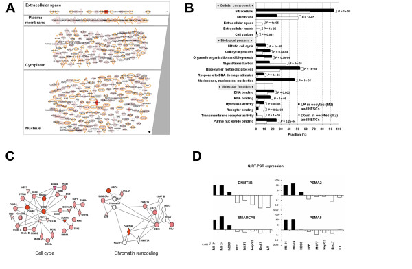
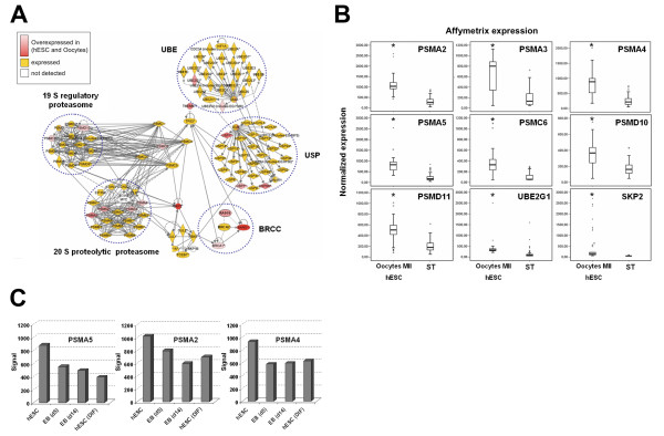
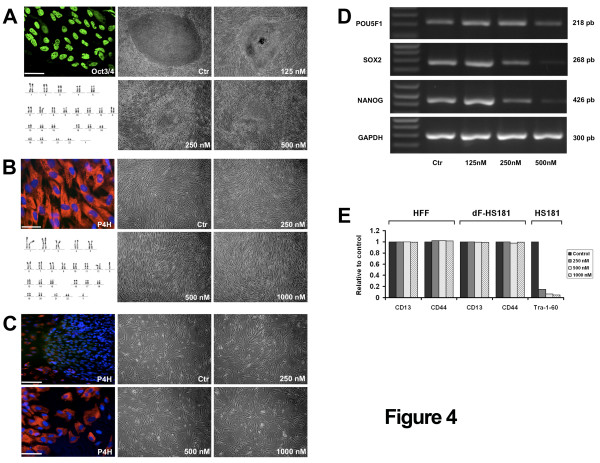
Results: Based on a microarray compendium of 205 samples, we compared the gene expression profile of mature MII oocytes and human ES cells (hESC) to that of somatic tissues. We identified a common oocyte/hESC gene expression profile, which included a strong cell cycle signature, genes associated with pluripotency such as LIN28 and TDGF1, a large chromatin remodelling network (TOP2A, DNMT3B, JARID2, SMARCA5, CBX1, CBX5), 18 different zinc finger transcription factors, including ZNF84, and several still poorly annotated genes such as KLHL7, MRS2, or the Selenophosphate synthetase 1 (SEPHS1). Interestingly, a large set of genes was also found to code for proteins involved in the ubiquitination and proteasome pathway. Upon hESC differentiation into embryoid bodies, the transcription of this pathway declined. In vitro, we observed a selective sensitivity of hESC to the inhibition of the activity of the proteasome.

Conclusion: These results shed light on the gene networks that are concurrently overexpressed by the two human cell types with somatic cell reprogramming properties.


Figures

Figure 1
Figure 1
Human mature MII oocytes and hESC display a common transcriptome signature. Principal component analysis (PCA) (A). A PCA of 9 mature oocytes (MII), 29 hESCs and 167 somatic tissue samples. CNS, central nervous system; PNS, peripheral nervous system. Hierarchical clustering (B). Each horizontal line represents a PS and each column represents a single sample. PS up and down-regulated were colored in red and purple, respectively. In white, PS which were neither over nor under-expressed. Clusters: a: hESCs genes; b: MII oocytes genes; c: the oocytes/hESC signature; d: genes expressed in CNS; e: genes expressed in hematopoietic cells. AD: adipocytes; hFF: human foreskin fibroblasts; SK: skin and keratinocytes; DT: digestive tract; Hemato: hematopoietic cells; PL: placenta; H: heart; LU: lung; TH: thyroid; UT: uterus; OV: ovary; M: muscle. Expression bar charts of selected genes (C). Expression bar charts of three oocytes specific genes (DAZL, SOX30, ZP2), three hESCs specific genes (POU5F1/OCT4, NANOG, and DPPA4), three genes up-regulated in hESC and oocytes MII (DNMT3B, TOP2A and ZNF84) and one ubiquitously expressed gene (RPL4) using our on-line expression atlas Amazonia! . Diff: non-specific differentiated hESC; MCF7: breast cancer cell line; HEPG2: hepatocarcinoma cell line. Venn diagram detailing shared and distinct gene expression among hESCs and MII human oocytes (D). The oocyte/hESC signature was defined as the intersection of the hESC signature (genes overexpressed in hESC compared to somatic tissues (ST)) and the MII oocytes signature (genes overexpressed in MII oocytes compared to ST). The oocytes/hESC signature was then further parted into a fraction sharing a cell cycle signature and a non-cell cycle part.
Figure 2
Figure 2
Biological annotations of genes overexpressed in human mature MII oocytes and hESC. Cellular compartment localisation according to Gene Ontology (GO) annotations (A). Statistical comparison of the distribution of GO annotations in the oocytes/hESC signature with the genes underexpressed in oocytes and hESC (B). Gene Ontology categories which differed significantly (p value ≤ 0.01) between oocytes and hESC are shown. Oocyte/hESC gene networks (C). We computed interaction networks from the oocyte/hESC signature. Genes included in the oocyte/hESC signature are in red (the color intensity is proportional to the oocyte/hESC to somatic samples fold change). Genes not found in the signature are in white. In each network, edge types are indicatives: a plain line indicates direct interaction, a dashed line indicates indirect interaction; a line without arrowhead indicates binding only; a line with an arrowhead indicates "acts on". Node types represent different types of molecules: diamond, enzyme; square, cytokine; triangle, phosphatase; and circle, other. Double line edge represents a group or a complex. Validation of microarray data (D). Gene expression of DNMT3A, SMARCA5, PSMA5, PSMA2 were assayed with quantitative RT-PCR (QRT-PCR). The expression of these four selected genes was compared in mature oocytes, hESC and various somatic samples: human fibroblasts (hFF), human breast adenocarcinoma cell line (MCF7), human hepatocellular liver carcinoma cell line (HepG2), endometrial cells (End7) and T-lymphocyte cells (TL) using QRT-PCR. All measurements were performed in duplicate in two separate runs. The relative levels of gene expression of target mRNA was normalized against GAPDH expression. Fold change values are plotted on a log10 scale.
Figure 3
Figure 3
Overexpression of the ubiquitination/proteasome pathway. Interaction network analysis of the ubiquitination/proteasome pathway in the oocytes/hESC signature. (A) The interaction network was generated with the Ingenuity software and shows a high number of genes from this cellular pathway overexpressed (red) or expressed (orange) in the oocytes/hESC signature. Transcripts that were not detected by the microarray are white. Notably, we found elements of the ubiquitin-activating enzyme (UBE), BRCA1- and BRCA2-containing complex (BRCC), the regulatory 19S proteasome, the core proteolytic 20S proteasome and the ubiquitin-specific protease (USP) modules in the oocyte/hESC signature. Gene expression measured by microarrays in 9 genes involved in the ubiquitin and proteasome pathway (B). Box-and-whisker plots comparing the expression level of PSMA2, PSMA3, PSMA4, PSMA5, PSMC6, PSMD10, PSMD11, UBE2G1 and SKP2 in mature oocytes and hESC (38 samples) versus somatic tissues (167 samples). The signal intensity for each gene is shown on the y axis as arbitrary units determined by the GCOS 1.2 software (Affymetrix). (*): P-value < 0.0001 using a Mann-Whitney statistical test. Down regulation of the proteasome pathway during hESC differentiation (C). U133A microarray signal values for PSMA2, PSMA4 and PSMA5 in 29 undifferentiated hESC samples (mean value) versus two embryoid bodies (EB) samples (EB day 5 and EB day 14) and 3 non-lineage differentiated hESC samples (mean value).
Figure 4
Figure 4
Blocking proteasome activity in hESC. Proteasome inhibition causes differentiation of hESCs (A). HS181 colonies (p56) were grown 4 days on hFF and then treated 40 hours with the proteasome inhibitor MG132. Control HS181 colonies express POU5F1 (scale bar 25 μ) and display a normal karyotype. Upon treatment with MG132, HS181 colonies differentiated. No effect of proteasome inhibition on hFF (B). HFF express the fibroblastic marker P4H and display a normal karyotype. HFF were cultured with MG132 during 40 hours. No morphological alteration was observed. No effect of proteasome inhibition on hES-dF-HD90 (C). The HD90 hESC line was differentiated into fibroblasts like cells (hES-dF) that display morphologic feature of dermal fibroblasts and express P4H: bottom left. An undifferentiated colony of HD90 P4H-, starts to differentiate at the edges into P4H+ hES-dF (upper left). hES-dF-HD90 cells were cultured with MG132 during 40 hours. No morphological alteration was observed. Down regulation of pluripotency transcription factor in hESC by proteasome blockage (D). The expression of POU5F1/OCT4, NANOG and SOX2 was measured by semiquantitative RT-PCR in HS181 hESC after 40 h culture with MG132. GAPDH was used as a control. Flow cytometry analysis after proteasome inhibition (E). HFF, hES-dF-HS181 and HS181 cells were treated 40 hours with MG132, and then the cell surface fibroblastic markers CD13 and CD44, and the pluripotent cell surface marker TRA-1-60 were measured by FACS. Markers for fibroblastic cells were not altered with MG132 treatment, whereas the marker for pluripotency dropped to barely detectable level. Y-axis: percentage of the control (untreated) sample.

Similar articles

Cited by

References

    1. Huang JC, Lei ZL, Shi LH, Miao YL, Yang JW, Ouyang YC, Sun QY, Chen DY. Comparison of histone modifications in in vivo and in vitro fertilization mouse embryos. Biochem Biophys Res Commun. 2007;354:77–83. doi: 10.1016/j.bbrc.2006.12.163. - DOI - PubMed
    1. Santos F, Hendrich B, Reik W, Dean W. Dynamic reprogramming of DNA methylation in the early mouse embryo. Dev Biol. 2002;241:172–182. doi: 10.1006/dbio.2001.0501. - DOI - PubMed
    1. Campbell KH, McWhir J, Ritchie WA, Wilmut I. Sheep cloned by nuclear transfer from a cultured cell line. Nature. 1996;380:64–66. doi: 10.1038/380064a0. - DOI - PubMed
    1. Gurdon JB, Byrne JA. The first half-century of nuclear transplantation. Proceedings of the National Academy of Sciences of the United States of America. 2003;100:8048–8052. doi: 10.1073/pnas.1337135100. - DOI - PMC - PubMed
    1. Sung LY, Gao S, Shen H, Yu H, Song Y, Smith SL, Chang CC, Inoue K, Kuo L, Lian J, et al. Differentiated cells are more efficient than adult stem cells for cloning by somatic cell nuclear transfer. Nature genetics. 2006;38:1323–1328. doi: 10.1038/ng1895. - DOI - PubMed

Publication types

MeSH terms

Substances