. 2009 Aug 1;57(10):1115-29.
doi: 10.1002/glia.20835.

Developmental and post-injury cortical gliogenesis: a genetic fate-mapping study with Nestin-CreER mice

Affiliations

Developmental and post-injury cortical gliogenesis: a genetic fate-mapping study with Nestin-CreER mice

Kevin A Burns et al. Glia. .

Abstract

The primary sources of cortical gliogenesis, either during development or after adult brain injury, remain uncertain. We previously generated Nestin-CreER mice to fate-map the progeny of radial glial cells (RG), a source of astrocytes and oligodendrocytes in the nervous system. Here, we show that Nestin-CreER mice label another population of glial progenitors, namely the perinatal subventricular zone (SVZ) glioblasts, if they are crossed with stop-floxed EGFP mice and receive tamoxifen in late embryogenesis (E16-E18). Quantification showed E18 tamoxifen-induction labeled more perinatal SVZ glioblasts than RG and transitional RG combined in the newborn brain (54% vs. 22%). Time-lapse microscopy showed SVZ-glioblasts underwent complex metamorphosis and often-reciprocal transformation into transitional RG. Surprisingly, the E10-dosed RG progenitors produced astrocytes, but no oligodendrocytes, whereas E18-induction fate-mapped both astrocytes and NG2+ oligodendrocyte precursors in the postnatal brain. These results suggest that cortical oligodendrocytes mostly derive from perinatal SVZ glioblast progenitors. Further, by combining genetic fate-mapping and BrdU-labeling, we showed that cortical astrocytes cease proliferation soon after birth (<P10) and only undergo nonproliferative gliosis (i.e., increased GFAP expression without cell-division) after stab-wound injury in adult brains. By contrast, 9.7% of cortical NG2+ progenitors remained mitotic at P29, and the ratio rose to 13.8% after stab-wound injury. Together, these results suggest NG2+ progenitors, rather than GFAP+ astrocytes, are the primary source of proliferative gliosis after adult brain injury.


Figures

Figure 1
Figure 1. Nestin-CreER mice lineage-trace radial glia and SVZ glioblasts
The identities of labeling progenitors were examined in Nestin-CreER/EGFP-Cre-Reporter (Bitransgenic) embryos at 2 days following tamoxifen (Tam)-induction at E10.5 (A-B, E-H) or E18.5 (I-L). A, B, E10Tam-E12 chase labeled many EGFP+ cells in the midbrain (MB) and the ventral telencephalon (VT), and a few cells in the developing cerebral cortex (Ctx). C, D Without Tam-induction, there were no EGFP+ cells in E14 bi-transgenic embryos. E, E10Tam-E12 chase labeled many EGFP+ cell profiles, including radial glia (RG) and short neural precursors (asterisk). V: ventricular surface. F, G, H, Double-labeling of E10Tam-E12 cortex shows RG and short neural precursors express nestin, whereas Cajal-Retzius cells (CR) near the pia do not. I-L, E18Tam-P1 chase labeled many EGFP+ cells in the subventricular zone (SVZ) and a few cells with curved radial fibers (arrowheads) which are labeled by nestin (L) Scale bar: 400μm in A, B; 50μm in C, D; 50μm in E-H; 100μm in I-K; 200μm in L.
Figure 2
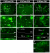
Figure 2. Genetic fate-mapping shows early divergent oligodendrocyte precursors
We examined EGFP+ cells in the cerebral cortex of P30 bitransgenic mice that were dosed with Tam on E10.5, E14.5, or E18.5. A, D, G, E10 Tam-induction labeled EGFP+ astrocytes (asterisk) and neurons in all layers of the cerebral cortex (A, D), but no oligodendrocytes in the cortex (Ctx) or the corpus callosum (CC) (G). B, E, H, E14-induction labeled astrocytes and neurons of upper cortical layers, as well as a few oligodendrocytes (Olig) in both Ctx and CC (H). C, F, I, J, E18-induction labeled astrocytes and oligodendrocytes, but no cortical neurons (C-I); however, perinatal-born hippocampal neurons were labeled by induction at E10, E14, and E18 (J). Scale bars: 300mm in A-C, J; 50mm in D-I.
Figure 3
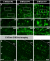
Figure 3. Late embryonic induction in Nestin-CreER mice labels perinatal SVZ glioblasts
A-C, Cortical gliogenesis was examined using Nestin-CreER/EGFP-Cre-Reporter mice dosed with Tam on E18 and sacrificed at P2, P4, or P7. a-c, Magnified images of boxed areas in A-C showed astrocytes in the cortical plate (CP) displayed increasingly branched cytoplasmic processes from P2 to P7. a′-c′, There was an increase of EGFP+ glioblasts in the subventricular zone (SVZ) from P2 to P4, then declined from P4 to P7. D, Time-lapse video microscope was used to image the proliferation and maturation of glial progenitors over 4 days following the E16Tam-E18 chase protocol (also see Supplementary Movie 1). CP: cortical plate, IZ: intermediate zone; SVZ/VZ: subventricular and ventricular zones. Scale bars: 300μm in A-C; 120μm in a-c and a′-c′.
Figure 4
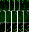
Figure 4. Live-imaging of SVZ glioblasts migration and proliferation in brain slices
Time-lapse video microscopy was used to image the migration and proliferation of glial progenitors in slices of E18 forebrain following E16 Tam-induction. A, Global view of glioblast proliferation and maturation over 4 days (see also supplementary movie 1). A, Translocation mode of glioblast migration from the SVZ/VZ across the intermediate zone (IZ) (Supplementary Movie 2). B, Locomotion mode of glioblasts migration in continuous waves (white and red arrows) from the SVZ towards the pial surface (Supplementary Movie 3). C, Simple glioblasts may undergo several rounds of division in the cortex (pial surface outlined) (Supplementary Movie 4). All times indicate hours in culture.
Figure 5
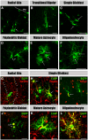
Figure 5. Classification of the descendants of RG and SVZ glioblast progenitors
Cortical glial cell types characterized at different postnatal stages include: A, Radial glia have characteristic radial processes. B, Transitional/bipolar cells have transitional morphologies shorter and thicker radial processes. C, Simple glioblasts have short leading processes. D, Polydendritic glioblasts have the basic architecture but lack the fine branching of mature astrocytes. E, Mature astrocytes have multiple processes with extensive branching. F, Oligodendrocytes have fine lacy processes aligned in spherical or parallel sheets. G-L, Radial glia express Nestin in their processes (arrows in G), simple glioblasts express Nestin (H) and Olig2 (I), polydendritic glioblasts express Olig2 (J), mature astrocytes express GFAP (K), and oligodendrocyte express RIP (L). Shown in G-L are the merged images. For single-channel images see Supplemental Figure 1. Scale bar: 50μm in A-F; 20μm in G-L.
Figure 6
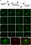
Figure 6. Simple glioblasts and transitional RG show reciprocal metamorphosis
A, Summary of morphological transformations between various forms of glioblasts in the perinatal cortex revealed by time-lapse microscopy. (see also supplementary movie 5). B, Simple glioblasts may extend processes similar to bipolar forms. C, Bipolar transitional forms can become simple or polydendritic glioblasts following mitosis. D, Transitional forms retract radially aligned processes and undergo branching to become mature glia. E, Simple glioblasts also undergo branching to become mature glia. F, The proliferation of polydendritic glioblasts is marked in vivo by incorporation of BrdU administered 30 minutes prior to sacrifice. Scale bars: 20μm in F.
Figure 7
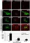
Figure 7. Oligodendrocytes have a longer time-frame of proliferation than astrocytes in the postnatal cerebral cortex
A-C, M, Administration of BrdU over a three-day dosing period (P2-4, P10-12, and P27-29) showed marked reduction of proliferation in the cortex in the first postnatal month. D-F, Astrocytes pre-labeled by E18 tamoxifen-dosing were readily labeled by P2-P4 BrdU injection, but their proliferation is curtailed by P10 (M). G-I, Oligodendrocyte precursors labeled by BrdU often appear as doublets. J-L, oligodendrocyte precursors predominantly express NG2. M, Quantification of BrdU+ cells among EGFP+ astrocytes and NG2+ precursors in the first postnatal month. Astrocytes showed a rapid decline of proliferative capacity, while 9.7% oligodendrocytes still incorporated BrdU at P27-29. Scale bars: 50μm A-C, 20μm D-L.
Figure 8
Figure 8. Stab-wound injury induces GFAP expression by astrocytes in adult brains
Bi-transgenic mice dosed with Tam at E18 were challenged by a needlestick wound at P45, and sacrificed on P49. A, B, The stab-wound robustly upregulated GFAPnear the needle insertion track (arrow). C, D, Following the unilateral needlestick stab-wound, EGFP+ astrocytes on the lesion side express a higher amount of GFAP in the proximal and distal branches (D), than on the contralateral side (C). E, The percentage of GFAP/EGFP double-labeled cells among all fate-mapped astrocytes was increased within the 1mm radius of the needlestick track in the cerebral cortex compared to the contralateral side (n=4). Scale bar: 200μm in A, B; 50μm in C, D.
Figure 9
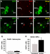
Figure 9. Oligodendrocyte progenitors, but not GFAP-astrocytes, respond to the stab-wound injury with proliferation
In the stab-wound experiment described in Figure 8, BrdU was injected daily from P42 to P48. BrdU/EGFP double-labeling showed the vast majority of EGFP+ astrocytes (arrowhead in A-C) did not incorporate BrdU after the stab-wound injury. In contrast, many EGFP+ oligodendrocyte precursors (arrows in D-I) incorporated BrdU after the stab-wound injury. G, The percentage of BrdU-incorporated astrocytes remained unchanged on the lesion side of the brain after stab-wound injury. H, However, the percentage of BrdU+/EGFP+ oligodendrocyte precursors (OPCs) trended upward on the lesion side (n=4) (p=0.16 by two-tailed student's t-test). Scale bars: 50μm
Figure 10
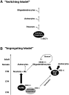
Figure 10. Two prototypic models to explain the lineage relationship among major cortical cell types
A, The “switching” model proposes a common progenitor pool that sequentially gives rise to neurons, astrocytes, and oligodendrocytes. This common progenitor population may be radial glia (RG). B, The “segregating” model proposes early divergence of neuron and macroglial lineages in the developing cerebral cortex. We suggest that a set of precursors expressing Nestin second-intron enhancer (NIE) around E10 is the principle neuronal progenitor (N) that may overlap with the RG lineage. The precursors of the subventricular zone glioblasts (SVZ-GB) are NIE-negative at E10 and gradually upregulate NIE in later development. The SVZ-GB progenitor pool eventually give rise to the bulk of postnatal astrocytes and NG2+ oligodendrocytes. In postnatal brains, NG2+ oligodendrocyte precursors maintain a renewal capacity and respond to injury.

Similar articles

Cited by

References

    1. Alonso G. NG2 proteoglycan-expressing cells of the adult rat brain: possible involvement in the formation of glial scar astrocytes following stab wound. Glia. 2005;49:318–338. - PubMed
    1. Anthony TE, Klein C, Fishell G, Heintz N. Radial glia serve as neuronal progenitors in all regions of the central nervous system. Neuron. 2004;41:881–890. - PubMed
    1. Baracskay KL, Kidd GJ, Miller RH, Trapp BD. NG2-positive cells generate A2B5 positive oligodendrocyte precursor cells. Glia. 2007;55:1001–1010. - PubMed
    1. Buffo A, Rite I, Tripathi P, Lepier A, Colak D, Horn AP, Mori T, Gotz M. Origin and progeny of reactive gliosis: A source of multipotent cells in the injured brain. Proc Natl Acad Sci U S A. 2008;105:3581–3586. - PMC - PubMed
    1. Burns KA, Ayoub AE, Breunig JJ, Adhami F, Weng WL, Colbert MC, Rakic P, Kuan CY. Nestin-CreER mice reveal DNA synthesis by nonapoptotic neurons following cerebral ischemia hypoxia. Cereb Cortex. 2007;17:2585–2592. - PubMed

Publication types

MeSH terms