Prominin 1 marks intestinal stem cells that are susceptible to neoplastic transformation
- PMID: 19092805
- PMCID: PMC2633030
- DOI: 10.1038/nature07589
Prominin 1 marks intestinal stem cells that are susceptible to neoplastic transformation
Abstract
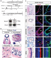
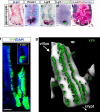
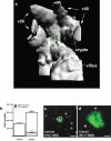
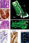
Cancer stem cells are remarkably similar to normal stem cells: both self-renew, are multipotent and express common surface markers, for example, prominin 1 (PROM1, also called CD133). What remains unclear is whether cancer stem cells are the direct progeny of mutated stem cells or more mature cells that reacquire stem cell properties during tumour formation. Answering this question will require knowledge of whether normal stem cells are susceptible to cancer-causing mutations; however, this has proved difficult to test because the identity of most adult tissue stem cells is not known. Here, using an inducible Cre, nuclear LacZ reporter allele knocked into the Prom1 locus (Prom1(C-L)), we show that Prom1 is expressed in a variety of developing and adult tissues. Lineage-tracing studies of adult Prom1(+/C-L) mice containing the Rosa26-YFP reporter allele showed that Prom1(+) cells are located at the base of crypts in the small intestine, co-express Lgr5 (ref. 2), generate the entire intestinal epithelium, and are therefore the small intestinal stem cell. Prom1 was reported recently to mark cancer stem cells of human intestinal tumours that arise frequently as a consequence of aberrant wingless (Wnt) signalling. Activation of endogenous Wnt signalling in Prom1(+/C-L) mice containing a Cre-dependent mutant allele of beta-catenin (Ctnnb1(lox(ex3))) resulted in a gross disruption of crypt architecture and a disproportionate expansion of Prom1(+) cells at the crypt base. Lineage tracing demonstrated that the progeny of these cells replaced the mucosa of the entire small intestine with neoplastic tissue that was characterized by focal high-grade intraepithelial neoplasia and crypt adenoma formation. Although all neoplastic cells arose from Prom1(+) cells in these mice, only 7% of tumour cells retained Prom1 expression. Our data indicate that Prom1 marks stem cells in the adult small intestine that are susceptible to transformation into tumours retaining a fraction of mutant Prom1(+) tumour cells.
Figures


Comment in
-
The stem of cancer.Cancer Cell. 2009 Feb 3;15(2):87-9. doi: 10.1016/j.ccr.2009.01.011. Cancer Cell. 2009. PMID: 19185843
Similar articles
-
Prominin-1/CD133 marks stem cells and early progenitors in mouse small intestine.Gastroenterology. 2009 Jun;136(7):2187-2194.e1. doi: 10.1053/j.gastro.2009.03.002. Epub 2009 Mar 24. Gastroenterology. 2009. PMID: 19324043
-
MET Signaling Mediates Intestinal Crypt-Villus Development, Regeneration, and Adenoma Formation and Is Promoted by Stem Cell CD44 Isoforms.Gastroenterology. 2017 Oct;153(4):1040-1053.e4. doi: 10.1053/j.gastro.2017.07.008. Epub 2017 Jul 14. Gastroenterology. 2017. PMID: 28716720
-
Prominin-1 (CD133) defines both stem and non-stem cell populations in CNS development and gliomas.PLoS One. 2014 Sep 3;9(9):e106694. doi: 10.1371/journal.pone.0106694. eCollection 2014. PLoS One. 2014. PMID: 25184684 Free PMC article.
-
CD133: molecule of the moment.J Pathol. 2008 Jan;214(1):3-9. doi: 10.1002/path.2283. J Pathol. 2008. PMID: 18067118 Review.
-
[Progress and prospects in cancer stem cell research for hepatocellular carcinoma].Ai Zheng. 2009 Sep;28(9):1004-8. doi: 10.5732/cjc.008.10835. Ai Zheng. 2009. PMID: 19728923 Review. Chinese.
Cited by
-
The stem cell marker Prom1 promotes axon regeneration by down-regulating cholesterol synthesis via Smad signaling.Proc Natl Acad Sci U S A. 2020 Jul 7;117(27):15955-15966. doi: 10.1073/pnas.1920829117. Epub 2020 Jun 17. Proc Natl Acad Sci U S A. 2020. PMID: 32554499 Free PMC article.
-
CD133, Selectively Targeting the Root of Cancer.Toxins (Basel). 2016 May 28;8(6):165. doi: 10.3390/toxins8060165. Toxins (Basel). 2016. PMID: 27240402 Free PMC article. Review.
-
Overlapping DNA methylation dynamics in mouse intestinal cell differentiation and early stages of malignant progression.PLoS One. 2015 May 1;10(5):e0123263. doi: 10.1371/journal.pone.0123263. eCollection 2015. PLoS One. 2015. PMID: 25933092 Free PMC article.
-
Generation and staining of intestinal stem cell lineage in adult midgut.Methods Mol Biol. 2012;879:47-69. doi: 10.1007/978-1-61779-815-3_4. Methods Mol Biol. 2012. PMID: 22610553 Free PMC article.
-
Heparin-binding EGF-like growth factor protects intestinal stem cells from injury in a rat model of necrotizing enterocolitis.Lab Invest. 2012 Mar;92(3):331-44. doi: 10.1038/labinvest.2011.167. Epub 2011 Dec 12. Lab Invest. 2012. PMID: 22157721 Free PMC article.
References
-
- Clarke MF, Fuller M. Stem cells and cancer: two faces of eve. Cell. 2006;124:1111–1115. - PubMed
-
- Barker N, et al. Identification of stem cells in small intestine and colon by marker gene Lgr5. Nature. 2007;449:1003–1007. - PubMed
-
- Ricci-Vitiani L, et al. Identification and expansion of human colon-cancer-initiating cells. Nature. 2007;445:111–115. - PubMed
-
- O'Brien CA, et al. A human colon cancer cell capable of initiating tumour growth in immunodeficient mice. Nature. 2007;445:106–110. - PubMed
-
- Sparks AB, et al. Mutational analysis of the APC/beta-catenin/Tcf pathway in colorectal cancer. Cancer Res. 1998;58:1130–1134. - PubMed
Publication types
MeSH terms
Substances
Grants and funding
- R01 CA129541/CA/NCI NIH HHS/United States
- P01CA96832/CA/NCI NIH HHS/United States
- R01 CA129541-02/CA/NCI NIH HHS/United States
- P30CA021765/CA/NCI NIH HHS/United States
- R01 CA129541-01/CA/NCI NIH HHS/United States
- P30 CA021765/CA/NCI NIH HHS/United States
- R01 MH079079/MH/NIMH NIH HHS/United States
- R01 MH079079-02/MH/NIMH NIH HHS/United States
- R01 MH079079-04/MH/NIMH NIH HHS/United States
- R01 MH079079-03/MH/NIMH NIH HHS/United States
- R01CA129541/CA/NCI NIH HHS/United States
- R01 MH079079-05/MH/NIMH NIH HHS/United States
- P01 CA096832-01A10003/CA/NCI NIH HHS/United States
- R01 MH079079-01A2/MH/NIMH NIH HHS/United States
- P01 CA096832/CA/NCI NIH HHS/United States
LinkOut - more resources
Full Text Sources
Other Literature Sources
Medical
Molecular Biology Databases
Research Materials
Miscellaneous

