The cell of origin of ovarian epithelial tumours
- PMID: 19038766
- PMCID: PMC4176875
- DOI: 10.1016/S1470-2045(08)70308-5
The cell of origin of ovarian epithelial tumours
Abstract
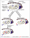
Although it is widely believed that ovarian epithelial tumours arise in the coelomic epithelium that covers the ovarian surface, it has been suggested that they could instead arise from tissues that are embryologically derived from the Müllerian ducts. This article revisits this debate by discussing recent epidemiological and molecular biological findings as well as evidence based on histopathological observations of surgical specimens from individuals with familial ovarian cancer predisposition. Morphological, embryological, and molecular biological characteristics of ovarian epithelial tumours that must be accounted for in formulating a theory about their cell of origin are reviewed, followed by comments about the ability of these two hypotheses to account for each of these characteristics. An argument is made that primary ovarian epithelial tumours, fallopian tube carcinomas, and primary peritoneal carcinomas are all Müllerian in nature and could therefore be regarded as a single disease entity. Although a substantial proportion of cancers currently regarded as of primary ovarian origin arise in the fimbriated end of the fallopian tube, this site cannot account for all of these tumours, some of which are most likely derived from components of the secondary Müllerian system.
Figures


Similar articles
-
Coming into focus: the nonovarian origins of ovarian cancer.Ann Oncol. 2013 Nov;24 Suppl 8(Suppl 8):viii28-viii35. doi: 10.1093/annonc/mdt308. Ann Oncol. 2013. PMID: 24131966 Free PMC article. Review.
-
Fallopian tube precursors of ovarian low- and high-grade serous neoplasms.Histopathology. 2013 Jan;62(1):44-58. doi: 10.1111/his.12046. Histopathology. 2013. PMID: 23240669 Review.
-
Expression of PAX2 in papillary serous carcinoma of the ovary: immunohistochemical evidence of fallopian tube or secondary Müllerian system origin?Mod Pathol. 2007 Aug;20(8):856-63. doi: 10.1038/modpathol.3800827. Epub 2007 May 25. Mod Pathol. 2007. PMID: 17529925
-
Intraperitoneal serous adenocarcinoma: a critical appraisal of three hypotheses on its cause.Am J Obstet Gynecol. 2004 Sep;191(3):718-32. doi: 10.1016/j.ajog.2004.02.067. Am J Obstet Gynecol. 2004. PMID: 15467531 Review.
-
[Significance and expression of PAX8, PAX2, p53 and RAS in ovary and fallopian tubes to origin of ovarian high grade serous carcinoma].Zhonghua Fu Chan Ke Za Zhi. 2017 Oct 25;52(10):687-696. doi: 10.3760/cma.j.issn.0529-567X.2017.10.008. Zhonghua Fu Chan Ke Za Zhi. 2017. PMID: 29060967 Chinese.
Cited by
-
Assessing the risk of cervical neoplasia in the post-HPV vaccination era.Int J Cancer. 2023 Mar 15;152(6):1060-1068. doi: 10.1002/ijc.34286. Epub 2022 Oct 10. Int J Cancer. 2023. PMID: 36093582 Free PMC article. Review.
-
Tubal ligation and risk of ovarian cancer subtypes: a pooled analysis of case-control studies.Int J Epidemiol. 2013 Apr;42(2):579-89. doi: 10.1093/ije/dyt042. Int J Epidemiol. 2013. PMID: 23569193 Free PMC article.
-
Exploring the Role of Fallopian Ciliated Cells in the Pathogenesis of High-Grade Serous Ovarian Cancer.Int J Mol Sci. 2018 Aug 24;19(9):2512. doi: 10.3390/ijms19092512. Int J Mol Sci. 2018. PMID: 30149579 Free PMC article. Review.
-
ARID1A mutations and PI3K/AKT pathway alterations in endometriosis and endometriosis-associated ovarian carcinomas.Int J Mol Sci. 2013 Sep 12;14(9):18824-49. doi: 10.3390/ijms140918824. Int J Mol Sci. 2013. PMID: 24036443 Free PMC article. Review.
-
Modeling high-grade serous ovarian carcinogenesis from the fallopian tube.Proc Natl Acad Sci U S A. 2011 May 3;108(18):7547-52. doi: 10.1073/pnas.1017300108. Epub 2011 Apr 18. Proc Natl Acad Sci U S A. 2011. PMID: 21502498 Free PMC article.
References
-
- Dubeau L. The cell of origin of ovarian epithelial tumors and the ovarian surface epithelium dogma: does the emperor have no clothes? Gynecol Oncol. 1999;72:437–442. - PubMed
-
- Altaras MM, Aviram R, Cohen I, Cordoba M, Weiss E, Beyth Y. Primary peritoneal papillary serous adenocarcinoma: clinical and management aspects. Gynecol Oncol. 1991;40:230–236. - PubMed
-
- August CZ, Mu rad TM, Newton M. Multiple focal extraovarian serous carcinoma. Int J Gynecol Pathol. 1985;4:11–23. - PubMed
-
- Dalrymple JC, Bannatyne P, Russell P, et al. Extraovarian peritoneal serous papillary carcinoma. Cancer. 1989;64:110–115. - PubMed
-
- Fromm G-L, Gershenson DM, Silva EG. Papillary serous carcinoma of the peritoneum. Obstet Gynecol. 1990;75:89–95. - PubMed
Publication types
MeSH terms
Grants and funding
LinkOut - more resources
Full Text Sources
Other Literature Sources
Medical

