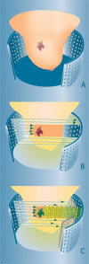
Optical imaging of the breast
- PMID: 19028613
- PMCID: PMC2590880
- DOI: 10.1102/1470-7330.2008.0032
Optical imaging of the breast
Abstract
This review provides a summary of the current state of optical breast imaging and describes its potential future clinical applications in breast cancer imaging. Optical breast imaging is a novel imaging technique that uses near-infrared light to assess the optical properties of breast tissue. In optical breast imaging, two techniques can be distinguished, i.e. optical imaging without contrast agent, which only makes use of intrinsic tissue contrast, and optical imaging with a contrast agent, which uses exogenous fluorescent probes. In this review the basic concepts of optical breast imaging are described, clinical studies on optical imaging without contrast agent are summarized, an outline of preclinical animal studies on optical breast imaging with contrast agents is provided, and, finally, potential applications of optical breast imaging in clinical practice are addressed. Based on the present literature, diagnostic performance of optical breast imaging without contrast agent is expected to be insufficient for clinical application. Development of contrast agents that target specific molecular changes associated with breast cancer formation is the opportunity for clinical success of optical breast imaging.
Figures


Similar articles
-
Developments toward diagnostic breast cancer imaging using near-infrared optical measurements and fluorescent contrast agents.Neoplasia. 2000 Sep-Oct;2(5):388-417. doi: 10.1038/sj.neo.7900118. Neoplasia. 2000. PMID: 11191107 Free PMC article. Review.
-
Oral Administration and Detection of a Near-Infrared Molecular Imaging Agent in an Orthotopic Mouse Model for Breast Cancer Screening.Mol Pharm. 2018 May 7;15(5):1746-1754. doi: 10.1021/acs.molpharmaceut.7b00994. Epub 2018 Apr 26. Mol Pharm. 2018. PMID: 29696981 Free PMC article.
-
[Contrast media for optical mammography].Radiologe. 1997 Sep;37(9):749-55. doi: 10.1007/s001170050277. Radiologe. 1997. PMID: 9424621 Review. German.
-
Polypyrrole nanoparticles: a potential optical coherence tomography contrast agent for cancer imaging.Adv Mater. 2011 Dec 22;23(48):5792-5. doi: 10.1002/adma.201103190. Epub 2011 Nov 21. Adv Mater. 2011. PMID: 22102372
-
[Functional and molecular imaging of breast tumors].Radiologe. 2010 Nov;50(11):1030-8. doi: 10.1007/s00117-010-2014-9. Radiologe. 2010. PMID: 20842342 German.
Cited by
-
Near infrared fluorescent imaging of choline kinase alpha expression and inhibition in breast tumors.Oncotarget. 2017 Mar 7;8(10):16518-16530. doi: 10.18632/oncotarget.14965. Oncotarget. 2017. PMID: 28157707 Free PMC article.
-
2'-Hydroxyflavanone: A novel strategy for targeting breast cancer.Oncotarget. 2017 Aug 24;8(43):75025-75037. doi: 10.18632/oncotarget.20499. eCollection 2017 Sep 26. Oncotarget. 2017. PMID: 29088842 Free PMC article.
-
p53-Independent apoptosis by benzyl isothiocyanate in human breast cancer cells is mediated by suppression of XIAP expression.Cancer Prev Res (Phila). 2010 Jun;3(6):718-26. doi: 10.1158/1940-6207.CAPR-10-0048. Epub 2010 May 18. Cancer Prev Res (Phila). 2010. PMID: 20484174 Free PMC article.
-
Benzyl isothiocyanate inhibits epithelial-mesenchymal transition in cultured and xenografted human breast cancer cells.Cancer Prev Res (Phila). 2011 Jul;4(7):1107-17. doi: 10.1158/1940-6207.CAPR-10-0306. Epub 2011 Apr 4. Cancer Prev Res (Phila). 2011. PMID: 21464039 Free PMC article.
-
Withaferin A inhibits activation of signal transducer and activator of transcription 3 in human breast cancer cells.Carcinogenesis. 2010 Nov;31(11):1991-8. doi: 10.1093/carcin/bgq175. Epub 2010 Aug 19. Carcinogenesis. 2010. PMID: 20724373 Free PMC article.
References
-
- Garcia M, Jemal A, Ward EM, et al. Atlanta, GA: American Cancer Society; 2007. Global cancer facts & figures 2007.
-
- Humphrey LL, Helfand M, Chan BK, Woolf SH. Breast cancer screening: a summary of the evidence for the US Preventive Services Task Force. Ann Intern Med. 2002;137:347–60. - PubMed
-
- Buist DS, Porter PL, Lehman C, Taplin SH, White E. Factors contributing to mammography failure in women aged 40–49 years. J Natl Cancer Inst. 2004;96:1432–40. - PubMed
-
- Carney PA, Miglioretti DL, Yankaskas BC, et al. Individual and combined effects of age, breast density, and hormone replacement therapy use on the accuracy of screening mammography. Ann Intern Med. 2003;138:168–75. - PubMed
MeSH terms
Substances
LinkOut - more resources
Full Text Sources
Medical
