. 2009 Jan 16;284(3):1505-13.
doi: 10.1074/jbc.M805113200. Epub 2008 Nov 18.

The heterogeneous nuclear ribonucleoprotein L is an essential component in the Ca2+/calmodulin-dependent protein kinase IV-regulated alternative splicing through cytidine-adenosine repeats

Affiliations

The heterogeneous nuclear ribonucleoprotein L is an essential component in the Ca2+/calmodulin-dependent protein kinase IV-regulated alternative splicing through cytidine-adenosine repeats

Jiankun Yu et al. J Biol Chem. .

Abstract

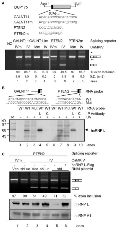
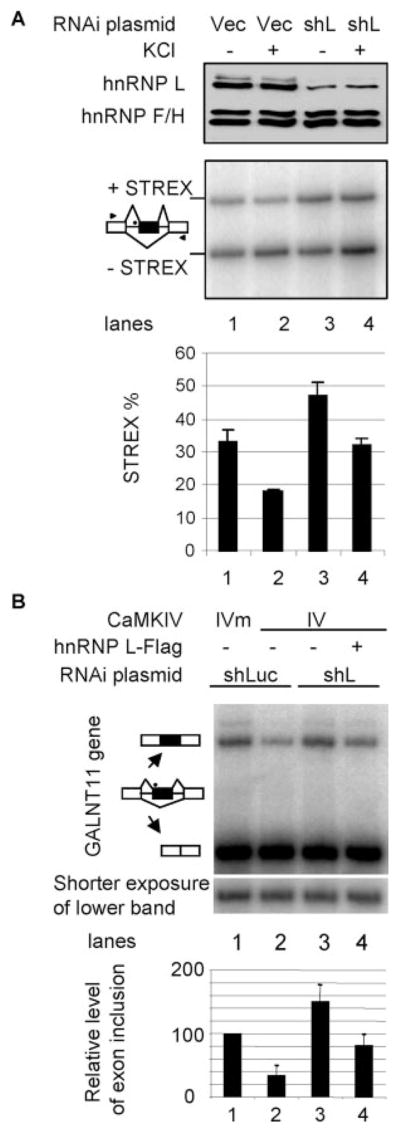
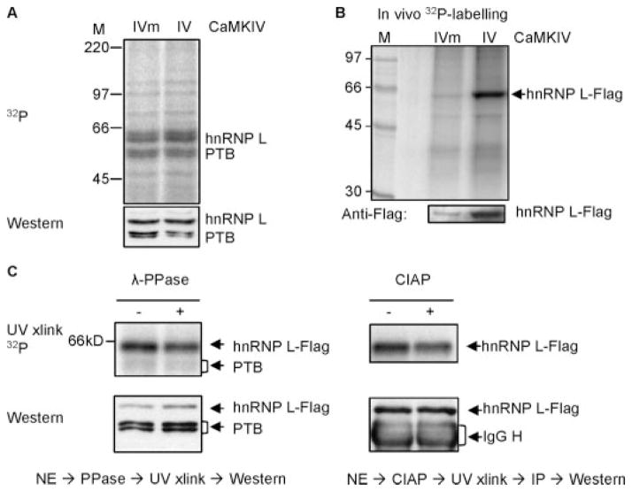
The regulation of gene expression through alternative pre-mRNA splicing is common in metazoans and is often controlled by intracellular signaling pathways that are important in cell physiology. We have shown that the alternative splicing of a number of genes is controlled by membrane depolarization and Ca2+/calmodulin-dependent protein kinase IV (CaMKIV) through CaMKIV-responsive RNA elements (CaRRE1 and CaRRE2); however, the trans-acting factors remain unknown. Here we show that the heterogeneous nuclear ribonucleoprotein (hnRNP) L is a CaRRE1 binding factor in nuclear extracts. An hnRNP L high affinity CA (cytidine-adenosine) repeat element is sufficient to mediate CaMKIV and hnRNP L repression of splicing in a location (3'-splice site proximity)-dependent way. Depletion of hnRNP L by RNA interference followed by rescue with coexpressed exogenous hnRNP L demonstrates that hnRNP L mediates the CaMKIV-regulated splicing through CA repeats in heterologous contexts. Depletion of hnRNP L also led to increased inclusion of the stress axis-regulated exon and a CA repeat-harboring exon under depolarization or with activated CaMKIV. Moreover, hnRNP L binding to CaRRE1 was increased by CaMKIV and, conversely, was reduced by pretreatments with protein phosphatases. Therefore, hnRNP L is an essential component of CaMKIV-regulated alternative splicing through CA repeats, with its phosphorylation likely playing a critical role.


Figures

FIGURE 1
FIGURE 1. Binding of hnRNP L to CaRRE1
A, left, diagram of the CaRRE1 RNA probe (wild type (WT)) and its mutant (Mut). The line represents the intron, and gray open box represents the downstream exon of DUP175ST. The CA repeat and upstream A/C-rich region is boxed, and the mutated nucleotides are underlined. Right, phosphorimages of SDS-PAGE gels of HeLa (lanes 1–2) or HEK293T (HEK, lanes 3–7) nuclear extract (NE) proteins UV-cross-linked to the wild type or mutant CaRRE1 RNA probes, with the protein kDa size markers indicated to the left. In lanes 5–7, 4, 8, or 4 times of UV-cross-linked nuclear extracts were used for immunoprecipitation (IP) with antibodies (Ab) against hnRNP L (L), hnRNP LL (LL), or PTB (PTB), respectively. B, left, UV cross-linking of the CaRRE1 wild type RNA probe in nuclear extracts of GH3 cells non-treated (NT) or treated with depolarizing concentrations of KCl (50 mM) for 12 h, similarly as in A. M, molecular size marker (kDa). Immunoprecipitation antibodies against hnRNP L (L), hnRNP F/H (F/H), or hnRNP LL (LL) are indicated above the lanes. Three (lanes 3–5) or eight times (lanes 6 and 7) of the cross-linked nuclear extracts were used for immunoprecipitation. Right, a bar graph of the ratio of hnRNP L/p55′ (mean ± S.E.) in GH3 nuclear extracts from non-treated or KCl-treated cells, with the p value and number of samples indicated (Student’s t test, paired non-treated and KCl-treated samples). C, specificity of the antibodies used against hnRNP L or LL in the experiments. The same blot of a GH3 nuclear extract was probed with hnRNP LL and hnRNP L antibodies, respectively, and aligned side by side with molecular markers. The hnRNP LL band is clearly lower than hnRNP L. *, a nonspecific band common for the anti-L, -LL, and -PTB antibodies (as seen in lanes 5–7 in A).
FIGURE 2
FIGURE 2. Effect of CaRRE1 replacement by an hnRNP L-high affinity CA repeat element in CaMKIV-regulated splicing
Upper, diagram of the exons (boxes) and introns (horizontal lines) of the splicing reporter mini-gene DUP175ST and its CaRRE1 in the upstream 3′-splice site of STREX (heavy line). In reporters L1 or L1m, the CaRRE1 was replaced with the hnRNP L high affinity CA repeats (L1) or low affinity sequence (L1m). Arrowheads, locations of PCR primers. Lower, an agarose gel of RT-PCR products from RNA samples of HEK293T cells co-transfected with each of the above splicing reporters and kinase-dead mutant (IVm) or constitutively active CaMKIV (IV), with average percentages (±S.D.) of exon inclusion under each lane, exon-included or -excluded products indicated to the right, and molecular size makers (M) to the indicated to the left. NC, PCR negative control. *, likely product from pre-mRNA. **, a cryptic 3′-splice product from 65 nt downstream in the middle exon.
FIGURE 3
FIGURE 3. The hnRNP L high affinity CA repeat element in CaMKIV- and hnRNP L-regulated splicing and its location dependence
A, the CA repeat element (or CG mutant as shown in sequence) is transferred to different positions of the middle exon or flanking introns in splicing reporters L2-L5 to replace the corresponding sequences of the vector DUP175 (except in reporter L2, the element was inserted upstream the branch point as indicated). Positions of the first nucleotide of the elements are indicated above the exon or introns, relative to the last nucleotide of upstream intron, first nucleotide of exon, or downstream intron, respectively. For comparison, the relative positions of L1 and L1m elements in DUP175ST are also indicated. B, an agarose gel of the RT-PCR products of the splicing reporters with co-expressed CaMKIVm, CaMKIV, or protein kinase A (PKA) in HEK293T cells, with average percentages (±S.D.) of exon inclusion under each lane and exon-included or -excluded products indicated to the right. NC, PCR negative control. *, same as in Fig. 2. C, effect of hnRNP L overexpression on splicing reporters L1-L5. Western blots (upper panel) of overexpressed hnRNP L-FLAG (anti-FLAG tag) and His-YB-1 (anti-His tag) from HEK293T cells cotransfected with reporters L1-L5 or mutants and His-YB-1 (Y) or hnRNP L-FLAG (L) plasmids. *, a nonspecific band recognized by the anti-His tag antibody. An agarose gel (lower panel) of RT-PCR products of the splicing reporters with the overexpressed hnRNP L or YB-1, similarly labeled as in B.
FIGURE 4
FIGURE 4. Role of hnRNP L in CaMKIV-regulated splicing through the hnRNP L high affinity CA repeats
A, effect of hnRNP L depletion by RNAi and rescue with exogenous hnRNP L on CaMKIV-regulated splicing. An agarose gel of RT-PCR products of the reporter L3 expressed in HEK293T cells infected with virus from lentiviral vector (Vec), shRNA against luciferase (shLuc) or hnRNP L (shL) and transfected with CaMKIV (IV), CaMKIV mutant (IVm), and hnRNP L-FLAG (+), with percentages of exon inclusion under each lane. Bottom, Western blots of hnRNP L and loading control hnRNP A1 (aligned with the agarose gel). Results are representative of 2–4 samples per lane. B, UV cross-linking of CA repeat or CG mutant (Mut) RNA probes with HeLa or HEK nuclear extracts (NE). WT, wild type. Upper, the probes containing the 3′-splice site of DUP175 and the CA repeat or CG mutant. Lower, a phosphorimage of HeLa (lanes 1–5) or HEK (lanes 6 –9) nuclear proteins cross-linked to the probes and resolved in a 4 –20% SDS-PAGE gel. Immunoprecipitating antibody (Ab) is against hnRNP L (L) or U2AF65 (C). A sixth of the cross-linking mix for immunoprecipitation (IP) was loaded in lanes 2 and 3 and a fourth in lanes 6 and 7.
FIGURE 5
FIGURE 5. Role of hnRNP L in CaMKIV-regulated splicing through short CA repeats from alternative exons
A, CA repeat elements from GALNT11 and PTEN2 genes and mutants replace the corresponding sequences of the vector DUP175 (upper, aligned nucleotides starting from the first nt of the exon). The reporter and mutant responses to CaMKIV in HEK293T cells are shown in agarose gels (lower) of RT-PCR products, with average percentages (±S.D.) of exon inclusion under each lane and exon-included or -excluded products indicated to the right. NC, PCR negative control. * and **, same as in Fig. 2. B, a phosphorimage of an SDS-PAGE gel of UV-cross-linked products of the GALNT11 and PTEN2 element RNA probes (as diagrammed above) of reporters (wild type (WT)) or mutants (Mut) and immunoprecipitated (IP) with anti-hnRNP L (L) or anti-U2AF65 antibody (as a control (C)). The hnRNP L band is indicated to the right. M, molecular weight markers. C, effect of hnRNP L depletion on the CaMKIV-regulated splicing of the PTEN2 reporter. Experiment and gel labeling are similar to that for reporter L3 in Fig. 4A.
FIGURE 6
FIGURE 6. Role of hnRNP L in depolarization- or CaMKIV-regulated endogenous exons
A, effect of hnRNP L depletion by RNAi on STREX exon inclusion and its regulation by depolarization in GH3 cells. Shown are a Western blot (upper panel) and a denaturing PAGE gel of RT-PCR products (middle panel, aligned with the blot) of rat GH3 cells infected with virus made from lentiviral vector (Vec) or plasmid expressing the specific shRNA against hnRNP L (shL) as in Figs. 4A and 5C and not treated (−) or treated (+) with KCl (50 mM) for 12 h. hnRNP F/H is a protein-loading control for the Western blots. The pre-mRNA splicing patterns for the products are indicated to the left, and treatments are indicated above the gels. The lower panel is a bar graph of the exon inclusion levels of the samples aligned with the above gels. The location of the CA repeat in the pre-mRNA intron (horizontal lines) is indicated as a black dot. Arrowheads, locations of PCR primers. B, effect of RNAi depletion of hnRNP L on the CaMKIV regulation of the GALNT11 exon in HEK293T cells as treated in Figs. 4A and 5C. Shown is a denaturing gel of RT-PCR products with the treatments indicated above and splicing patterns to the left of the gel. A shorter exposure of the lower band is shown below. The bar graph at the bottom is the level of exon inclusion (average ± S.D., n = 3) relative to the first lane (taken as 100%). This normalization reduces the effect from varying transfection efficiencies.
FIGURE 7
FIGURE 7. hnRNP L is a phosphoprotein with its CaRER1 binding activity regulated by CaMKIV and sensitive to treatment with phosphatases
A, a phosphorimage (upper) of the CaRRE1 probe cross-linked to HEK nuclear extracts of cells transfected with CaMKIVm or CaMKIV, with the protein levels of hnRNP L in the same gel shown below in Western blots after exposure to the phosphorimaging plate, rehydrated, blotted to a polyvinylidene difluoride membrane and probed with anti-hnRNP L antibody. The nuclear extract was made from green cells cotransfected with CaMKIV and enhanced green fluorescent protein plasmids and collected in flow cytometer. M, molecular weight markers. B, a phosphorimage (upper gel) of immunoprecipitated hnRNP L-FLAG from HEK cells coexpressing CaMKIVm or CaMKIV and in vivo labeled with [32P]orthophosphoric acid. At the bottom is the hnRNP L-FLAG protein level in a Western blot of the same gel. C, pretreatment of the nuclear extracts with λ-protein phosphatase (λ-PPase, left panel) or calf intestine alkaline phosphatase (CIAP, right panel) reduces hnRNP L-FLAG binding to the CaRRE1 probe in UV-cross-linking assays. HEK nuclear extracts (NE) from cells cotransfected with hnRNP L-FLAG and CaMKIV was treated (+) or not (−) with the phosphatases and cross-linked with the CaRRE1 RNA probe (upper gels). Western blots of the hnRNP L-FLAG in the same above-corresponding gels are shown (lower gels). Endogenous PTB is also shown in the left panel. The calf intestinal alkaline phosphatase-treated samples were immunoprecipitated (IP) with anti-FLAG antibody after cross-linking to avoid interference from the 65-kDa calf intestinal alkaline phosphatase monomer in gels. The activities of the phosphatases were verified by their ability to reduce the 32P signal of [α-32P]ATP-labeled protein bands in nuclear extracts. Each gel is representative of two to three experiments except the calf intestinal alkaline phosphatase gel (once).

Similar articles

Cited by

References

    1. Black DL. Annu Rev Biochem. 2003;72:291–336. - PubMed
    1. Black DL. Cell. 2000;103:367–370. - PubMed
    1. Modrek B, Resch A, Grasso C, Lee C. Nucleic Acids Res. 2001;29:2850–2859. - PMC - PubMed
    1. Johnson JM, Castle J, Garrett-Engele P, Kan Z, Loerch PM, Armour CD, Santos R, Schadt EE, Stoughton R, Shoemaker DD. Science. 2003;302:2141–2144. - PubMed
    1. Graveley BR. Trends Genet. 2001;17:100–107. - PubMed

Publication types

MeSH terms

Substances

LinkOut - more resources