. 2009 Mar 1;418(2):345-67.
doi: 10.1042/BJ20081422.

An essential role for the Glut1 PDZ-binding motif in growth factor regulation of Glut1 degradation and trafficking

Affiliations

An essential role for the Glut1 PDZ-binding motif in growth factor regulation of Glut1 degradation and trafficking

Heather L Wieman et al. Biochem J. .

Abstract

Cell surface localization of the Glut (glucose transporter), Glut1, is a cytokine-controlled process essential to support the metabolism and survival of haemopoietic cells. Molecular mechanisms that regulate Glut1 trafficking, however, are not certain. In the present study, we show that a C-terminal PDZ-binding motif in Glut1 is critical to promote maximal cytokine-stimulated Glut1 cell surface localization and prevent Glut1 lysosomal degradation in the absence of growth factor. Disruption of this PDZ-binding sequence through deletion or point mutation sharply decreased surface Glut1 levels and led to rapid targeting of internalized Glut1 to lysosomes for proteolysis, particularly in growth factor-deprived cells. The PDZ-domain protein, GIPC (G(alpha)-interacting protein-interacting protein, C-terminus), bound to Glut1 in part via the Glut1 C-terminal PDZ-binding motif, and we found that GIPC deficiency decreased Glut1 surface levels and glucose uptake. Unlike the Glut1 degradation observed on mutation of the Glut1 PDZ-binding domain, however, GIPC deficiency resulted in accumulation of intracellular Glut1 in a pool distinct from the recycling pathway of the TfR (transferrin receptor). Blockade of Glut1 lysosomal targeting after growth factor withdrawal also led to intracellular accumulation of Glut1, a portion of which could be rapidly restored to the cell surface after growth factor stimulation. These results indicate that the C-terminal PDZ-binding motif of Glut1 plays a key role in growth factor regulation of glucose uptake by both allowing GIPC to promote Glut1 trafficking to the cell surface and protecting intracellular Glut1 from lysosomal degradation after growth factor withdrawal, thus allowing the potential for a rapid return of intracellular Glut1 to the cell surface on restimulation.


Figures
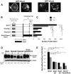
Figure 1
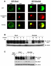
Figure 1. The Glut1 C-terminal tail is required for maximal surface Glut1 localization
(A) FL5.12 cells were transiently transfected with GFP-Glut1 or GFP-Glut3 for 18 hours and then cultured in the presence or absence of IL3 for an additional six hours. Cells were visualized with confocal microscopy. (B) Schematic of FLAG-tagged Glut1 full length, C-terminal tail truncation (FLAG-Glut1ΔC), C-terminal four amino acid truncation (FLAG-Glut1Δ4) and Glut1 chimeric protein with C-terminus of Glut3 (Glut1/CtermGlut3). (C and D) Cells stably expressing FLAG-Glut1 constructs as shown in B, were cultured in the presence or absence of IL3 for six hours. (C) Surface levels of FLAG-Glut1 were measured by flow cytometry. (D) Total FLAG-Glut1 levels were measured by immunoblot with actin loading control. (E) Cell surface FLAG-Glut1 levels were normalized to total FLAG expression. Mean and standard deviations of triplicate samples are shown. Asterisks (*) indicate p≤ 0.001 within the experiment. Representative results are shown for three or more experiments.
Figure 2
Figure 2. Akt can prevent degradation of Glut1Δ4
(A) Total levels of FLAG-Glut1 and phospho-Akt (ser473) were measured by immunoblot with actin loading control. (B) Surface FLAG-Glut1 levels were measured by flow cytometry. Mean and standard deviations of triplicate samples are shown. Asterisks (*) indicate p≤ 0.001 within the experiment. Representative results are shown for three or more experiments.
Figure 3
Figure 3. Glut1 Lysine 456 and the C-terminal PDZ-binding motif regulate Glut1 degradation
Cells stably expressing FLAG-Glut1, FLAG-Glut1Δ4, FLAG-Glut1K456A, and FLAG-Glut1K456AΔ4 were cultured in the presence or absence of IL3 for six hours. (A) Total FLAG expression levels were measured by immunoblot with actin as a loading control. (B) Surface FLAG levels were analyzed by flow cytometry. (C) Glucose uptake was measured. Mean and standard deviations of triplicate samples are shown. Asterisks (*) indicate p≤ 0.005 and (**) p≤ 0.01 within the experiment. Representative results are shown for three or more experiments.
Figure 4
Figure 4. Targeting of Glut1 to lysosomes is regulated by the Glut1 C-terminal four amino acids
(A) FL5.12 cells were co-transfected with either GFP-Glut1 or GFP-Glut1Δ4 and hBcl-xL to maintain cell viability for 18 hours then cultured in the presence or absence of IL3 for an additional eight hours. Lysotracker RED was added to culture in the final 30 minutes followed by fixation and visualization with fluorescence microscopy. (B) FLAG-Glut1 and FLAG-Glut1Δ4 cells were withdrawn from IL3 for six hours and vehicle control, Chloroquine (CQ) or NH4Cl (N) was added to cells for the duration of the IL3-withdrawal. Total FLAG expression levels were measured by immunoblot with actin as a loading control. (C) FLAG-Glut1 and FLAG-Glut1Δ4 cells were transiently transfected with control shRNAi or Rab7 shRNAi for 24 hours followed by a six hour growth factor withdrawal. Total FLAG and Rab7 protein levels were measured via immunoblot with actin as a loading control. Representative results are shown for three or more experiments.
Figure 5
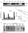
Figure 5. The Glut1 PDZ-binding motif is critical to regulate Glut1 total and surface levels
(A, B, and C) Cells were transiently transfected with FLAG-Glut1 constructs containing point mutations in the PDZ-binding motif, as noted in bold print, for 18 hours and then cells were cultured in the presence or absence of IL3 for an additional six hours. (A) Total FLAG-Glut1 was measured by immunoblot with actin as a loading control. (B) Flow cytometric analysis was utilized to measure surface FLAG-Glut1 levels. FL5.12 cells without FLAG-Glut1 were used for a negative stain control. (C) Cells were stained with anti-FLAG antibody, washed and cultured at 37 °C for indicated times prior to staining with fluorescent secondary antibody to determine the fraction of surface label remaining after various times incubation. (D) Cells were transiently transected with FLAG-Glut1, FLAG-Glut1Δ4 and FLAG-Glut1point mutants (DSQA, DAQV, DSAV ASQV) constructs for 24 hours and lysates were prepared. Lysates were then incubated with purified uncoated or nickel-bound His6GIPC beads. Anti- 6XHis immunoblot compares uncoated beads to nickel-bound His6GIPC beads used for pull-down. After washing, beads were boiled with sample buffer and FLAG levels were measured via immunoblot. U = uncoated beads, G = nickel-bound His6GIPC beads, I = input cell lysate, P = pull-down with HIS6GIPC beads. Mean and standard deviations of triplicate samples are shown. Asterisks (*) indicate p< 0.0005 and (**) p≤ 0.005 within the experiment. Representative results are shown for three or more experiments.
Figure 6
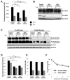
Figure 6. GIPC-deficiency decreases glucose uptake and surface Glut1 levels but does not cause Glut1 degradation
(A and B) FL5.12 cells were transiently transfected with either control shRNAi, GIPC shRNAi1or GIPC shRNAi2 constructs for 48 hours and then were cultured in the presence or absence of IL3 for six hours. (A) Glucose uptake was measured. (B) Total endogenous Glut1 and GIPC levels were measured by immunoblot with actin as a loading control. (C, D, E) FLAG-Glut1 and FLAG-Glut1Δ4 expressing cells were transiently transfected with control shRNAi, GIPC shRNAi1 or GIPC shRNAi2 constructs for 48 hours followed by culture in the presence or absence of IL3 for six hours. (C) Total FLAG-Glut1 and GIPC levels were measured by immunoblot with actin as a loading control. (D) Total surface FLAG-Glut1 levels were measured by flow cytometry and surface levels were normalized to total FLAG expression levels. (E) Surface FLAG-Glut1Δ4 levels were measured with flow cytometry. (F) FLAG-Glut1 cells were transfected with control shRNAi or GIPC shRNAi1 constructs for 48 hours and cultured in the presence of IL3. Cells were stained with anti-FLAG antibody, washed and cultured at 37 °C for indicated times prior to staining with fluorescent secondary antibody. Mean and standard deviations of triplicate samples are shown. Asterisks (*) indicate p≤ 0.005 and (**) p<0.05 within the experiment. Representative results are shown for three or more experiments.
Figure 7
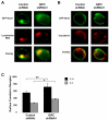
Figure 7. Disruption of GIPC suggests that Glut1 can exist in a recycling pool distinct from TfR
(A and B) FL5.12 cells were transfected with GFP-Glut1 and control shRNAi or GIPC shRNAi1 constructs for 48 hours and were incubated with (A) 50 nM Lysotracker RED for 30 minutes or (B) 50μg/ml Alexa Fluor 568-tagged Transferrin for one hour followed by fixation and visualization with fluorescence microscopy. (C) Cells were transfected with control shRNAi or GIPC shRNAi constructs for 48 hours and then were cultured in the presence or absence of IL3 for an additional six hours. Cells were stained with anti-TfR antibody and surface levels of TfR were measured via flow cytometry. Mean and standard deviations of triplicate samples are shown. Asterisks (*) indicate p< 0.0005 and (**) p≤ 0.05 within the experiment. Representative results are shown for three or more experiments.
Figure 8
Figure 8. Inhibition of Glut1 degradation does not lead to high surface Glut1 levels but allows a portion of Glut1 to rapidly recycle following growth factor stimulation
(A) FLAG-Glut1 cells were transiently transfected with control shRNAi or Rab7 shRNAi plasmids for 24 hours and cultured for an additional six hours in the presence or absence of IL3. Surface FLAG-Glut1 levels were analyzed by flow cytometry. (B) FLAG-Glut1 cells stably expressing hBcl-xL were transfected with control shRNAi or Rab7 shRNAi for 24 hours followed by a 24 hour growth factor withdrawal. Surface FLAG-Glut1 levels were measured by flow cytometry after 0, 15, 30 and 60 minutes of IL3 re-addition. (C) FLAG-Glut1 cells stably expressing hBcl-xL were withdrawn from IL3 for 24 hours with either vehicle or 40μM Chloroquine added during the last 6 hours of starve before re-addition of IL3. Surface FLAG-Glut1 levels were measured by flow cytometry 15 minutes after re-addition of IL3. Mean and standard deviations of triplicate samples are shown. Asterisks (*) indicate p< 0.005 within the experiment. Representative results are shown for three or more experiments.

Similar articles

Cited by

References

    1. Kan O, Baldwin SA, Whetton AD. Apoptosis is regulated by the rate of glucose transport in an interleukin 3 dependent cell line. J Exp Med. 1994;180:917–923. - PMC - PubMed
    1. Rathmell JC, Farkash EA, Gao W, Thompson CB. IL-7 enhances the survival and maintains the size of naive T cells. J Immunol. 2001;167:6869–6876. - PubMed
    1. Bentley J, Itchayanan D, Barnes K, McIntosh E, Tang X, Downes CP, Holman GD, Whetton AD, Owen-Lynch PJ, Baldwin SA. Interleukin-3-mediated cell survival signals include phosphatidylinositol 3-kinase-dependent translocation of the glucose transporter GLUT1 to the cell surface. J Biol Chem. 2003;278:39337–39348. - PubMed
    1. Rathmell JC, Vander Heiden MG, Harris MH, Frauwirth KA, Thompson CB. In the absence of extrinsic signals, nutrient utilization by lymphocytes is insufficient to maintain either cell size or viability. Mol Cell. 2000;6:683–692. - PubMed
    1. Rathmell JC, Fox CJ, Plas DR, Hammerman PS, Cinalli RM, Thompson CB. Akt-directed glucose metabolism can prevent Bax conformation change and promote growth factor-independent survival. Mol Cell Biol. 2003;23:7315–7328. - PMC - PubMed

Publication types

MeSH terms

Substances

LinkOut - more resources