. 2008 Nov 17;183(4):737-49.
doi: 10.1083/jcb.200805113.

p120 catenin induces opposing effects on tumor cell growth depending on E-cadherin expression

Affiliations

p120 catenin induces opposing effects on tumor cell growth depending on E-cadherin expression

Edwin Soto et al. J Cell Biol. .

Abstract

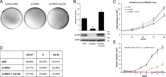
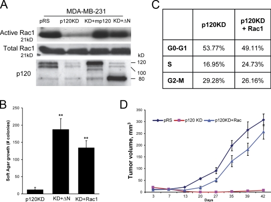
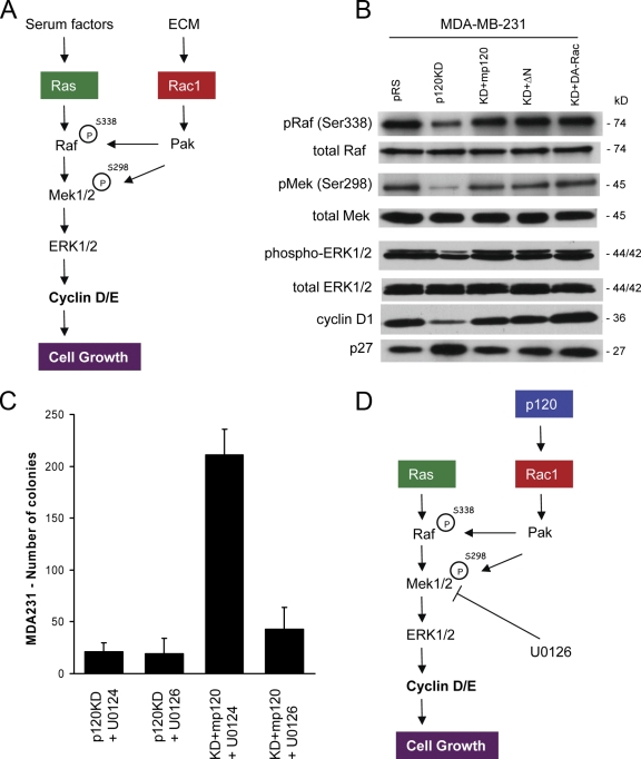
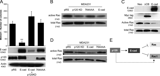
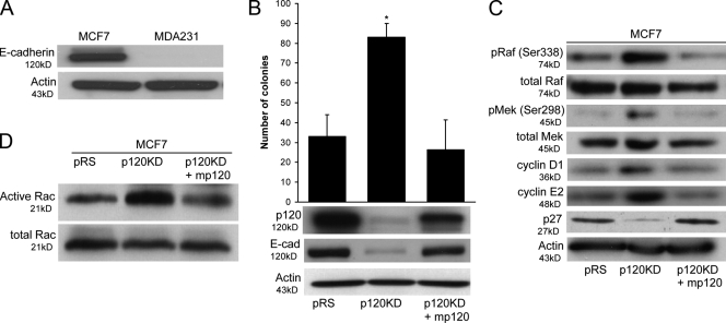
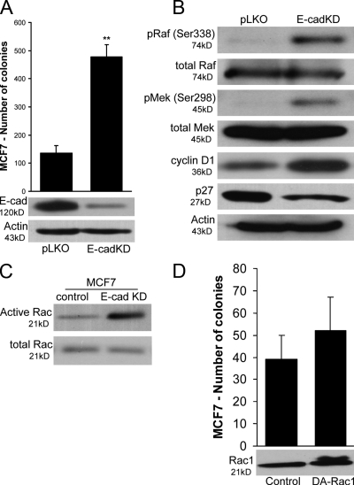
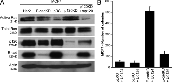
p120 catenin regulates the activity of the Rho family guanosine triphosphatases (including RhoA and Rac1) in an adhesion-dependent manner. Through this action, p120 promotes a sessile cellular phenotype when associated with epithelial cadherin (E-cadherin) or a motile phenotype when associated with mesenchymal cadherins. In this study, we show that p120 also exerts significant and diametrically opposing effects on tumor cell growth depending on E-cadherin expression. Endogenous p120 acts to stabilize E-cadherin complexes and to actively promote the tumor-suppressive function of E-cadherin, potently inhibiting Ras activation. Upon E-cadherin loss during tumor progression, the negative regulation of Ras is relieved; under these conditions, endogenous p120 promotes transformed cell growth both in vitro and in vivo by activating a Rac1-mitogen-activated protein kinase signaling pathway normally activated by the adhesion of cells to the extracellular matrix. These data indicate that both E-cadherin and p120 are important regulators of tumor cell growth and imply roles for both proteins in chemoresistance and targeted therapeutics.


Figures

Figure 1.
Figure 1.
p120 promotes the transformed growth of MDA-MB-231 cells. (A and B) MDA-MB-231 cells were infected with control pRS and LZRS-neo retroviruses (pRS), p120 shRNA virus together with LZRS-neo (p120KD), or p120 shRNA virus together with a retrovirus expressing murine p120 isoform 1A (p120KD + mp120). Stable polyclonal cell populations were generated, and the ability of the cells to grow in soft agar was tested. p120-depleted cells (p120KD) were less able to grow in soft agar when compared with control (pRS) cells or cells reexpressing p120 (***, P < 0.0001; analysis of variance [ANOVA]). Data represent the mean ± SEM (error bars) of three independent experiments performed in triplicate. (C) The ability of the same cell lines to grow on plastic was also determined over time. 1.5 × 10⁴ cells per well were plated in 24-well plates, and their growth was examined over time by cell counting. p120-depleted cells grew slower on plastic (**, P < 0.001 at 96 h; ANOVA). Mean ± SD (error bars; n = 9). (D) Cells were fixed in ethanol, treated with RNase, stained with propidium iodide, and analyzed with flow cytometry for their distribution in the cell cycle. Cells without p120 exhibit a G1 and G2 arrest that is rescued by mp120 expression. (E) 106 cells were injected into each flank of nude mice, and tumor growth was monitored over time by measuring tumor volume. p120 depletion inhibited MDA-MB-231 tumor growth in nude mice. Results represent the mean ± SD (error bars) of 10 independent determinations.
Figure 2.
Figure 2.
p120 promotes MDA-MB-231 tumor growth via activation of Rac1. (A) Levels of total and GTP-bound (active) Rac1 were determined in MDA-MB-231 cells infected with control pRS and LZRS-neo viruses (pRS), p120-depleted cells (p120KD) or cells reexpressing full-length murine p120 (KD + mp120), or a RhoA-uncoupled p120 mutant (Yanagisawa et al., 2008) lacking 323 N-terminal amino acids (KD + ΔN). The bottom panel shows levels of endogenously expressed and exogenous p120. p120-depleted cells exhibited low levels of basal Rac1 activity. Expression of either murine p120 or the ΔN p120 mutant rescued Rac1 activation. (B) The ability of p120-depleted cells (p120KD) or p120-depleted cells stably expressing either the RhoA-uncoupled ΔN p120 mutant (KD + ΔN) or constitutively active V12-Rac1 (KD + Rac1) to grow in soft agar was tested. Data represent the mean ± SEM (error bars) of three independent determinations performed in triplicate (**, P < 0.001 as compared with p120KD control; Student's t test). (C) The ability of constitutively active Rac1 (p120KD + Rac1) to rescue the cell cycle distribution of p120-depleted cells (p120KD) was also examined using flow cytometry, as described for Fig. 1 D. (D) The ability of p120-depleted cells ectopically expressing constitutively active Rac1 (p120KD + Rac1) to grow tumors was examined in nude mice. Results represent the mean ± SD (error bars) of 10 independent determinations. Note that activated Rac1 is able to rescue the growth of p120-depleted MDA-MB-231 cells both in vitro and in mice.
Figure 3.
Figure 3.
The MAPK pathway is essential for p120-mediated growth effects. (A) One of the proposed mechanisms by which the ECM is thought to promote cell growth is via the Rac1/PAK-mediated constitutive activation of the MAPK pathway. Phosphorylation of Raf (S338) and MEK (S298) at specific serines by the Rac1 effector PAK induces complex formation and promotes the constitutive activation of the MAPK–ERK pathway in mitogen-stimulated cells, leading to the increased expression of cyclin D. (B) Western blots of phosphorylated and total Raf, MEK, and ERK, cyclin D1, and p27 levels in control MDA-MB-231 cells (pRS), p120-depleted cells (p120KD), p120-depleted cells expressing murine p120 (KD + mp120), N-terminally deleted p120 (KD + ΔN), or constitutively active Rac1 (KD + DA-Rac). p120 depletion in MDA-MB-231 cells results in reduced phosphorylation of activating sites in Raf (Ser338), MEK (Ser298), and ERK1/2 (Thr202/Tyr204), reduced cyclin D1, and increased p27 levels. These effects are reversed by p120 expression or expression of activated Rac1. (C) p120-depleted cells (p120KD) and cells reexpressing p120 (KD + mp120) were grown in soft agar in the presence of either 10 μM of the MEK inhibitor U0126 or 10 μM of its inactive isomer U0124. The inhibition of MEK by U0126 blocks p120-mediated growth in soft agar. Mean ± SEM (error bars; n = 9). (D) Proposed model of p120 action in MDA-MB-231 cells. In MDA-MB-231 cells, p120 promotes growth by activating a Rac1–MAPK signaling cascade.
Figure 4.
Figure 4.
E-cadherin expression reverses p120 oncogenic effects and suppresses growth, Rac1, and Ras activity in a p120-dependent manner. (A) MDA-MB-231 cells expressing control pRS virus (pRS) or p120-specific shRNA were infected with an LZRS retrovirus expressing wild-type E-cadherin (E-cad and E-cad + p120KD, respectively). Control pRS cells were also infected with a virus expressing a p120-uncoupled E-cadherin mutant (764AAA). Polyclonal cell populations stably expressing these cadherin constructs were generated after G418 selection. The ability of these cell lines to grow in soft agar was tested. E-cadherin expression potently inhibits growth (***, P < 0.0001; ANOVA; n = 9). p120 is required for this because depletion of endogenous p120 reversed the E-cadherin phenotype, whereas p120-uncoupled E-cadherin failed to inhibit the growth of MDA-MB-231 cells. Mean ± SEM (error bars; n = 9). (B) Levels of total and GTP-bound (active) Rac1 were determined in MDA-MB-231 cells infected with control pRS and LZRS-neo viruses (pRS), p120-depleted cells (p120KD), or cells reexpressing wild-type (E-cad) or p120-uncoupled E-cadherin (764AAA). E-cadherin–expressing cells exhibited lower levels of Rac1 activation when compared with control (pRS) or 764AAA-expressing cells. (C) Levels of total and active Rac1 were also tested in MDA-MB-231 cells infected with control LZRS-neo virus (neo), a virus expressing the ΔCB E-cadherin mutant, or a virus expressing wild-type E-cadherin (E-cad). ΔCB is a Myc-tagged truncation mutant of the E-cadherin cytoplasmic domain, which binds p120 but not β-catenin. Cells expressing ΔCB exhibited reduced levels of Rac1 activation when compared with LZRS-neo virus control. (D) Levels of total and GTP-bound (active) Ras were also determined in the same cells. E-cadherin–expressing cells (E-cad) exhibit lower Ras activity than control cells (pRS), p120KD cells, or cells expressing the p120-uncoupled E-cadherin mutant (764AAA). (E) Proposed model of action. In the absence of E-cadherin (dotted arrow), p120 activates Rac1. In MDA-MB-231 cells expressing exogenous E-cadherin (solid arrow), p120 promotes the suppressive function of E-cadherin on both Rac1 and Ras activities.
Figure 5.
Figure 5.
p120 increases E-cadherin levels and suppresses growth in MCF7 cells. (A) Total lysates of MCF7 and MDA-MB-231 cells were immunoblotted for E-cadherin and actin as a loading control. MCF7 cells express endogenous E-cadherin. (B) MCF7 cells were infected with control pRS and LZRS-neo retroviruses (pRS), p120 shRNA virus together with LZRS-neo (p120KD), or p120 shRNA virus together with a retrovirus expressing murine p120 isoform 1A (p120KD + mp120). Stable cell populations were generated, and the ability of the cells to grow in soft agar was tested. p120-depleted MCF7 cells (p120KD) exhibited low levels of endogenous E-cadherin (bottom) and grew better than control cells (pRS) or cells expressing murine p120 (p120KD + mp120). Data are mean ± SEM (error bars; *, P < 0.01; ANOVA; n = 6). (C) Lysates of the same cell lines were also subjected to immunoblots for Raf, MEK, cyclins D1 and E2, and actin (control). p120-depleted MCF7 cells exhibited higher levels of known PAK1 phosphorylation sites in Raf (Ser338) and MEK (Ser298) than in control (pRS) or murine p120 reconstituted cells (p120KD + mp120). In accordance with this finding, the levels of cyclins D1 and E2 were also elevated in these cells. (D) Finally, the levels of total and GTP-bound (active) Rac1 activity were also determined using a specific pull-down assay. Consistent with the previous data, p120-depleted cells have higher Rac1 activity than control cells or cells expressing murine p120.
Figure 6.
Figure 6.
Depletion of endogenous E-cadherin in MCF7 cells increases anchorage-independent growth. (A) Control MCF7 cells were infected with control pLKO lentivirus or lentivirus expressing E-cadherin–specific shRNA. After the selection of stable polyclonal cell lines in puromycin, the ability of cell lines to form colonies in soft agar was tested. Cells expressing low levels of E-cadherin (E-cadKD) grow better in soft agar than control cells (pLKO). Data are mean ± SEM (error bars; **, P < 0.001; Student's t test; n = 6). Identical results were also obtained with a separate shRNA construct targeting E-cadherin (not depicted). (B) Lysates of control and E-cadherin–depleted cells (E-cadKD) were immunoblotted for E-cadherin, active and total Raf and MEK, and cyclin D1 as well as actin as a loading control. E-cadherin–depleted cells exhibited increased phosphorylation of activating sites on both Raf (Ser338) and MEK (Ser298). In agreement with these results, cyclin D1 levels were also higher. (C) Levels of GTP-bound (active) and total Rac1 were also determined. Consistent with the previous data, E-cadherin–depleted cells exhibited higher levels of activated Rac1. (D) To test the hypothesis that increased Rac1 activation is responsible for the increased ability of E-cadherin–deficient cells to grow in soft agar, we infected control MCF7 cells with a retrovirus expressing a constitutively active V12-Rac1 mutant (DA-Rac1). The data show that Rac1 activation is not sufficient to increase the ability of MCF7 cells to grow in soft agar. Mean ± SEM (error bars; n = 9).
Figure 7.
Figure 7.
E-cadherin suppresses growth by blocking Ras-MAPK activation in a p120-dependent manner. (A) Levels of total and GTP-bound (active) Ras were determined in MCF7 cells infected with control pRS and LZRS-neo viruses (pRS), p120-depleted cells (p120KD), p120-depleted cells expressing murine p120 (p120KD + mp120), E-cadherin–depleted cells (E-cadKD), or cells overexpressing the RTK Her2 (as a positive control). Both E-cadherin– and p120-depleted cells exhibited high levels of Ras activation, which are comparable with the levels induced by overexpression of Her2. The expression of murine p120 in p120-depleted cells potently blocks Ras activation. Levels of p120 and E-cadherin as well as actin loading controls are shown in the bottom panels. Note that upon p120 depletion, E-cadherin levels are drastically reduced but reinstated by exogenous expression of murine p120. (B) E-cadherin–depleted cells (E-cadKD) and cells expressing control pLKO lentivirus (pLKO) were grown in soft agar in the presence of either 10 μM of the MEK inhibitor U0126 or 10 μM of its inactive isomer U0124. Inhibition of MEK by U0126 blocks the ability of E-cadherin–deficient MCF7 cells to grow in soft agar, suggesting the involvement of the MAPK pathway. Mean ± SEM (error bars; n = 9).
Figure 8.
Figure 8.
Proposed model of p120 action. The presence of E-cadherin causes the potent inhibition of Ras and Rac1, inhibits MAPK/ERK signaling, reduces cyclin D1, and promotes increased levels of p27, thus blocking cell growth. Under these conditions, p120 promotes the stabilization of E-cadherin complexes and their ability to suppress Ras. p120 recruitment by E-cadherin also decreases mesenchymal cadherin stability and function, resulting in reduced Rac1 activation. However, upon E-cadherin loss during tumor progression, the negative regulation of Ras is relieved. Under these conditions, endogenous p120 induces transformed cell growth by activating Rac1 and inducing the Rac1-dependent, PAK-mediated phosphorylation of Raf and MEK, which result in the constitutive, anchorage-independent activation of the Ras–MAPK–ERK signaling pathway. Dotted arrows denote potential Ras-mediated signaling events leading to anchorage-independent growth.

Similar articles

Cited by

References

    1. Anastasiadis, P.Z. 2007. p120-ctn: a nexus for contextual signaling via Rho GTPases. Biochim. Biophys. Acta. 1773:34–46. - PubMed
    1. Anastasiadis, P.Z., S.Y. Moon, M.A. Thoreson, D.J. Mariner, H.C. Crawford, Y. Zheng, and A.B. Reynolds. 2000. Inhibition of RhoA by p120 catenin. Nat. Cell Biol. 2:637–644. - PubMed
    1. Beeser, A., Z.M. Jaffer, C. Hofmann, and J. Chernoff. 2005. Role of group A p21-activated kinases in activation of extracellular-regulated kinase by growth factors. J. Biol. Chem. 280:36609–36615. - PubMed
    1. Behrens, J., M.M. Mareel, F.V. Roy, and W. Birchmeier. 1989. Dissecting tumor cell invasion: epithelial cells acquire invasive properties after the loss of uvomorulin-mediated cell-cell adhesion. J. Cell Biol. 108:2435–2447. - PMC - PubMed
    1. Betson, M., E. Lozano, J. Zhang, and V.M. Braga. 2002. Rac activation upon cell-cell contact formation is dependent on signaling from the epidermal growth factor receptor. J. Biol. Chem. 277:36962–36969. - PubMed

Publication types

MeSH terms