. 2009 Jan 16;284(3):1884-95.
doi: 10.1074/jbc.M803641200. Epub 2008 Nov 13.

The substrate recognition domains of the N-end rule pathway

Affiliations

The substrate recognition domains of the N-end rule pathway

Takafumi Tasaki et al. J Biol Chem. .

Abstract

The N-end rule pathway is a ubiquitin-dependent system where E3 ligases called N-recognins, including UBR1 and UBR2, recognize type-1 (basic) and type-2 (bulky hydrophobic) N-terminal residues as part of N-degrons. We have recently reported an E3 family (termed UBR1 through UBR7) characterized by the 70-residue UBR box, among which UBR1, UBR2, UBR4, and UBR5 were captured during affinity-based proteomics with synthetic degrons. Here we characterized substrate binding specificity and recognition domains of UBR proteins. Pull-down assays with recombinant UBR proteins suggest that 570-kDa UBR4 and 300-kDa UBR5 bind N-degron, whereas UBR3, UBR6, and UBR7 do not. Binding assays with 24 UBR1 deletion mutants and 31 site-directed UBR1 mutations narrow down the degron-binding activity to a 72-residue UBR box-only fragment that recognizes type-1 but not type-2 residues. A surface plasmon resonance assay shows that the UBR box binds to the type-1 substrate Arg-peptide with Kd of approximately 3.4 microm. Downstream from the UBR box, we identify a second substrate recognition domain, termed the N-domain, required for type-2 substrate recognition. The approximately 80-residue N-domain shows structural and functional similarity to 106-residue Escherichia coli ClpS, a bacterial N-recognin. We propose a model where the 70-residue UBR box functions as a common structural element essential for binding to all known destabilizing N-terminal residues, whereas specific residues localized in the UBR box (for type 1) or the N-domain (for type 2) provide substrate selectivity through interaction with the side group of an N-terminal amino acid. Our work provides new insights into substrate recognition in the N-end rule pathway.


Figures
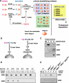
FIGURE 1.
FIGURE 1.
A, the mammalian N-end rule pathway. N-terminal residues are indicated by single-letter abbreviations for amino acids. Yellow ovals denote the rest of a protein substrate. C* denotes oxidized N-terminal Cys, either Cys-sulfinic acid [CysO2(H)] or Cys-sulfonic acid [CysO3(H)]. The Cys oxidation requires nitric oxide and oxygen (O2) or its derivatives. The oxidized Cys is arginylated by ATE1 Arg-tRNA-protein transferase (R-transferase). N-recognins also recognize internal (non-N-terminal) degrons in other substrates of the N-end rule pathway. B, the X-peptide pull-down assay. Left, a 12-mer peptide bearing N-terminal Arg (type 1), Phe (type 2), Trp (type 2), or Gly (stabilizing control) residue was cross-linked through its C-terminal Cys residue to Ultralink Iodoacetyl beads. Right, the otherwise identical 12-mer peptide, bearing C-terminal biotinylated Lys instead of Cys, was conjugated, via biotin, to the streptavidin-Sepharose beads. C, the X-peptide pull-down assay of endogenous UBR proteins using testes extracts. Extracts from mouse testes were mixed with bead-conjugated X-peptides bearing N-terminal Phe (F), Gly (G), or Arg (R). After centrifugation, captured proteins were separated and subjected to anti-UBR immunoblotting. Mo, a pull-down reaction with mock beads. D, the X-peptide pull-down assays using rat testis extracts were performed in the presence of varying concentrations of NaCl. After incubation and washing, bound proteins were eluted by 10 mm Tyr-Ala for Phe-peptide, 10 mm Arg-Ala for Arg-peptide, and 5 mm Tyr-Ala and 5 mm Arg-Ala for Val-peptide. Eluted proteins were subjected to immunoblotting for UBR1 and UBR5. E, cytoplasmic fractions of wild-type (+/+), Ubr1-/-, Ubr2-/-, Ubr1-/-Ubr2-/-, and Ubr1-/-Ubr2-/-Ubr4RNAi MEFs were subjected to X-peptide pull-down assay. Precipitated proteins were separated and analyzed by immunoblotting for UBR1 and UBR4.
FIGURE 2.
FIGURE 2.
The binding properties of the UBR box family members to type-1 and type-2 destabilizing N-terminal residues. A, the X-peptide pull-down assay with overexpressed, full-length UBR proteins: UBR2, UBR3 (in S. cerevisiae cells), UBR4, UBR5 (in COS7 cells), and UBR6 and UBR7 (in the wheat germ lysates). Precipitates were analyzed by immunoblotting (for UBR2, UBR3, UBR4, and UBR5) with tag-specific antibodies as indicated in B or autoradiography (for UBR6 and UBR7). B, the structures of UBR box proteins. Shown are locations of the UBR box, the N-domain, and other E3-related domains. UBR, UBR box; RING, RING finger; UAIN, UBR-specific autoinhibitory domain; CRD, cysteine-rich domain; PHD, plant homeodomain; HECT, HECT domain.
FIGURE 3.
FIGURE 3.
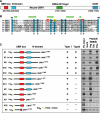
The UBR box is the substrate recognition domain of UBR1. A, locations of the UBR box, the N-domain, the RING domain, and the UAIN domain in mouse UBR1. B, sequence alignment of UBR boxes in mouse UBR proteins, in which conserved Cys and His residues are highlighted (cyan). Indicated by yellow highlight is the Cys residue of mouse UBR1 deduced from the Cys residue of Arabidopsis BIG/UBR4 whose missence mutation perturbs auxin transport (42). Indicated by red highlight are the residues of mouse UBR1 deduced from those of S. cerevisiae UBR1 that were identified to be essential for degradation of type-1 N-end rule substrates (41). Predicted secondary structure elements of the UBR box of mouse UBR1 (arrow, β-sheet) are shown above the sequence alignment. C, the X-peptide pull-down assay with C-terminal deleted UBR1 fragments expressed in the CECF-based wheat germ extracts. The identification numbers for UBR1 fragments are shown to the left. Most UBR1 fragments are N-terminal His6-tagged. The binding activity of each fragment was recorded as either positive (+) or negative (-). The autoradiography for the X-peptide pull-down assay is shown to the right. UAIN, UBR-specific autoinhibitory domain.
FIGURE 4.
FIGURE 4.
The X-peptide pull-down assay of deletion mutants of UBR1 and UBR2. A, the X-peptide pull-down assay with N-terminal deleted and other related UBR1 fragments that were expressed in the CECF-based wheat germ extracts. The identification numbers for UBR1 fragments are shown to the left. Most UBR1 fragments are N-terminal His6-tagged. The binding activity of each fragment was recorded as either positive (+) or negative (-). The autoradiography for the X-peptide pull-down assay is shown to the right. B, the X-peptide pull-down assay with UBR2 fragments expressed in the wheat germ extracts.
FIGURE 5.
FIGURE 5.
The binding properties of E. coli ClpS to mammalian N-end rule substrates. A, the sequence alignment of the ∼80-residue N-domain of eukaryotic N-recognins with E. coli ClpS with a size of 106 amino acids. Indicated by red highlight are the residues of mouse UBR1 deduced from those of S. cerevisiae UBR1 that were identified to be essential for degradation of type-2 N-end rule substrates (41). Predicted secondary structure elements of the N-domain of mouse UBR1 (arrow, β-sheet; cylinder, α-helix) are shown above the sequence alignment. B, the X-peptide pull-down assay with E. coli ClpS expressed in the wheat germ extracts. E. coli ClpS binds to type-2, but not type-1, N termini in the context of a mammalian N-end rule substrate. *, a putative cleavage product of His6-ClpS.
FIGURE 6.
FIGURE 6.
Surface plasmon resonance (Biacore) analysis for the interaction between MBP-UBR1-(91-191)-His6 and X-peptides (X = Arg, Phe, or Gly). Biotinylated X-peptides, adjusted to 20 nm in the binding buffer (see “Experimental Procedures”), were immobilized to a sensor chip. A, the structures of MBP-UBR1-(91-191)-His6 and MBP-His6. B, purified MBP-UBR1-(91-191)-His6 and MBP-His6 were separated on SDS-PAGE and stained with Coomassie Brilliant Blue R-250 (Bio-Rad). C, the X-peptide pull-down assay. Purified MBP-UBR1-(91-191)-His6 or MBP-His6 was mixed with X-peptide beads, followed by anti-His6 immunoblotting of precipitated proteins. D, Biacore sensorgram illustrating the binding of MBP-UBR1-(91-191)-His6 to Arg-peptide but not to Phe-peptide or Gly-peptide. MBP-UBR1-(91-191)-His6, adjusted to 5 μm in the binding buffer, was injected over immobilized X-peptides at a flow rate of 40 μl/min. E, Biacore sensorgram with MBP-His6 showing no detectible affinity to X-peptides. F, kinetic analysis of the affinity of MBP-UBR1-(91-191)-His6 to Arg-peptide. MBP-UBR1-(91-191)-His6 was serially diluted in the binding buffer and subsequently injected over immobilized Arg-peptide at a flow rate of 40 μl/min. Numbers (1-10) represent, respectively, the concentrations (3.13, 2.5, 1.25, 0.63, 0.31, 0.16, 0.08, 0.04, 0.02, and 0.01 μm) of injected MBP-UBR1-(91-191)-His6.
FIGURE 7.
FIGURE 7.
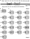
Site-directed mutagenesis analysis of the UBR box of mouse UBR1. A, diagram of the UBR1-(1-453) fragment showing residues that are mutated into alanine. Group 1 is 11 residues (8 Cys, 2 His, and 1 Asp), which are localized within the 70-residue UBR box and conserved in all known UBR proteins. Group 2 is 9 residues that are incompletely conserved within the UBR box. B, the X-peptide pull-down assay with wild-type UBR1-(1-453), expressed in the wheat germ lysate, and bead-conjugated X-peptides bearing N-terminal Arg (R), Gly (G), or Phe (F). The levels of signals compared with 5% input signal are shown at the bottom. The lane m represents a pull-down reaction with mock beads. C and D, the X-peptide pull-down assays with Group-1 (C) and Group-2 (D) UBR1-(1-453) mutants. Note that Group-1 mutations show clear tendency to completely abolish the type-1 substrate binding activity.
FIGURE 8.
FIGURE 8.
Site-directed mutagenesis analysis of the N-domain. A, the diagram of UBR1-(1-453) showing residues that are mutated into Ala. Group 3 is 5 residues within the N-domain, and Group 4 is 6 residues localized outside the UBR box and the N-domain. B and C, the X-peptide pull-down assay with Group 3 (B) and Group 4 (C) UBR1-(1-453) mutants and bead-conjugated X-peptides bearing N-terminal Arg (R), Gly (G), or Phe (F). The levels of signals compared with 5% input signal are shown to the bottom. Lane m represents a pull-down reaction with mock beads.
FIGURE 9.
FIGURE 9.
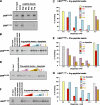
Selective inhibition of the interaction between the UBR box and the X-peptide by dipeptides. A, the X-peptide pull-down assay with UBR1-(34-405) (top) and UBR1-(91-191) (bottom), expressed in the CECF-based wheat germ extracts, using X-peptides that bear N-terminal Arg, Phe, Trp, or Gly. B, inhibition of the interaction between UBR1-(34-405) and Arg-peptide by dipeptides bearing various N-terminal residues. The X-peptide pull-down assay was performed in the presence of varying concentrations of dipeptides. RA, Arg-Ala; KA, Lys-Ala; AR, Ala-Arg; FA, Phe-Ala; WA, Trp-Ala. C, quantitation of B. D, inhibition of the interaction between UBR1-(34-405) and Phe-peptide by dipeptides bearing various N-terminal residues. E, quantitation of D. F, inhibition of the interaction between UBR1-(91-191) and Arg-peptide by dipeptides bearing various N-terminal residues. G, quantitation of F.

Similar articles

Cited by

References

    1. Bachmair, A., and Varshavsky, A. (1989) Cell 56 1019-1032 - PubMed
    1. Varshavsky, A. (1996) Proc. Natl. Acad. Sci. U. S. A. 93 12142-12149 - PMC - PubMed
    1. Kwon, Y. T., Reiss, Y., Fried, V. A., Hershko, A., Yoon, J. K., Gonda, D. K., Sangan, P., Copeland, N. G., Jenkins, N. A., and Varshavsky, A. (1998) Proc. Natl. Acad. Sci. U. S. A. 95 7898-7903 - PMC - PubMed
    1. Kwon, Y. T., Kashina, A. S., and Varshavsky, A. (1999) Mol. Cell. Biol. 19 182-193 - PMC - PubMed
    1. Kwon, Y. T., Lévy, F., and Varshavsky, A. (1999) J. Biol. Chem. 274 18135-18139 - PubMed

Publication types