. 2008 Dec;36(22):7136-45.
doi: 10.1093/nar/gkn880. Epub 2008 Nov 7.

Efficient processing of TFO-directed psoralen DNA interstrand crosslinks by the UvrABC nuclease

Affiliations

Efficient processing of TFO-directed psoralen DNA interstrand crosslinks by the UvrABC nuclease

Laura A Christensen et al. Nucleic Acids Res. 2008 Dec.

Abstract

Photoreactive psoralens can form interstrand crosslinks (ICLs) in double-stranded DNA. In eubacteria, the endonuclease UvrABC plays a key role in processing psoralen ICLs. Psoralen-modified triplex-forming oligonucleotides (TFOs) can be used to direct ICLs to specific genomic sites. Previous studies of pyrimidine-rich methoxypsoralen-modified TFOs indicated that the TFO inhibits cleavage by UvrABC. Because different chemistries may alter the processing of TFO-directed ICLs, we investigated the effect of another type of triplex formed by purine-rich TFOs on the processing of 4'-(hydroxymethyl)-4,5',8-trimethylpsoralen (HMT) ICLs by the UvrABC nuclease. Using an HMT-modified TFO to direct ICLs to a specific site, we found that UvrABC made incisions on the purine-rich strand of the duplex approximately 3 bases from the 3'-side and approximately 9 bases from the 5'-side of the ICL, within the TFO-binding region. In contrast to previous reports, the UvrABC nuclease cleaved the TFO-directed psoralen ICL with a greater efficiency than that of the psoralen ICL alone. Furthermore, the TFO was dissociated from its duplex binding site by UvrA and UvrB. As mutagenesis by TFO-directed ICLs requires nucleotide excision repair, the efficient processing of these lesions supports the use of triplex technology to direct DNA damage for genome modification.


Figures

Figure 1.
Figure 1.
The target duplex and psoralen-modified TFO. (A) Space-filling model of a psoralen-modified TFO bound to its target duplex. The psoralen moiety is shown in yellow and the TFO is shown in red bound to the purine-rich strand of the target duplex (blue) in the major groove. The pyrimidine-rich strand is shown in green (21). (B) Psoralen-modified TFO and TFO binding site. The TFO binding site is shown in red, the psoralen crosslinking site is shown in yellow and the pyrimidine-rich (py) and purine-rich (pu) strands are depicted in green and blue, respectively. The sequence of the TFO binding site is shown in bold capital letters, the psoralen crosslinking site is underlined and psoAG30, the psoralen-conjugated TFO, is depicted in an antiparallel orientation relative to the purine-rich strand of the target duplex. The MluI restriction site was used to remove the 3′-radiolabel on the pyrimidine strand. Figure 1A reprinted with permission from Vasquez et al., Biochemistry, 35, 10712–10719, Copyright 1996 American Chemical Society.
Figure 2.
Figure 2.
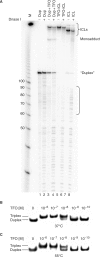
Triplex formation occurs under conditions of the UvrABC incision assay. (A) DNase footprint to verify presence of the triplex structure. Substrate labeled on the 5′-end of the purine-rich strand was incubated −/+ DNase I as indicated. Lanes 1 and 2 contain duplex, lanes 3 and 4 contain unpurified TFO-ICL substrate, lanes 5 and 6 contain gel-purified TFO-ICL and lanes 7 and 8 contain gel-purified ICL only. A bracket indicates the protected region. (B) and (C) TFO-binding assay. Target duplex DNA was end-labeled with [γ-32P]ATP and incubated with increasing concentrations of TFO as indicated. Samples were either incubated in standard triplex binding buffer at 37°C and subjected to native PAGE on a 12% TBM gel (B) or were incubated in UvrABC buffer at 55°C for 30 min prior to loading (C). Triplex formation occurs under both conditions (binding affinity of ∼10−8 M), indicating that the reaction conditions did not affect TFO binding.
Figure 3.
Figure 3.
UvrABC incision assay with the radiolabel on the purine-rich strand of the target duplex. (A) UvrABC incision assay with 5′-end-labeled purine-rich duplex target strand. Substrate DNA was incubated with purified UvrA (20 nM), UvrB (100 nM) and UvrC (50 nM) as described in the Materials and Methods section and subjected to denaturing PAGE on a 6% gel. Lanes 1 and 2 contain purified psoralen crosslinked duplex substrate treated to remove the TFO (labeled as ICL) −/+ UvrABC, lanes 3 and 4 contain nondamaged duplex DNA (labeled as Dup) −/+ UvrABC and lanes 5 and 6 contain purified TFO-directed psoralen ICL substrate with the TFO covalently attached (labeled as TFO-ICL) −/+ UvrABC. The bar (−) indicates lanes that have been removed for clarity of presentation. (B) Expected incision sites for the UvrABC nuclease on ICL and TFO-ICL substrates. The expected incision product is shown in red, the TFO is depicted in blue and incision sites are marked by arrows. A green star indicates the position of the radiolabel. The pyrimidine-rich and purine-rich strands of the target duplex are labeled py and pu, respectively. The incision product migrating between 80 and 90 bases corresponds with cleavage on the purine-rich strand ∼9 phosphodiester bonds on the 5′-side of the ICL, within the triplex binding site. (C) UvrABC incision assay with the purine-rich strand of the target duplex 3′-end-labeled. Lanes 1 and 2 contain TFO-ICL +/− UvrABC. The incision product migrating between 20 and 30 bases corresponds with cleavage on the purine-rich strand ∼3 phosphodiester bonds on the 3′-side of the ICL. (D) Expected UvrABC incision sites on the TFO-ICL substrate. The figure is marked as in (B). (E) UvrABC incision assay with fluorescein-adducted positive control (F2650) 5′-end-labeled on the fluorescein-adducted strand. Lanes 1 and 2 contain F2650 −/+ UvrABC. (F) Histogram indicating the average incision (as a percent of the total substrate) observed for ICL and TFO-ICL substrates by the UvrABC nuclease. Incision assays were repeated at least three times. Data presented are ±SD, P < 0.05 using Student's t-test.
Figure 4.
Figure 4.
UvrABC incision assay with the radiolabel on the pyrimidine-rich strand of the target duplex. (A) Samples were incubated with 20 nM UvrA, 100 nM UvrB and 50 nM UvrC as described in the Materials and methods section and subjected to denaturing PAGE on a 6% gel. Lanes 1 and 2 contain TFO-ICL −/+ UvrABC. The insert contains a magnified view of the incision product. The incision product migrating just above 20 bases corresponds to cutting on the pyrimidine-rich strand of the target duplex. (B) Expected incision sites for UvrABC on the TFO-ICL substrate. The expected incision product is shown in red, the TFO is depicted in blue and incision sites are marked by arrows. A green star indicates the position of the radiolabel. The pyrimidine-rich and purine-rich strands of the target duplex are labeled py and pu, respectively. (C) UvrABC incision assay with the ICL-only substrate 5′-end-labeled on the pyrimidine-rich strand of the target duplex. Lanes 1 and 2 contain the F2650 positive control, and lanes 3 and 4 contain ICL-only substrate. (D) Expected incision sites for ICL-only substrate. The figure is marked as in (B). (E) UvrABC incision assay with substrate 3′-end-labeled on both strands of the target duplex. Lanes 1 and 2 contain TFO-ICL −/+ UvrABC. (F). Expected incision sites for the UvrABC nuclease on the TFO-ICL substrate. The figure is marked as in (B).
Figure 5.
Figure 5.
UvrA or UvrA+UvrB protein binding assay. UvrA or UvrA+UvrB protein binding assays with 5′-end-labeled pyrimidine-rich duplex target strand. (A) Substrate DNA was incubated with purified UvrA (20 nM) or UvrA (20 nM) and UvrB (100 nM) as described in the Materials and Methods section and subjected to 4% native PAGE gel. Lanes 1–3 contain F2650 positive control with no protein, UvrA or UvrA+UvrB, respectively. Lanes 4–6 contain nondamaged duplex DNA with no protein, UvrA or UvrA+UvrB, respectively. Lanes 7–9 contain purified ICL only with no protein, UvrA or UvrA+UvrB, respectively. Lanes 10–12 contain purified TFO-ICL substrate with no protein, UvrA or UvrA+UvrB, respectively. UvrB binding was similar with ICL and TFO-ICL substrates (∼40%, lanes 9 and 12). An ‘altered substrate’ was observed when the TFO-ICL substrate was incubated with UvrA+UvrB (lane 12). Asterisks mark an artifact occurring in lanes containing crosslinked substrate. (B) UvrA or UvrA+UvrB protein-binding assay to test for possible structural modifications of TFO-ICL resulting in an ‘altered substrate’ following incubation with UvrA or UvrA+UvrB. Substrate in the presence or absence of UvrA and/or UvrB was either loaded directly on the gel following incubation at 55°C or heated to 95°C prior to loading. The ‘altered substrate’ disappeared with heat denaturation, consistent with structural modifications of the TFO-ICL. (C) UvrA or UvrA+UvrB protein-binding assay to test for displacement of noncrosslinked TFO. Noncrosslinked triplex substrate (pso-TFO + duplex in the absence of UVA irradiation) was incubated with UvrA or UvrA+UvrB as described and subjected to native PAGE (4% gel). Triplex substrate was denatured to duplex form after incubation with UvrA+UvrB, indicating displacement of the TFO from its target duplex binding site.
Figure 6.
Figure 6.
UvrA concentration-dependent incision efficiency of ICL and TFO-ICL substrates. ICL only (A) and TFO-ICL (B) substrates were labeled on the 5′-end of the purine-rich strand. Substrates were incubated with increasing concentrations of UvrA (0–70 nM as indicated for each lane), 100 nM UvrB and 50 nM UvrC for 30 min at 55°C. (C) Trend lines for ICL and TFO-ICL incision efficiencies with increasing concentrations of UvrA. Relative incision values were determined by calculating the amount of product as a percent of total sample and normalizing to the incision efficiency of ICL-only substrate at 20 nM UvrA.

Similar articles

Cited by

References

    1. Stern RS. Psoralen and ultraviolet a light therapy for psoriasis. N Engl J. Med. 2007;357:682–690. - PubMed
    1. Song PS, Tapley K.J., Jr Photochemistry and photobiology of psoralens. Photochem. Photobiol. 1979;29:1177–1197. - PubMed
    1. Cimino GD, Gamper HB, Isaacs ST, Hearst JE. Psoralens as photoactive probes of nucleic acid structure and function: organic chemistry, photochemistry, and biochemistry. Annu. Rev. Biochem. 1985;54:1154–1193. - PubMed
    1. Shi Y-B, Hearst JE. Wavelength dependence for the photoreactions of DNA-psoralen monoadducts. 2. Photo-cross-linking of monoadducts. Biochemistry. 1987;26:3792–3798. - PubMed
    1. Van Houten B, Gamper H, Hearst JE, Sancar A. Construction of DNA substrates modified with psoralen at a unique site and study of the action mechanism of ABC excinuclease on these uniformly modified substrates. J. Biol. Chem. 1986;261:14135–14141. - PubMed

Publication types

MeSH terms