. 2008 Dec 26;283(52):36586-91.
doi: 10.1074/jbc.M805676200. Epub 2008 Nov 3.

Plasmin activates epithelial Na+ channels by cleaving the gamma subunit

Affiliations

Plasmin activates epithelial Na+ channels by cleaving the gamma subunit

Christopher J Passero et al. J Biol Chem. .

Abstract

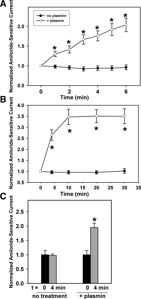
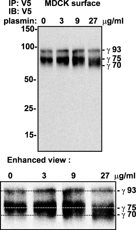
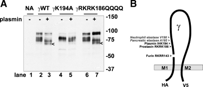
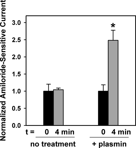
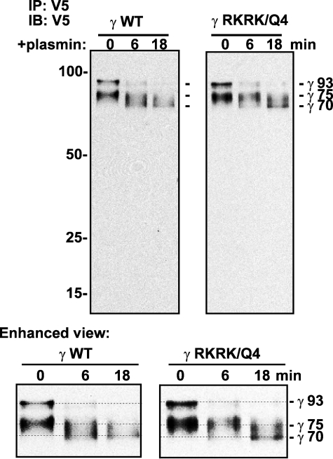
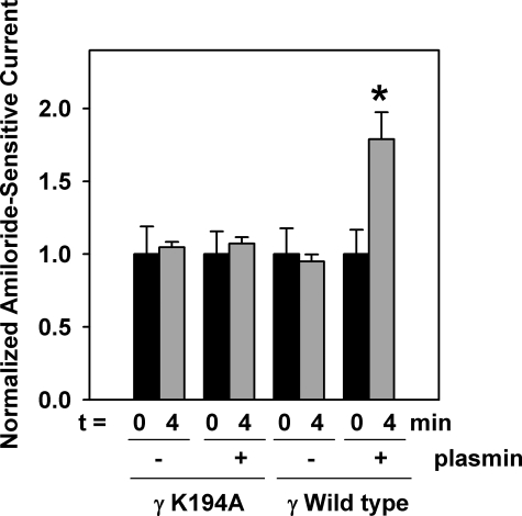
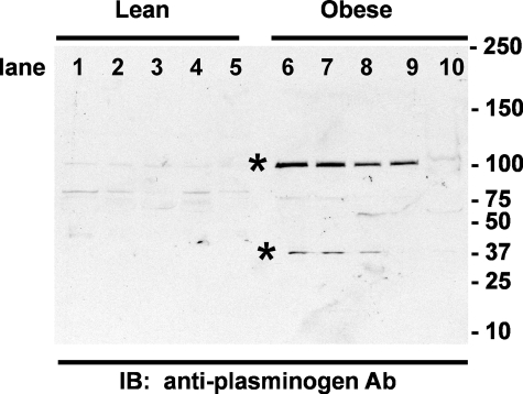
Proteolytic processing of epithelial sodium channel (ENaC) subunits occurs as channels mature within the biosynthetic pathway. The proteolytic processing events of the alpha and gamma subunits are associated with channel activation. Furin cleaves the alpha subunit ectodomain at two sites, releasing an inhibitory tract and activating the channel. However, furin cleaves the gamma subunit ectodomain only once. A second distal cleavage in the gamma subunit induced by other proteases, such as prostasin and elastase, is required to release a second inhibitory tract and further activate the channel. We found that the serine protease plasmin activates ENaC in association with inducing cleavage of the gamma subunit at gammaLys194, a site distal to the furin site. A gammaK194A mutant prevented both plasmin-dependent activation of ENaC and plasmin-dependent production of a unique 70-kDa carboxyl-terminal gamma subunit cleavage fragment. Plasmin-dependent cleavage and activation of ENaC may have a role in extracellular volume expansion in human disorders associated with proteinuria, as filtered plasminogen may be processed by urokinase, released from renal tubular epithelium, to generate active plasmin.


Figures

FIGURE 1.
FIGURE 1.
Plasmin activates mouse ENaC expressed in Xenopus oocytes. Oocytes injected with cRNAs for non-tagged α and β and HA-γ-V5 were analyzed by TEV before and after exposure to plasmin (10 μg/ml) for the indicated times. A and B, time course for activation of amiloride-sensitive Na+ currents in oocytes treated with external plasmin (10 μg/ml) is shown. A, for control oocytes, amiloride-sensitive currents at base line were-2330 ± 390 nA (mean and S.E., n = 17). For oocytes exposed to 10 μg/ml plasmin, amiloride-sensitive currents at base line were -2020 ± 300 nA (mean and S.E., n = 16-17). Normalized currents are also shown. *, p < 0.005, plasmin-treated versus control normalized currents at each time point following the initiation of plasmin treatment. B, for control oocytes, amiloride-sensitive currents at base line were -1560 ± 310 nA (mean and S.E., n = 8-10). For oocytes exposed to 10 μg/ml plasmin, amiloride-sensitive currents at base line were -1320 ± 330 nA (mean and S.E., n = 11). Normalized currents are also shown. *, p < 0.005, plasmin-treated versus control normalized currents at each time point following the initiation of plasmin treatment. C, for control oocytes, amiloride-sensitive currents at base line were -1700 ± 260 nA (mean and S.E., n = 27). For oocytes exposed to 10 μg/ml plasmin, amiloride-sensitive currents at baseline were -1810 ± 260 nA (mean and S.E., n = 23). Normalized currents are also shown. *, p < 0.005, plasmin-treated versus control normalized currents at each time point following the initiation of plasmin treatment. Treatment of oocytes for 4 min with plasmin (10 μg/ml) increased amiloride-sensitive sodium currents by 1.95-fold.
FIGURE 2.
FIGURE 2.
Plasmin cleaves the γ subunit at a site distal to the furin cleavage site when ENaC is expressed in MDCK cells. MDCK cells were transiently transfected with α-Myc, β-FLAG, and HA-γ-V5. The following day, cells were exposed to 0, 3, 9, or 27 μg/ml plasmin for 6 min prior to biotinylation of surface proteins. Biotinylated γ was recovered with streptavidin-conjugated beads from anti-V5 immunoprecipitates (IP) and analyzed by immunoblotting (IB) with anti-V5 antibodies. Mobilities of the non-cleaved 93-kDa γ (γ 93), furin-cleaved 75-kDa γ (γ 75), and the new plasmin-dependent 70-kDa γ (γ 70) fragments are indicated to the right of the gel. An enhanced version of the banding pattern is shown in the lower panel and includes three parallel lines that denote γ93, γ75, and γ70. Numbers to the left of the gel represent the mobility of Bio-Rad Precision Plus protein standards in kDa on a 7.5% gel. The blot is representative of three independent experiments.
FIGURE 3.
FIGURE 3.
Plasmin cleaves at γLys194 when ENaC is expressed in Xenopus oocytes. A, Xenopus oocytes were injected with cRNA for wild-type α and β and either wild-type γ (γWT) or γ with mutation of the prostasin cleavage site (γRKRK186QQQQ) or the plasmin cleavage site predicted at γIHK194 (γK194A). NA, no addition CRNA. All γ constructs contained N-terminal HA and C-terminal V5 epitope tags. The next day, oocytes were treated with plasmin (10 μg/ml) for 4 min prior to surface biotinylation. Biotinylated proteins were precipitated with streptavidin beads and eluted for analysis by immunoblotting with anti-V5 antibodies. Appearance of the 70-kDa band (<) due to plasmin cleavage is blocked by the γK194A mutation. B, model of γENaC denoting the cleavage sites determined for furin (γRKRR143), prostasin (γRKRK186), pancreatic elastase (γAla195), neutrophil elastase (γVal198), and now, plasmin (γIHK194) (4, 5, 7, 8). See “Results” for details. Numbers to the right of the gel represent the mobility of Bio-Rad Precision Plus protein standards in kDa on a 10% gel.
FIGURE 4.
FIGURE 4.
Plasmin activates ENaC containing a γ subunit with a prostasin cleavage site mutation. Oocytes injected with cRNAs for non-tagged α and β and mutant HA-γRKRK186/QQQQ-V5 were analyzed by TEV before and after exposure to plasmin (10 μg/ml) for 4 min. For control oocytes, amiloride-sensitive base-line currents were -2410 ± 490 nA (mean and S.E., n = 12). For oocytes exposed to plasmin, amiloride-sensitive currents at base line were -1740 ± 320 nA (mean and S.E., n = 15). Application of extracellular plasmin (10 μg/ml) for 4 min increased mutant ENaC currents by 2.48-fold. *, p < 0.005, plasmin-treated versus control normalized currents at 4 min.
FIGURE 5.
FIGURE 5.
Plasmin cleaves the γ subunit at a site distinct from the prostasin cleavage site. MDCK cells were transiently transfected with α-Myc, β-FLAG, and either wild-type HA-γ-V5 (γ WT) or the HA-γRKRK186/QQQQ-V5 mutant. ENaC was immunoprecipitated (IP) with anti-V5 antibodies from cell extracts, incubated with 15 μl of 20 μg/ml plasmin for 0, 6, or 18 min, and analyzed by immunoblotting (IB) with anti-V5 antibodies. Mobilities of the non-cleaved 93-kDa γ(γ93), furin-cleaved 75-kDaγ(γ75), and the new plasmin-dependent 70-kDa γ (γ 70) fragments are indicated to the right of the gels. An enhanced version of the banding pattern is shown in the lower panel and includes three parallel lines that denote γ93, γ75, and γ70. Numbers to the left of the gels represent the mobility of Bio-Rad Precision Plus protein standards in kDa. The blot is representative of two independent experiments.
FIGURE 6.
FIGURE 6.
ENaC containing γK194A is not activated by plasmin exposure. Oocytes injected with cRNAs for non-tagged α and β and either the HA-γK194A-V5 mutant or wild-type HA-γ-V5 were analyzed by TEV before and after exposure to plasmin (10 μg/ml) for 4 min. Oocytes expressing the αβγK194A mutant or wild-type αβγ had stable amiloride-sensitive currents over 4 min of TEV recording in both the absence and presence of 10 μg/ml plasmin. For control oocytes expressing the αβγK194A mutant, amiloride-sensitive currents at base line were -2130 ± 400 nA (mean and S.E., n = 19). For oocytes exposed to plasmin, amiloride-sensitive currents at base line were -3380 ± 530 nA (mean and S.E., n = 23). Extracellular plasmin (10 μg/ml) for 4 min did not increase αβγK194A ENaC currents. Treatment of oocytes expressing wild-type αβγENaC for 4 min with plasmin (10 μg/ml) was used as a positive control. Amiloride-sensitive currents for control oocytes (-plasmin) at baseline were -2020 ± 360 nA (mean and S.E., n = 18). For oocytes exposed to plasmin, amiloride-sensitive currents at base line were -2020 ± 340 nA (mean and S.E., n = 17). Extracellular plasmin (10 μg/ml) for 4 min did increase wild-type αβγENaC currents by 1.8-fold. *, p < 0.005, plasmin treated versus control normalized currents at 4 min.
FIGURE 7.
FIGURE 7.
Increased levels of plasminogen and plasmin are present in the urine of rats with metabolic syndrome. Urine aliquots (0.15 ml) from five different control lean (lanes 1-5) and obese (lanes 6-10) rats were separately concentrated 10-fold and subjected to SDS-PAGE under reducing conditions for analysis by immunoblotting (IB) with anti-human plasminogen antibodies (Ab) as described under “Experimental Procedures.” Note the plasminogen (∼100 kDa) and plasmin L-chain (∼37 kDa) in the urine of the obese rats (asterisks). Numbers to the right of the gel indicate the mobility of Bio-Rad Precision Plus protein standards in kDa on a 4-15% gel.

Similar articles

Cited by

References

    1. Sheng, S., Johnson, J. P., and Kleyman, T. R. (2008) in The Kidney, Physiology and Pathophysiology (Alpern, R. J., and Hebert, S. C., eds) 4th Ed., pp. 743-68 Elsevier Publishing, Burlington, MA
    1. Kleyman, T. R., Myerburg, M. M., and Hughey, R. P. (2006) Kidney Int. 70 1391-1392 - PubMed
    1. Hughey, R. P., Carattino, M. D., and Kleyman, T. R. (2007) Curr. Opin. Nephrol. Hypertens. 16 444-450 - PubMed
    1. Hughey, R. P., Bruns, J. B., Kinlough, C. L., Harkleroad, K. L., Tong, Q., Carattino, M. D., Johnson, J. P., Stockand, J. D., and Kleyman, T. R. (2004) J. Biol. Chem. 279 18111-18114 - PubMed
    1. Bruns, J. B., Carattino, M. D., Sheng, S., Maarouf, A. B., Weisz, O. A., Pilewski, J. M., Hughey, R. P., and Kleyman, T. R. (2007) J. Biol. Chem. 282 6153-6160 - PubMed

Publication types

LinkOut - more resources