. 2008 Dec;82(24):12365-73.
doi: 10.1128/JVI.01321-08. Epub 2008 Oct 15.

Measles virus V protein is a decoy substrate for IkappaB kinase alpha and prevents Toll-like receptor 7/9-mediated interferon induction

Affiliations

Measles virus V protein is a decoy substrate for IkappaB kinase alpha and prevents Toll-like receptor 7/9-mediated interferon induction

Christian K Pfaller et al. J Virol. 2008 Dec.

Abstract

The central role of plasmacytoid dendritic cells (pDC) in activating host immune responses stems from their high capacity to express alpha interferon (IFN-alpha) after stimulation of Toll-like receptors 7 and 9 (TLR7 and -9). This involves the adapter MyD88 and the kinases interleukin-1 receptor-associated kinase 1 (IRAK1), IRAK4, and IkappaB kinase alpha (IKKalpha), which activates IFN regulatory factor 7 (IRF7) and is independent of the canonical kinases TBK1 and IKKepsilon. We have recently shown that the immunosuppressive measles virus (MV) abolishes TLR7/9/MyD88-dependent IFN induction in human pDC (Schlender et al., J. Virol. 79:5507-5515, 2005), but the molecular mechanisms remained elusive. Here, we have reconstituted the pathway in cell lines and identified IKKalpha and IRF7 as specific targets of the MV V protein (MV-V). Binding of MV-V to IKKalpha resulted in phosphorylation of V on the expense of IRF7 phosphorylation by IKKalpha in vitro and in living cells. This corroborates the role of IKKalpha as the kinase phosphorylating IRF7. MV-V in addition bound to IRF7 and to phosphomimetic IRF7 and inhibited IRF7 transcriptional activity. Binding to both IKKalpha and IRF7 required the 68-amino-acid unique C-terminal domain of V. Inhibition of TLR/MyD88-dependent IFN induction by MV-V is unique among paramyxovirus V proteins and should contribute to the unique immunosuppressive phenotype of measles. The mechanisms employed by MV-V inspire strategies to interfere with immunopathological TLR/MyD88 signaling.


Figures
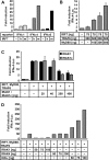
FIG. 1.
FIG. 1.
Reconstitution of MyD88-dependent TLR7/9 signaling in cell lines. (A) 293T cells were transfected with the indicated plasmids, and activation of IFN-α promoters was determined by dual-luciferase assay. Overexpression of mIRF7 (m) and hIRF7 (h) (450 ng of plasmid) leads to efficient induction of the IFNα4 and IFNα6 promoters and moderate induction of IFNα1. (B) TRAF6 and MyD88 expression increases the activity of mIRF7 in a dose-dependent manner. (C) Expression of IRAK1 or IRAK1c in addition to IRF7, MyD88, and TRAF6 leads to a dose-dependent inhibitory effect on IFNα4 promoter-driven FL activity. (D) Effect of indicated amounts of plasmids encoding for IRAK1, IRAK4, and IKKα. A stimulatory and dose-dependent positive effect is only observed for IKKα. Data are derived from three independent experiments and are represented as means + standard deviations.
FIG. 2.
FIG. 2.
MV-P gene products and cDNA constructs. The P gene of MV is located at the second position of the 16-kb MV negative-sense ssRNA [(−)ssRNA] genome. The P mRNA consists of 1,685 bases; insertion of an additional guanosine between bases 751 and 752 by RNA editing gives rise to the V mRNA. V and P proteins share a common N-terminal protein sequence (PVN, aa 1 to 231) but have unique C termini (PC, aa 232 to 507; VC, aa 232 to 299). cDNAs encoding authentic or tagged P, V, PVN, PC, and VC were constructed by cloning into different vectors for eukaryotic or prokaryotic expression. Expression of the C protein, which is encoded in an alternative ORF, was abolished in the P/V cDNA constructs by site-directed mutagenesis (for details see Materials and Methods) or was expressed from a separate cDNA.
FIG. 3.
FIG. 3.
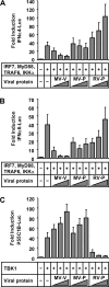
MV-V protein inhibits MyD88-dependent induction of IFN-α promoters. (A and B) Induction of IFNa4 (A) and IFNa6 (B) promoters by IRF7 (10 ng) and MyD88, TRAF6, and IKKα (50 ng each) is inhibited by cotransfection of MV-V (250, 500, and 750 ng) in a dose-dependent manner, in contrast to MV-P and RV-P. (C) Induction of the p55C1B part of the IFN-β promoter by overexpression of TBK1 (150 ng of plasmid) is antagonized by expression of RV-P, but not by MV-V or MV-P. Data are derived from three independent experiments and are represented as means + standard deviations.
FIG. 4.
FIG. 4.
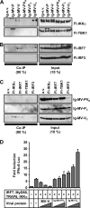
Interaction of MV-V with IKKα and IRF7. (A and B) pIg-MV-P, -MV-V, and -MV-C or empty vector (pIg-e.v.) was cotransfected with pFl-IKKα, -IKKβ, -TBK1, -IKKɛ, -IRF7, or -IRF3 in 293T cells as indicated. Cells were lysed 24 h posttransfection under native conditions, and Ig-tagged proteins were pulled down with protein A-conjugated Sepharose beads and subjected to Western blot analysis. MV-V coprecipitated (co-IP) IKKα, but not IKKβ, TBK1, or IKKɛ (A), and in addition, IRF7, but not IRF3 (B). Precipitation of any of the kinases or transcription factors by MV-P or MV-C was not detected. (C and D) Native cell lysates of transfected 293T cells were subjected to anti-Flag affinity gel purification and analyzed by Western blotting. Flag-tagged IKKα, IRF7, TBK1, and IRF3 were efficiently pulled down (D). Only MV-V was coprecipitated by IKKα and IRF7, but not with TBK1 or IRF3 (C).
FIG. 5.
FIG. 5.
VC mediates interaction with IKKα and IRF7. (A and B) The indicated plasmids or empty vector (e.v.) was cotransfected with pFl-IKKα, -TBK1, -IRF7, or -IRF3 into 293T cells. Ig-tagged proteins were pulled down under native conditions and analyzed by Western blotting for interaction partners. Full-length Ig-MV-V as well as Ig-VC was identified to efficiently bind IKKα, but not TBK1 (A). In addition, IRF7, but not IRF3, was precipitated by Ig-MV-VC as efficiently as by full-length Ig-MV-V (B). (C) Native cell lysates of transfected 293T cells were subjected to anti-Flag affinity gel purification and analyzed by Western blotting for interaction of Fl-IKKα, -IRF7, -TBK1, or -IRF3 with Ig-MV-PVN, -MV-PC, or -MV-VC. Ig-MV-VC was coprecipitated efficiently by both IKKα and IRF7. Ig-MV-PVN and Ig-MV-PC did not show interaction with any of the tested proteins. (D) Luciferase reporter gene assay using overexpression of IRF7, MyD88, TRAF6, and IKKα for induction of IFNα6-Luc in 293T cells. In contrast to full-length MV-V, which inhibits induction of the IFNα6 promoter in a dose-dependent manner (250, 500, and 750 ng), Ig-MV-VC or MV-PVN was ineffective. Data are derived from three independent experiments and are represented as means + standard deviations.
FIG. 6.
FIG. 6.
MV-V is able to bind and inhibit activated IRF7. (A to C) Induction of IFNα6 promoter was stimulated in Huh7.5 cells by expression of IRF7 (10 ng plasmid) and MyD88, TRAF6, and IKKα (50 ng each) (A) or by individual expression of IRF7 (100 ng) (B) or constitutively active IRF7-2D (100 ng) (C). Cotransfection of increasing amounts of MV-V (250, 500, 750 ng) revealed a dose-dependent inhibition in all experiments. (D) Plasmids encoding Ig-tagged MV-P, MV-V, MV-PVN, MV-PC, and MV-VC or empty vector were cotransfected with phosphomimetic Fl-IRF7-2D, and Fl-IRF3-5D into 293T cells. Full-length Ig-MV-V as well as Ig-VC was identified to bind IRF7, but not IRF3. Note that precipitated protein bands representing Ig-MV-V, -PVN, and -PC appear in the coimmunoprecipitation (Co-IP) figure (left panel) as well.
FIG. 7.
FIG. 7.
MV-V is a substrate of IKKα and competes with IRF7 phosphorylation. His-tagged IRF7, MV-V, and MV-P were expressed in prokaryotic cells and purified by Ni-NTA affinity chromatography under native conditions. (A and B) Equal amounts of IRF7 (500 ng/reaction) and increasing amounts of MV-V (0 to 1,600 pmol per reaction) were subjected to an in vitro kinase assay together with commercial His-IKKα (100 ng/reaction) and [γ-32P]ATP. Probes were subjected to Western blot analysis after incubation at indicated time points. Phosphorylation of proteins was detected by autoradiography, while total protein amounts were determined by Western blotting (WB). (A) MV-V is a substrate for efficient phosphorylation by IKKα in vitro. Increasing amounts of MV-V reduce levels of phospho-IRF7, while total protein levels are equal in all lanes. α-His, anti-His. (B) Quantification of phospho-IRF7 over time with dependence on increasing amounts of MV-V. IRF7 phosphorylation is completely abolished in the presence of large amounts of MV-V (800 and 1,600 pmol), while smaller amounts of MV-V (200 and 400 pmol) delay IRF7 phosphorylation. (C and D) Five hundred nanograms of His-IRF7 (C) or 100 ng of GST-IκB-α (D) was subjected to an in vitro kinase assay in either the absence (lanes 1) or presence (lanes 2 to 4) of 100 ng of His-IKKα and 100 pmol of His-MV-V (lanes 3) or His-MV-P (lanes 4). Reactions were stopped after indicated time points and subjected to Western blot analysis. (E and F) Quantification of phospho-IRF7 (E) or pIκB-α (F) reveals specificity of MV-V. Phosphorylation of IRF7 is inhibited only by MV-V, but not MV-P. Phosphorylation of IκB-α is not inhibited by either viral protein. (G) Reduction of activated IRF7 in living cells. Flag-tagged IKKα, IRF7, and MV-V were pulled down from 293T cells transfected with the indicated plasmids at 24 h posttransfection and analyzed by Western blotting using anti-Flag M2 antibody. A band representing activated IRF7 (marked by a star) appeared only after coexpression of IKKα and IRF7. A significant reduction of this band was observed in the presence of MV-V.

Similar articles

Cited by

References

    1. Andrejeva, J., K. S. Childs, D. F. Young, T. S. Carlos, N. Stock, S. Goodbourn, and R. E. Randall. 2004. The V proteins of paramyxoviruses bind the IFN-inducible RNA helicase, mda-5, and inhibit its activation of the IFN-beta promoter. Proc. Natl. Acad. Sci. USA 10117264-17269. - PMC - PubMed
    1. Barchet, W., M. Cella, B. Odermatt, C. Asselin-Paturel, M. Colonna, and U. Kalinke. 2002. Virus-induced interferon alpha production by a dendritic cell subset in the absence of feedback signaling in vivo. J. Exp. Med. 195507-516. - PMC - PubMed
    1. Bellini, W. J., G. Englund, S. Rozenblatt, H. Arnheiter, and C. D. Richardson. 1985. Measles virus P gene codes for two proteins. J. Virol. 53908-919. - PMC - PubMed
    1. Brzózka, K., S. Finke, and K.-K. Conzelmann. 2005. Identification of the rabies virus alpha/beta interferon antagonist: phosphoprotein P interferes with phosphorylation of interferon regulatory factor 3. J. Virol. 797673-7681. - PMC - PubMed
    1. Brzózka, K., S. Finke, and K. K. Conzelmann. 2006. Inhibition of interferon signaling by rabies virus phosphoprotein P: activation-dependent binding of STAT1 and STAT2. J. Virol. 802675-2683. - PMC - PubMed

Publication types

MeSH terms