Review
. 2008;15(24):2456-71.
doi: 10.2174/092986708785909094.

Computational models of neuronal biophysics and the characterization of potential neuropharmacological targets

Affiliations
Review

Computational models of neuronal biophysics and the characterization of potential neuropharmacological targets

Michele Ferrante et al. Curr Med Chem. 2008.

Abstract

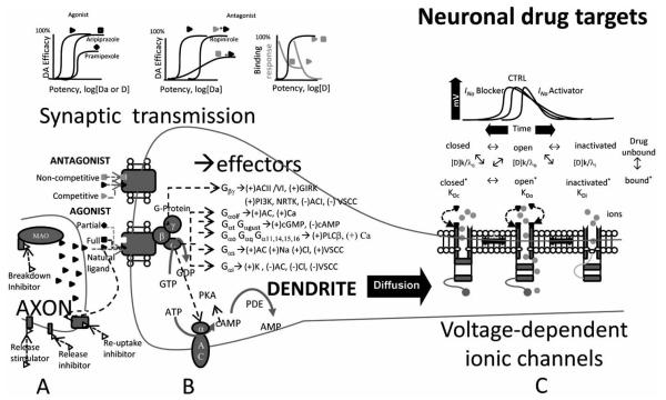
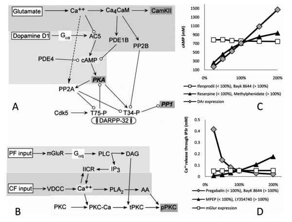
The identification and characterization of potential pharmacological targets in neurology and psychiatry is a fundamental problem at the intersection between medicinal chemistry and the neurosciences. Exciting new techniques in proteomics and genomics have fostered rapid progress, opening numerous questions as to the functional consequences of ligand binding at the systems level. Psycho- and neuro-active drugs typically work in nerve cells by affecting one or more aspects of electrophysiological activity. Thus, an integrated understanding of neuropharmacological agents requires bridging the gap between their molecular mechanisms and the biophysical determinants of neuronal function. Computational neuroscience and bioinformatics can play a major role in this functional connection. Robust quantitative models exist describing all major active membrane properties under endogenous and exogenous chemical control. These include voltage-dependent ionic channels (sodium, potassium, calcium, etc.), synaptic receptor channels (e.g. glutamatergic, GABAergic, cholinergic), and G protein coupled signaling pathways (protein kinases, phosphatases, and other enzymatic cascades). This brief review of neuromolecular medicine from the computational perspective provides compelling examples of how simulations can elucidate, explain, and predict the effect of chemical agonists, antagonists, and modulators in the nervous system.


Figures

Fig. (1)
Fig. (1)
From neuroimaging experiments to cellular and sub-cellular electrophysiological and enzymatic models. (A) Functional Magnetic Resonance Imaging represents neural activity measured through hemodynamic responses (circle indicates the position of the hippocampus), connecting function/activity to brain structures (adapted from [232], with permission granted by “Nature Publishing Group”). (B-D) Ultramicroscopy is a new neuroimaging tool allowing visualization of biological neural networks at different resolution level (adapted from [233], with permission granted by “Nature Publishing Group”) (B) Whole hippocampus imaged from multiple optical sections (scale bar: 500 μm). (C) 3D visualization of the CA1 region of the hippocampus (scale bar: 200 μm), with observable cell bodies and main branches of the dendritic trees. (D) Spines and dendritic arborization of CA1 pyramidal neurons (scale bar: 5 μm). (E) Computational/mathematical models of both electrophysiological and enzymatic activity are implemented to predict, reproduce, interpret, and explain experimental results.
Fig. (2)
Fig. (2)
Neuronal drug targets. (A) A drug can target the presynaptic neuron acting at many levels. It can stimulate or inhibit neurotransmitter release, reuptake or breakdown. Once released, the natural ligand diffuses in the synaptic cleft and binds to specific receptors. (B) The pharmacological effects of postsynaptic receptor-drug interactions (full/partial, agonist/antagonist) are characterized in terms of potency, efficacy and binding, or response. These interactions can be competitive or non-competitive depending on whether or not the natural ligand and drug share the same binding site. Metabotropic receptors are associated with a GTP-binding protein complex composed of three subunits, which detach upon activation and target various ion channels or enzymes. (C) A drug can also interact directly with voltage-dependent ionic channels. Depending on the membrane potential and on their kinetic properties, ionic channels can be described by at least two (open and closed) and up to four (same plus inactivated and deinactivated) different conformational and functional states. A specific drug (D) can bind to each of these states with specific association (k) and dissociation (γ) constants and affinities (K).
Fig. (3)
Fig. (3)
Molecular structures and functions of drugs interacting with the simulated mechanisms. (A) Lamotrigine (C9H7Cl2N5) is an anticonvulsant and mood stabilizer used to treat epilepsy and bipolar disorder. (B) Diazepam (C16H13ClN2O) is a benzodiazepine commonly prescribed in the treatment of anxiety, insomnia, seizures, alcohol withdrawal, and muscle spasms. (C) Flindokalner (C16H10ClF4NO2) is a drug candidate for post-stroke neuroprotection. It is an activator of large conductance calcium dependent K+ conductances, but the therapeutic mechanism of action is still under investigation. (D) Reserpine (C33H40N2O9) is a bulky molecule used as an antipsychotic. It acts by blocking the norepinephrine, serotonin, and dopamine transporters. (E) Methylphenidate (C14H19NO2) is a stimulant used to treat attention-deficit hyperactivity disorder, chronic fatigue syndrome, and daytime drowsiness due to narcolepsy. It works by binding the dopamine transporter and inhibiting its reuptake. (F) Bupropion (C13H18ClNO) is an atypical antidepressant acting mainly as a dopamine reuptake inhibitor. It is also a weak nicotinic antagonist and a norepinephrine reuptake inhibitor. (G) Ifenprodil (C21H27NO2), a candidate drug in schizophrenia, is a selective non-competitive NMDA antagonist, which reduces cell excitability by blocking calcium influx. (H) LY354740 (C8H11NO4), also known as eglumegad, is a drug candidate for the treatment of anxiety and drug addiction. It is a highly selective agonist of metabotropic glutamate receptors (mGluR2/3). (I) Pregabalin (C8H17NO2) is effective in treating neuropathic pain, generalized anxiety disorder and as an adjunct therapy for partial seizures. It inhibits P/Q type Ca++ channels by binding to their α2δ subunit. (J) Bayk 8644 (C16H15F3N2O4), is an experimental drug candidate known to act as a mood regulator. It is an L-type Ca++ channel activator. (K) MPEP (C14H11N) is an extensively studied drug candidate to treat epilepsy, pain, neurodegenerative diseases, and anxiety. It is a highly selective and potent mGluR and AMPA antagonist.
Fig. (4)
Fig. (4)
Computational neuropharmacology and electrophysiology. (A) Raster plot describing how various antiepileptic drugs targeting different neuronal mechanisms can affect spike patterns and sequences (signal generation/integration). (B) All these simulated treatment conditions (black lines) significantly abate the instantaneous frequencies. (C) Compared to control (gray histograms), individual drugs can preferentially favor certain frequency ranges, cutting off others. (D) Even if they have equal average output frequencies (top bars), two drugs (3 vs. 4) can differentially modulate membrane voltage (bottom bars). (E) Drug effects on the timing of the first and second spike.
Fig. (5)
Fig. (5)
Two intracellular signaling models of molecular dynamics involved in plasticity. (A) Dopaminergic and glutamatergic activated signaling pathways of medium spiny projection neurons involved in the production of cyclic adenosine monophosphate (cAMP). Glutamate stimulation leads to calcium elevation; Ca++ binding to calmodulin (CaM) activates calcium - Calmodulin kinase 2 (CaMKII), protein phosphatase 2B (PP2B) and phosphodiesterase 1B (PDE1B), the latter of which degrades cAMP. Dopamine (D1) receptor stimulation activates the protein kinase A (PKA) pathway via adenylate cyclase type V (AC5) and cAMP. PKA activation increases phosphorylation of DARPP-32, which then inhibits protein phosphatase 1 (PP1). (B) The Purkinje cell model incorporates three separate biochemical pathways. The first pathway describes parallel fiber (PF) activation and consequent diacylglycerol (DAG), and inositol trisphosphate (IP3) production via activation of G-proteins and phospholipase C (PLC) and subsequent intracellular Ca2+ release. Another biochemical pathway depicts how Ca++ elevation due to CF stimulation activates voltage dependent calcium channels (VDCC) and leads to arachidonic acid (AA) production. The last set of reactions describes how calcium, DAG, and AA elevation influence the activity of the persistently active form of protein kinase C (pPKC). (C) Dose-dependent effects on cAMP of different drugs modulating dopamine reuptake (Reserpine and methylphenidate), dopamine receptor density (DaR expression, a possible target for genetic therapies) and neuronal calcium influx (ifenprodil and Bayk 8644). (D) Simulated effect of different dosages of drugs modulating glutamate release (MPEP and LY354740), glutamate receptor density (mGluR expression), and intracellular Ca++ concentration (pregabalin and Bayk 8644) on the IP3 receptor dependent release of intracellular calcium from the endoplasmic reticulum.

Similar articles

Cited by

References

    1. http://www.caregiver.org/caregiver/jsp/content_node.jsp?nodeid=4 38.
    1. Malphrus AD, Wilfong AA. Curr. Treat. Options Neurol. 2007;9:256. - PubMed
    1. Youdim MB, Buccafusco JJ. J. Neural. Transm. 2005;112:519. - PubMed
    1. Migliore M, Shepherd GM. Nat. Rev. Neurosci. 2002;3:362. - PubMed
    1. Lindskog M, Kim M, Wikström MA, Blackwell KT, Kotaleski JH. PLoS Comput. Biol. 2006;2:119. - PMC - PubMed

Publication types

LinkOut - more resources