. 2008 Nov 5;27(21):2829-38.
doi: 10.1038/emboj.2008.206. Epub 2008 Oct 9.

Laminin and biomimetic extracellular elasticity enhance functional differentiation in mammary epithelia

Affiliations

Laminin and biomimetic extracellular elasticity enhance functional differentiation in mammary epithelia

Jordi Alcaraz et al. EMBO J. .

Abstract

In the mammary gland, epithelial cells are embedded in a 'soft' environment and become functionally differentiated in culture when exposed to a laminin-rich extracellular matrix gel. Here, we define the processes by which mammary epithelial cells integrate biochemical and mechanical extracellular cues to maintain their differentiated phenotype. We used single cells cultured on top of gels in conditions permissive for beta-casein expression using atomic force microscopy to measure the elasticity of the cells and their underlying substrata. We found that maintenance of beta-casein expression required both laminin signalling and a 'soft' extracellular matrix, as is the case in normal tissues in vivo, and biomimetic intracellular elasticity, as is the case in primary mammary epithelial organoids. Conversely, two hallmarks of breast cancer development, stiffening of the extracellular matrix and loss of laminin signalling, led to the loss of beta-casein expression and non-biomimetic intracellular elasticity. Our data indicate that tissue-specific gene expression is controlled by both the tissues' unique biochemical milieu and mechanical properties, processes involved in maintenance of tissue integrity and protection against tumorigenesis.


Figures

Figure 1
Figure 1
The ‘physiological' or ‘biomimetic' elasticity of MECs can be defined in culture. (A) Schematic representation of the experimental setup: either mammary organoids (MO) or single MECs (SCp2 or EpH4) were plated on top of an ECM gel. An AFM probe was used to quantify the elasticity of either the cell or the underlying ECM gel. (B) Morphology of primary mammary organoids isolated from mice in early pregnancy and cultured on top of a LM1-rich ECM gel (Matrigel) in differentiation medium for 24 h. The elastic modulus of the substrata as measured by AFM is indicated at the bottom of each image. Most mammary organoids had either small or medium sizes, corresponding to roughly a dozen or a few dozen cells, respectively. (C) Elastic moduli of cells within either small- or medium-sized primary mammary organoids cultured as in (B). Dashed horizontal lines correspond to the lower and upper values defined by the elasticity of cells within mammary organoids, used as a reference for biomimetic cell elastic moduli throughout this study. All scale bars correspond to 20 μm throughout the figures unless otherwise indicated.
Figure 2
Figure 2
Laminin-111 signalling and biomimetic extracellular elasticity, but not cell shape per se, induce robust β-casein expression and a cellular elasticity comparable with cells within mammary organoids. Two MECs (SCp2 and EpH4) were cultured in the presence (Matrigel) or absence (poly-HEMA) of ECM signalling. (AC) Both culture conditions produced a similarly round morphology in SCp2 (A) and EpH4 cells (data not shown), but differed dramatically in their effects on β-casein, as measured by quantitative RT–PCR (B) and cellular elasticity (C). Note that fold β-casein/18S rRNA data are plotted on a logarithmic scale throughout the figures. **P<0.05 and ***P<0.01 were determined by two-tailed Student's t-test with respect to (w.r.t.) Matrigel or the corresponding control throughout this work unless otherwise indicated.
Figure 3
Figure 3
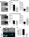
Increasing extracellular elasticity beyond normal mammary tissue values inhibits β-casein expression and promotes spreading and stiffening in MECs. We used two independent culture assays to increase extracellular elasticity beyond normal mammary tissue values while maintaining biochemical composition constant. The elastic modulus of the substrata as measured by AFM is indicated at the bottom of each image. (AC) Floating gel assay: EpH4 cells were cultured on top of LM1 mixed with COL-I (3 g/l) (40:60% v/v). Four hours after plating, differentiation media was added and half of the gels were gently detached from the container. The elastic modulus of the floating gel was comparable to normal tissue (A, top image), whereas that of the attached gel was three-fold stiffer (A, bottom image). Corresponding β-casein expression (B) and cell stiffness (C) in these culture conditions. (DH) Polyacrylamide gel assay: EpH4 cells were cultured on top of gels coated with equal fibronectin concentration but exhibiting a stiffness comparable with normal tissue (referred to as ‘soft') or within the range of mammary tumours (referred to as ‘stiff'). Cell morphology (D) and elasticity (F) 24 h after plating on either soft or stiff polyacrylamide gels. (E) β-Casein expression 48 h after overlaying cells with 2% Matrigel diluted in differentiation media. (G) Phase contrast (top) and corresponding CFP fluorescence intensity images (bottom panels) of EpH4 cells stably transfected with 16 concatenated copies of the β-casein promoter fused to CFP cultured as in (E). (H) Corresponding quantification of CFP intensity of cells cultured as in (E). The two assays consistently reported a downregulation of β-casein expression as well as non-biomimetic cell shape and elasticity in gels with non-biomimetic elastic moduli (for more details see the Discussion). *P<0.1 and ***P<0.01 were determined by two-tailed Student's t-test.
Figure 4
Figure 4
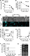
Loss of LM1 signalling downregulates β-casein expression and induces non-biomimetic cellular elasticity and/or morphology in SCp2 and EpH4 MECs. (A) β-Casein expression of MECs cultured on top of COL-I (2 g/l) gels mixed with decreasing concentrations of LM1. (B and C) Visualization (C) and corresponding quantification (B) of the activity of the β-casein promoter in EpH4 cells cultured as in (A). (D) Elastic moduli of MECs cultured as in (A). Although both cell lines exhibited non-biomimetic elasticity for LM1 concentrations below 10%, the elastic moduli of SCp2 was lower than the physiological range (dashed lines), whereas that of EpH4 was higher. (E) Elasticity of LM1–COL-I mixed gels. (F) Elasticity of SCp2 cells as a function of gel elasticity. Note the non-linear relationship between these two properties. (G) Messenger RNA levels of LM1- and COL-I-specific integrin receptors in SCp2 and EpH4 MECs cultured in 2D assessed by RT–PCR.
Figure 5
Figure 5
Receptors containing the β1-integrin subunit are major candidates for mediating LM1-induced cell biomimetic elasticity in MECs. (A and B) Elastic moduli of SCp2 cells cultured on top of Matrigel in the presence of function blocking antibodies against β1- (A) and α6-integrins (B) or corresponding isotype controls. (C) Elasticity of dystroglycan-expressing cells (DG+/+) or DG-negative (DG−/−). Both cell types remained fairly round under all conditions.
Figure 6
Figure 6
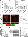
Laminin-111-induced cell biomimetic elasticity is partially mediated through downregulation of both actin polymerization and myosin II activity. (A, B) Comparison of the elasticity of SCp2 cells cultured for 24 h on 2D (A) or on top of LM1-rich gel (Matrigel) (B) after 30 min treatment with either vehicle (DMSO) or inhibitors of actin polymerization (latrunculin B), ROCK (Y26732) and myosin II ATPase (blebbistatin) activity using concentrations described in Materials and methods. *P<0.1, **P<0.05 and ***P<0.01 were determined by two-tailed Student's t-test w.r.t. untreated cells. (C) F-actin (red) and diphosphorylated MLC-II (green) organization in SCp2 cells spread on 2D substrata or rounded on top of Matrigel studied with confocal microscopy. Images correspond to the maximum intensity value projected on either the xy plane (top images) or the zx plane (bottom images). Both horizontal and vertical scale bars indicate 5 μm. (D) Box plot of cell spreading of SCp2 cells cultured as in (C). **P<0.05 was determined using Mann–Whitney rank sum test. (E) Quantification of the average fluorescence intensity of F-actin phalloidin staining per cell. (F) Immunoblot of total and diphosphorylated MLC-II in MECs cultured as in (C); (G) corresponding quantification by densitometry analysis of the ratio between diphosphorylated and total MLC-II. 2D substrata corresponds to either untreated borosilicate glass or tissue culture polystyrene dish.

Similar articles

Cited by

References

    1. Akhtar N, Streuli CH (2006) Rac1 links integrin-mediated adhesion to the control of lactational differentiation in mammary epithelia. J Cell Biol 173: 781–793 - PMC - PubMed
    1. Alcaraz J, Buscemi L, Grabulosa M, Trepat X, Fabry B, Farre R, Navajas D (2003) Microrheology of human lung epithelial cells measured by atomic force microscopy. Biophys J 84: 2071–2079 - PMC - PubMed
    1. Alcaraz J, Nelson CM, Bissell MJ (2004) Biomechanical approaches for studying integration of tissue structure and function in mammary epithelia. J Mammary Gland Biol Neoplasia 9: 361–374 - PMC - PubMed
    1. Barcellos-Hoff MH, Aggeler J, Ram TG, Bissell MJ (1989) Functional differentiation and alveolar morphogenesis of primary mammary cultures on reconstituted basement membrane. Development 105: 223–235 - PMC - PubMed
    1. Bissell MJ, Hall HG, Parry G (1982) How does the extracellular matrix direct gene expression? J Theor Biol 99: 31–68 - PubMed

Publication types

MeSH terms