. 2008 Dec;19(12):5309-26.
doi: 10.1091/mbc.e08-07-0712. Epub 2008 Oct 8.

The AP-2 adaptor beta2 appendage scaffolds alternate cargo endocytosis

Affiliations

The AP-2 adaptor beta2 appendage scaffolds alternate cargo endocytosis

Peter A Keyel et al. Mol Biol Cell. 2008 Dec.

Abstract

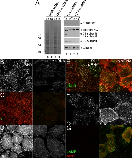
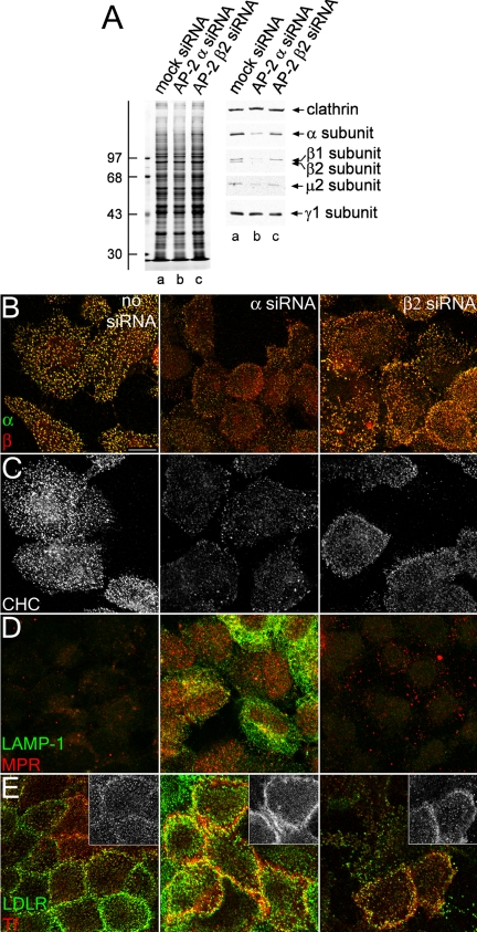
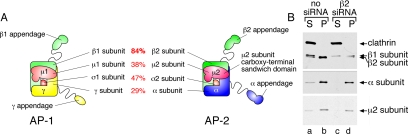
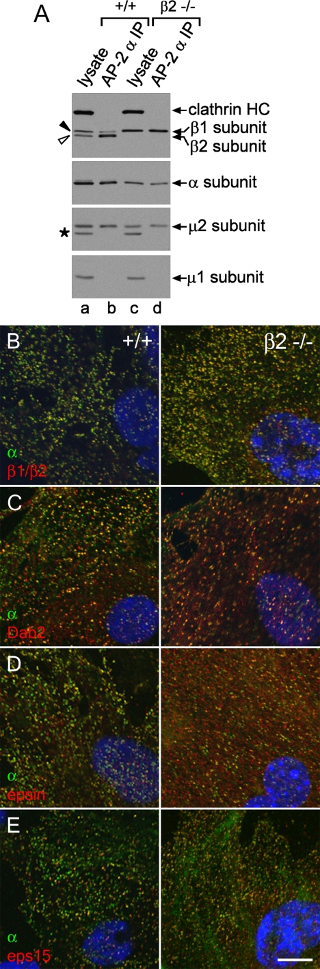
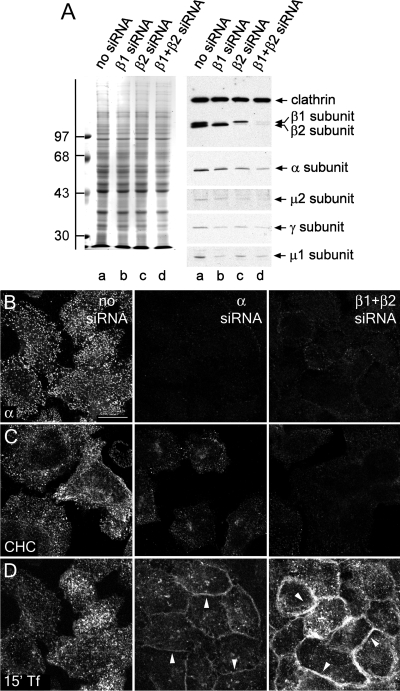
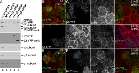
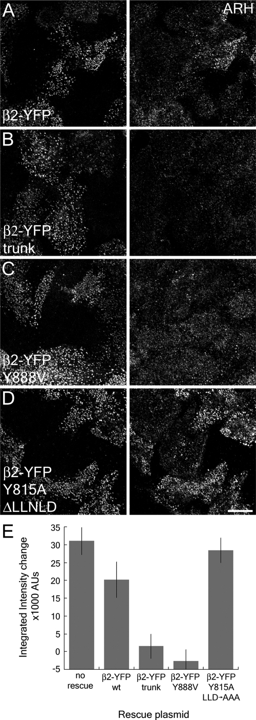
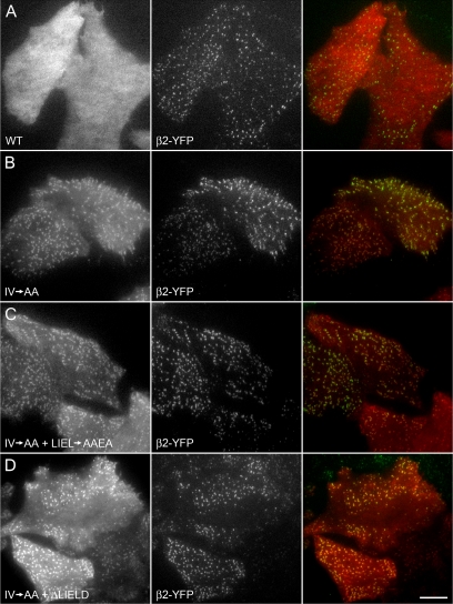
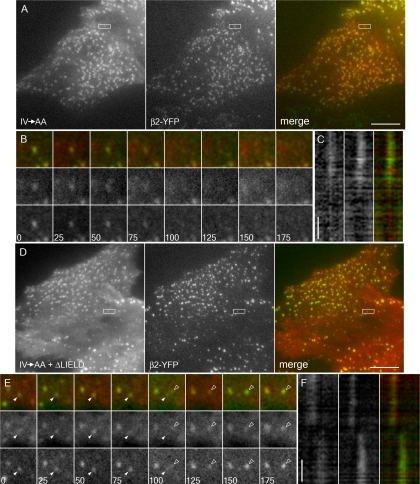
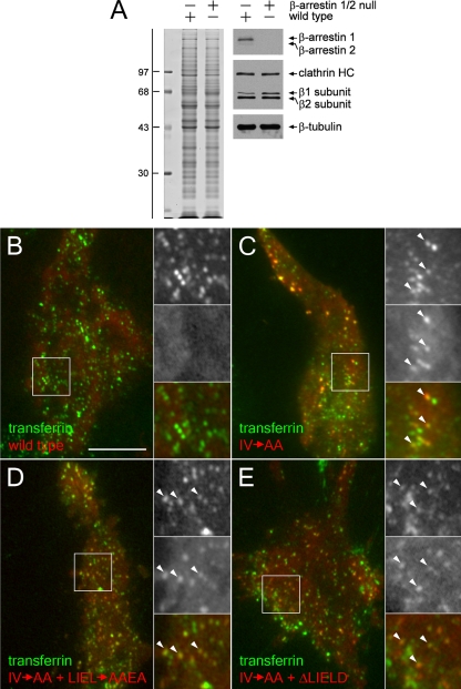
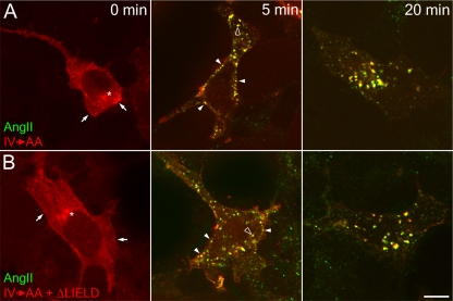
The independently folded appendages of the large alpha and beta2 subunits of the endocytic adaptor protein (AP)-2 complex coordinate proper assembly and operation of endocytic components during clathrin-mediated endocytosis. The beta2 subunit appendage contains a common binding site for beta-arrestin or the autosomal recessive hypercholesterolemia (ARH) protein. To determine the importance of this interaction surface in living cells, we used small interfering RNA-based gene silencing. The effect of extinguishing beta2 subunit expression on the internalization of transferrin is considerably weaker than an AP-2 alpha subunit knockdown. We show the mild sorting defect is due to fortuitous substitution of the beta2 chain with the closely related endogenous beta1 subunit of the AP-1 adaptor complex. Simultaneous silencing of both beta1 and beta2 subunit transcripts recapitulates the strong alpha subunit RNA interference (RNAi) phenotype and results in loss of ARH from endocytic clathrin coats. An RNAi-insensitive beta2-yellow fluorescent protein (YFP) expressed in the beta1 + beta2-silenced background restores cellular AP-2 levels, robust transferrin internalization, and ARH colocalization with cell surface clathrin. The importance of the beta appendage platform subdomain over clathrin for precise deposition of ARH at clathrin assembly zones is revealed by a beta2-YFP with a disrupted ARH binding interface, which does not restore ARH colocalization with clathrin. We also show a beta-arrestin 1 mutant, which engages coated structures in the absence of any G protein-coupled receptor stimulation, colocalizes with beta2-YFP and clathrin even in the absence of an operational clathrin binding sequence. These findings argue against ARH and beta-arrestin binding to a site upon the beta2 appendage platform that is later obstructed by polymerized clathrin. We conclude that ARH and beta-arrestin depend on a privileged beta2 appendage site for proper cargo recruitment to clathrin bud sites.


Figures

Figure 1.
Figure 1.
siRNA-mediated gene silencing of the AP-2 α subunit. (A) Lysates of HeLa SS6 cells untreated (lanes a and b) or treated with AP-2 α subunit siRNA (lanes c and d) were resolved by SDS-PAGE and either Coomassie stained or transferred to nitrocellulose. Sections of the blots were probed with anti-AP-2 α subunit mAb clone 8, anti-clathrin heavy chain (HC) mAb TD.1, anti-β1/β2 subunit mAb 100/1, anti-AP-2 μ2 antiserum, or anti-tubulin mAb E7, and only the relevant portion of each blot shown. The position of molecular mass standards (in kilodaltons) is indicated on the left. (B–D) HeLa SS6 cells untreated (left) or treated with AP-2 α subunit siRNA (right) were treated with 10 μg/ml brefeldin A for 15 min, permeabilized on ice for 1 min before fixation, and prepared for immunofluorescence using the anti-AP-2 α subunit mAb AP.6 (B), anti-AP-1 γ subunit mAb (C; green), anti-β1/β2 subunit antibody GD/2 (C; red), or anti-clathrin heavy chain mAb X22 (D). (E–G) HeLa SS6 cells untreated (left) or treated with AP-2 α subunit siRNA (right) were incubated on ice for 1 h with either anti-LDL receptor mAb C7 (E; green), Tf568 (E, red and F), anti-LAMP-1 mAb (G; green), or anti-MPR antibody (G; red) and fixed (E and G) or washed and warmed to 37°C for 15 min and fixed (F) followed by indirect immunofluorescence. Bar, 10 μm.
Figure 2.
Figure 2.
AP-2 knocked down cell ultrastructure. HeLa SS6 cells either mock transfected (A) or transfected with α subunit siRNA oligonucleotides (B–D) were briefly sonicated to prepare cell cortices, fixed, labeled with anti-transferrin receptor mAb H68.4, and then 15-nm gold-conjugated anti-mouse secondary antibodies to visualize extra-lattice receptors. Arrows indicate gold-labeled receptors.
Figure 3.
Figure 3.
AP-2 β2 subunit gene silencing. (A) Lysates of HeLa SS6 cells untreated (lane a) or treated with either AP-2 α or β2 subunit siRNA (lanes b and c) were resolved by SDS-PAGE and either Coomassie stained or transferred to nitrocellulose. Sections of the blots were probed with anti-clathrin HC mAb TD.1, anti-AP-2 α subunit mAb clone 8, anti-β1/β2 subunit mAb 100/1, anti-AP-2 μ2 antiserum, or anti-AP-1 γ subunit antibody AE/1, and only the relevant portion of the blots is shown. The position of molecular mass standards (in kilodaltons) is indicated on the left. (B and C) HeLa SS6 cells untreated (left) or treated with AP-2 α- (middle), or β2 subunit siRNA (right) were treated with 10 μg/ml brefeldin A for 15 min, permeabilized on ice for 1 min before fixation, and prepared for immunofluorescence using the anti-AP-2 α subunit mAb AP.6 (B, green), anti-β1-/β2 subunit antibody GD/2 (B; red), or anti-clathrin HC mAb X22 (C). (D and E) HeLa SS6 cells untreated (left) or treated with AP-2 α (middle) or β2 subunit (right) siRNA were incubated on ice for 1 h with either anti-LAMP-1 mAb (D; green) and anti-MPR antibody (D; red), or anti-LDL receptor mAb C7 (E; green) and Tf568 (E; red), fixed, and prepared for indirect immunofluorescence. The inset in E shows Tf568 immunofluorescence from each sample.
Figure 4.
Figure 4.
β1 subunit rescues β2 subunit knockdown of AP-2. (A) Schematic diagram of AP-1 and AP-2 complex composition and subunit homology. (B) Lysates from either untreated or β2 siRNA-treated HeLa SS6 cells were prepared and AP-2 immunoprecipitated using the anti-α subunit mAb AP.6. Aliquots of the supernatant and precipitate fractions from both control and β2 RNAi samples were resolved by SDS-PAGE and transferred to nitrocellulose. Blots were probed with the anti-AP-2 α subunit mAb clone 8, anti-β1/β2 subunit antibody mAb 100/1, anti-clathrin HC antibody mAb TD.1, or anti-AP-2 μ2 antiserum, and only the relevant portion of each blot is shown.
Figure 5.
Figure 5.
Stable incorporation of the β1 subunit into AP-2 in β2 subunit-null mouse fibroblasts. (A) Aliquots of lysates from either control +/+ (lane a) or β2-null −/− (lane c) mouse embryonic fibroblasts and of mAb AP.6 immunoprecipitates from the +/+ (lane b) or −/− (lane d) lysates were resolved by SDS-PAGE and transferred to nitrocellulose. Blots were probed with anti-clathrin HC mAb TD.1 and affinity purified anti-β1/β2 subunit polyclonal GD/1, anti-AP-2 α subunit mAb clone 8, anti-AP-2 μ2 subunit antiserum, or affinity-purified anti-μ1 subunit polyclonal RY/1, and only the relevant portion of each blot is shown. The asterisk indicates a nonspecific band. Loss of β2-chain expression results in a compensatory increase in the steady-state level of the β1 subunit in the cultured fibroblasts, although the level of AP-2 is nevertheless reduced compared with wild-type fibroblasts. (B–E) Wild-type (+/+; left column) or β2-null (−/−; right column) mouse embryonic fibroblasts were fixed and labeled with Hoechst 33258, anti-α subunit mAb AP.6 and either anti-β1/β2 subunit GD/1 (B), anti-Dab2 (C), anti-epsin (D), or anti-eps15 (E) polyclonal antibodies. Representative confocal sections of the ventral plasma membrane are shown. In all cases, absence of β2 does not obviously perturb the punctate patterning of the α subunit or CLASPs. Bar, 10 μm.
Figure 6.
Figure 6.
β1 + β2 subunit RNAi recapitulates α subunit RNAi phenotype. (A) Lysates of HeLa SS6 cells untreated (lane a) or treated with either AP-1 β1 (lane b), AP-2 β2 (lane c), or β1 + β2 subunit siRNAs (lane d) were resolved by SDS-PAGE and either Coomassie stained or transferred to nitrocellulose. Sections of the blots were probed with the anti-clathrin HC mAb TD.1 and anti-β1/β2 subunit mAb 100/1, anti-AP-2 α subunit mAb clone 8, anti-AP-2 μ2 subunit antiserum, anti-AP-1 γ subunit antibody AE/1, or anti-AP-1 μ1 subunit antibody RY/1 polyclonal antibodies, and only the relevant portions are shown. The position of molecular mass standards (in kilodaltons) is indicated on the left. (B–D) HeLa SS6 cells were either untreated (left), treated with either AP-2 α subunit siRNA (middle), or both AP-1 β1 and AP-2 β2 subunit siRNA (right). Next, they were either fixed and probed with anti-AP-2 α subunit mAb AP.6 (B), brefeldin treated and permeabilized on ice before fixation and probed with anti-clathrin HC mAb X22 (C), or serum starved for 1 h, given 25 μg/ml Tf568 continuously for 15 min at 37°C, and fixed (D). Arrowheads indicate accumulation of transferrin on the cell surface in contrast to the control cells. Bar, 10 μm.
Figure 7.
Figure 7.
Rescue of AP-2 knockdown with β2-YFP. (A) Lysates of HeLa SS6 cells untreated (lane a) or treated with either AP-2 α subunit (lane b), both AP-1 β1 subunit and AP-2 β2 subunit siRNA (lane c) along with full-length β2-YFP (lane d), or β2-YFPTRNK (lane e) were resolved by SDS-PAGE and transferred to nitrocellulose. Sections of the blots were probed with the anti-clathrin HC mAb TD.1 and anti-β1/β2 subunit mAb 100/1, anti-GFP antibody, anti-AP-2 α subunit mAb clone 8, anti-AP-2 μ2 subunit antiserum, or anti-AP-1 γ subunit antibody AE/1 and only the relevant portions show. (B–F) Mock-transfected (B and E) or β1 + β2 subunit siRNA-transfected (C, D, and F) HeLa SS6 cells were transfected with either full-length β2-YFP (B–D) or β2-YFPTRNK (E and F) (green in merged images), and either surface labeled with Tf568 (B, C, E, and F) (red in merged images) and fixed, or incubated with Tf568 continuously for 15 min at 37°C, fixed, and probed with anti-AP-2 α subunit mAb AP.6 (D) (blue in merged image). Note that β2-YFP–expressing cells cluster transferrin at the surface (C) and do not have the typical circumferential band of transferrin at 15 min (D; arrowheads) and that the β2-YFPTRNK restores clustering of surface transferrin receptors (F) as well. Bar, 10 μm.
Figure 8.
Figure 8.
ARH requires the β2 appendage platform for plasma membrane localization. HeLa SS6 cells transfected with β1 + β2 subunit siRNAs and either full-length β2-YFP (A), β2-YFP trunk (B), or full-length β2-YFP bearing Y888V (C), or dual Y815A and ΔLLNLD (D) mutations (left column) were incubated with 50 μg/ml Tf633 for 15 min at 37°C, permeabilized, fixed and stained with polyclonal anti-ARH antibodies (right column). (E) A comparison of the fluorescence intensity between β2-YFP–transfected cells and knockdown cells (as determined by lack of Tf633 internalization) shows the differences between β2-YFP fusions that can recruit ARH to the membrane and those that cannot. No rescue indicates the difference in ARH intensity between wild-type and β1 + β2 knocked down cells.
Figure 9.
Figure 9.
Clathrin-binding deficient β-arrestin 1 associates with AP-2. HeLa SS6 cells stably expressing β2-YFP were transiently transfected with wild-type (WT) tdRFP-β-arrestin 1 (A), tdRFP-β-arrestin 1 with the IV→AA activating mutation (B), or tdRFP-β-arrestin 1 (IV→AA) + clathrin box mutation LIEL→AAEA (C) or + clathrin box deletion ΔLIELD (D). Approximately 18 h after transfection, cells were imaged by TIR-FM to selectively observe the ventral surface. Representative images show tdRFP-β-arrestin 1 (left column), β2-YFP (middle column), and merged (right column) signals. Note that despite a considerable cytosolic pool, there is near complete colocalization of β2-YFP puncta with the tdRFP-β-arrestin 1 (IV→AA) alone or with either clathrin box disruption. Bar, 10 μm.
Figure 10.
Figure 10.
Clathrin-binding deficient β-arrestin 1 dynamically associates with AP-2. HeLa SS6 cells stably expressing β2-YFP were transiently transfected with tdRFP-β-arrestin 1 (IV→AA) (A–C) or tdRFP-β-arrestin 1 (IV→AA+ΔLIELD) (D–F). Approximately 18 h after transfection, cells were imaged by TIR-FM at one frame every 5 s. (A) Representative frames from the tdRFP-β-arrestin 1 (IV→AA)–expressing cells. β-arrestin 1 (left frame) and β2-YFP (middle frame) show extensive colocalization (right frame). (B) A sequence of frames at 25-s intervals shows the simultaneous disappearance of tdRFP-β-arrestin 1 (IV→AA) (middle row) and β2-YFP (bottom row) from the evanescent field. The spot is the same as that boxed in A. (C) A line drawn along the axis of the box in A was used to create a kymograph. tdRFP-β-arrestin 1 IV→AA (left column) and β2-YFP (middle column) leave the evanescent field together (right column). (D) tdRFP-β-arrestin 1 (IV→AA+ΔLIELD)–expressing cells likewise show significant overlap with β2-YFP as in depicted in A. (E) The boxed spot in D containing tdRFP-β-arrestin 1 (IV→AA+ΔLIELD) (middle row) and β2-YFP (bottom row) disappears from the field (solid arrowhead) at 100 s as a second spot containing both proteins appears (open arrowhead). (F) A line drawn along the axis of the box in D was used to create a kymograph. tdRFP-β-arrestin 1 (IV→AA+ΔLIELD) (left column) and β2-YFP (middle column) disappear together, whereas the appearance of arrestin in the second spot is slightly preceded by β2-YFP. All horizontal bars, 10 μm (A and D); vertical time bars, 60 s (C and F).
Figure 11.
Figure 11.
Clathrin-binding deficient β-arrestin 1 associates with clathrin-coated structures in the absence of endogenous β-arrestins. Wild-type and β-arrestin 1/2 null mouse embryonic fibroblasts were lysed and resolved by SDS-PAGE (A; left) and transferred to nitrocellulose (A; right). Portions of the blots were probed with an anti-β-arrestin polyclonal antibody, anti-clathrin HC mAb TD.1, and anti-β1/β2 subunit mAb 100/1, or with anti-tubulin mAb E7. The position of the molecular mass standards (in kilodaltons) is noted on the left. Alternatively, β-arrestin 1/2 null mouse embryonic fibroblasts were transiently transfected with tdRFP-β-arrestin 1 (red) wild type (B), the tdRFP-β-arrestin 1 (IV→AA)-activating mutation (C), IV→AA + clathrin box mutation LIEL→AAEA (D), or IV→AA + clathrin box deletion ΔLIELD (E). At 18 h after transfection, cells were incubated with 25 μg/ml Tf488 (green) on 4°C on ice for 1 h to label transferrin receptor in clathrin-coated structures, and then they were rapidly imaged at 17°C by TIR-FM. Under these conditions, transferrin is found both at clathrin coated structures on the cell surface and already in peripheral endosomes. Magnified insets are denoted by white boxes; the IV→AA and IV→AA + clathrin box disruption β-arrestin mutants (middle inset) both colocalize with Tf488 (top inset), as denoted by arrowheads. Bar, 10 μm.
Figure 12.
Figure 12.
Down-regulation of the type 1 angiotensin II receptor by both tdRFP-β-arrestin 1 (IV→AA) and β-arrestin (IV→AA+ΔLIELD). HEK293 cells stably expressing FLAG-tagged type I angiotensin II receptor were transiently transfected with tdRFP-β-arrestin 1 (IV→AA) (A), or tdRFP-β-arrestin 1 (IV→AA+ΔLIELD) (B). Cells were starved for 1 h in starvation medium and then stimulated with 100 nM angiotensin II conjugated to Alexa488 (AngII; green) for 0, 5, or 20 min. Cells were washed, fixed, and examined by confocal microscopy. Representative images of medial focal planes are shown. Before stimulation, β-arrestin is largely diffuse in the cytosol but also present at the plasma membrane (arrows) or at a juxtanuclear location (asterisks). After 5 min of stimulation, β-arrestin clusters with angiotensin II at the plasma membrane (closed arrowheads), and on endosomes (open arrowheads). At 20 min after stimulation, nearly all β-arrestin and angiotensin II colocalize on larger endosomes. Bar. 10 μm.

Similar articles

Cited by

References

    1. Aguilar R. C., Ohno H., Roche K. W., Bonifacino J. S. Functional domain mapping of the clathrin-associated adaptor medium chains μ1 and μ2. J. Biol. Chem. 1997;272:27160–27166. - PubMed
    1. Ahle S., Mann A., Eichelsbacher U., Ungewickell U. Structural relationships between clathrin assembly proteins from the Golgi and the plasma membrane. EMBO J. 1988;7:919–929. - PMC - PubMed
    1. Bairstow S. F., Ling K., Su X., Firestone A. J., Carbonara C., Anderson R. A. Type Iγ 661 phosphatidylinositol phosphate kinase directly interacts with AP2 and regulates endocytosis. J. Biol. Chem. 2006;281:20632–20642. - PubMed
    1. Barriere H., Nemes C., Lechardeur D., Khan-Mohammad M., Fruh K., Lukacs G. L. Molecular basis of Ub-dependent internalization of membrane proteins in mammalian cells. Traffic. 2006;7:282–297. - PubMed
    1. Blondeau F., et al. Tandem MS analysis of brain clathrin-coated vesicles reveals their critical involvement in synaptic vesicle recycling. Proc. Natl. Acad. Sci. USA. 2004;101:3833–3838. - PMC - PubMed

Publication types

MeSH terms