. 2008 Dec;100(6):3209-24.
doi: 10.1152/jn.90539.2008. Epub 2008 Oct 8.

Effects of axotomy on cultured sensory neurons of Aplysia: long-term injury-induced changes in excitability and morphology are mediated by different signaling pathways

Affiliations

Effects of axotomy on cultured sensory neurons of Aplysia: long-term injury-induced changes in excitability and morphology are mediated by different signaling pathways

Supinder S Bedi et al. J Neurophysiol. 2008 Dec.

Abstract

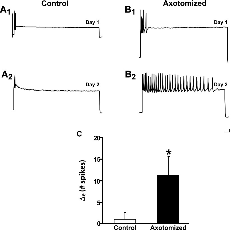
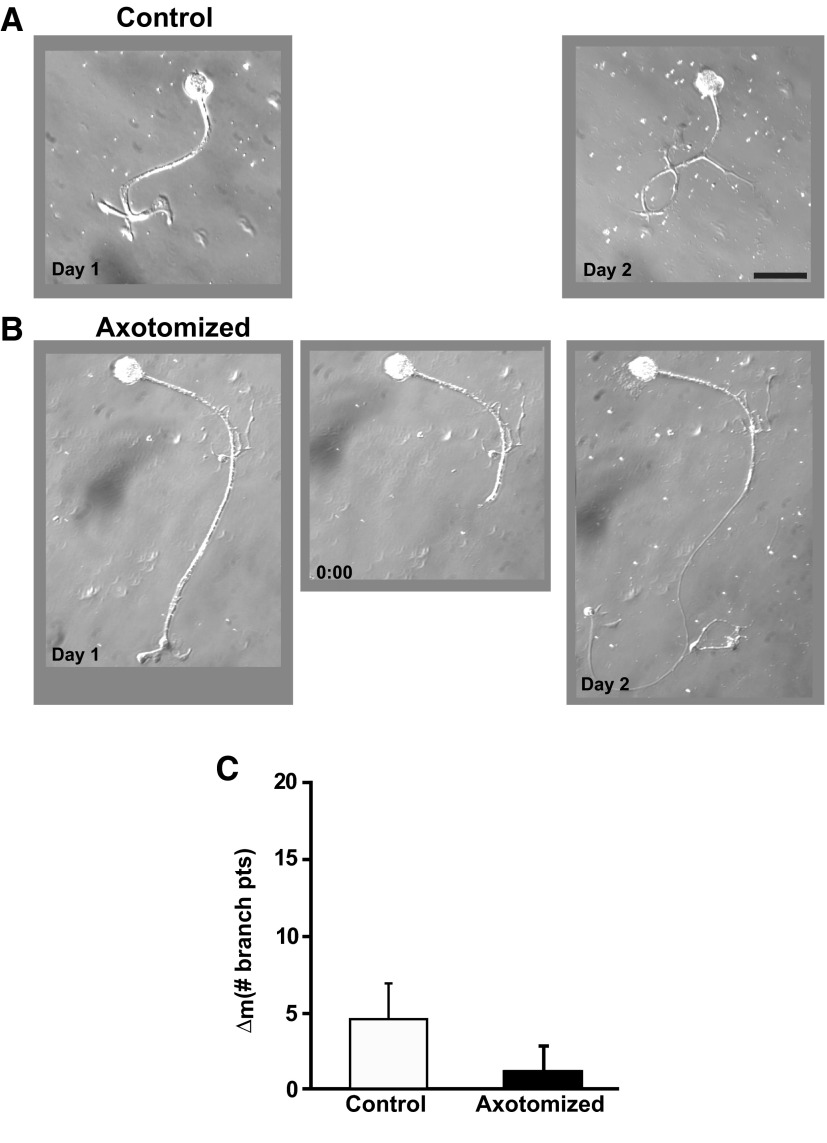
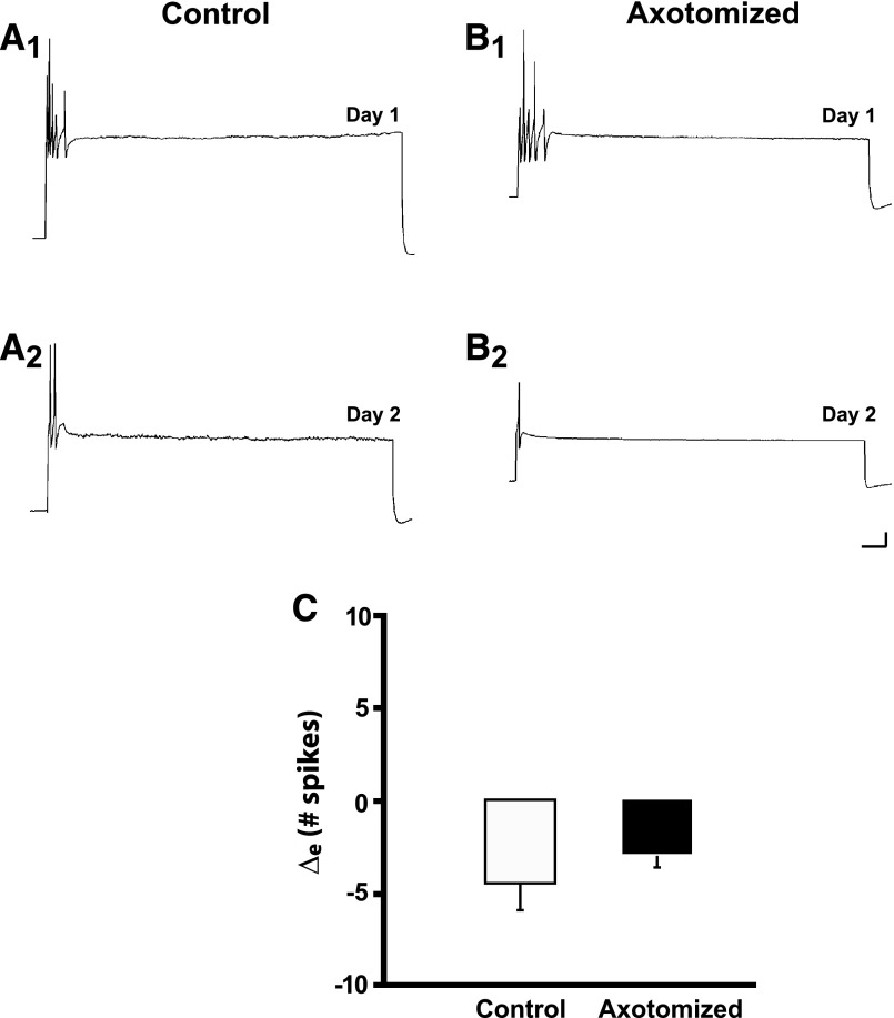
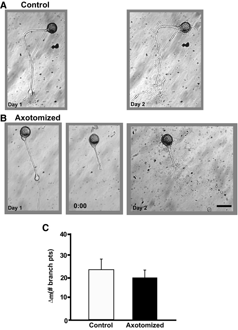
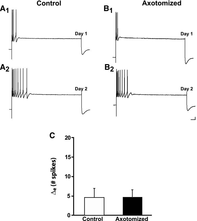
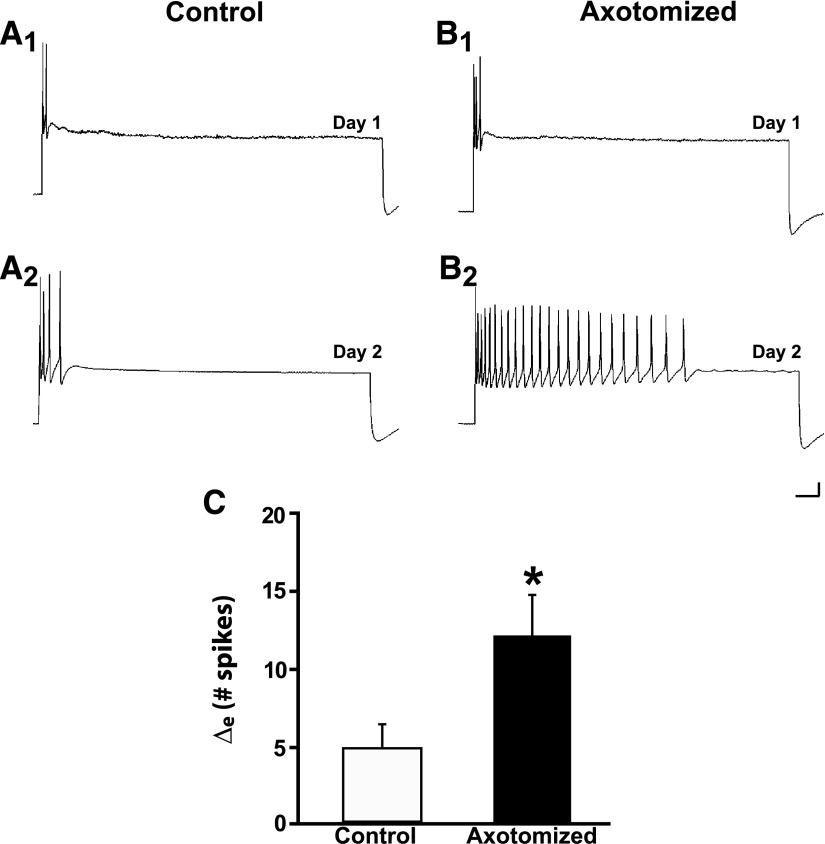
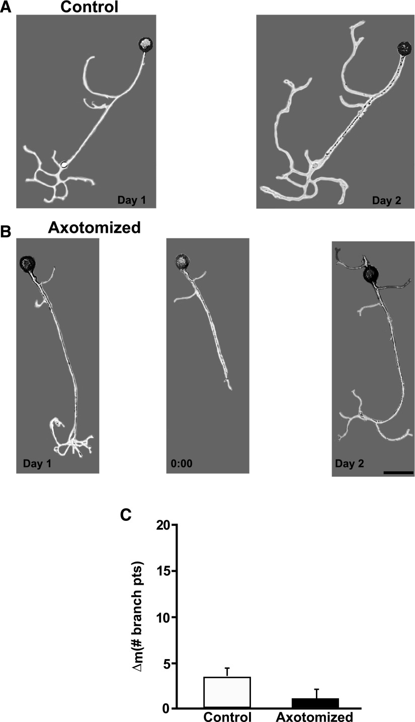
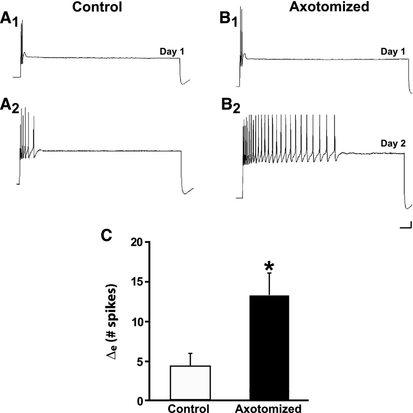
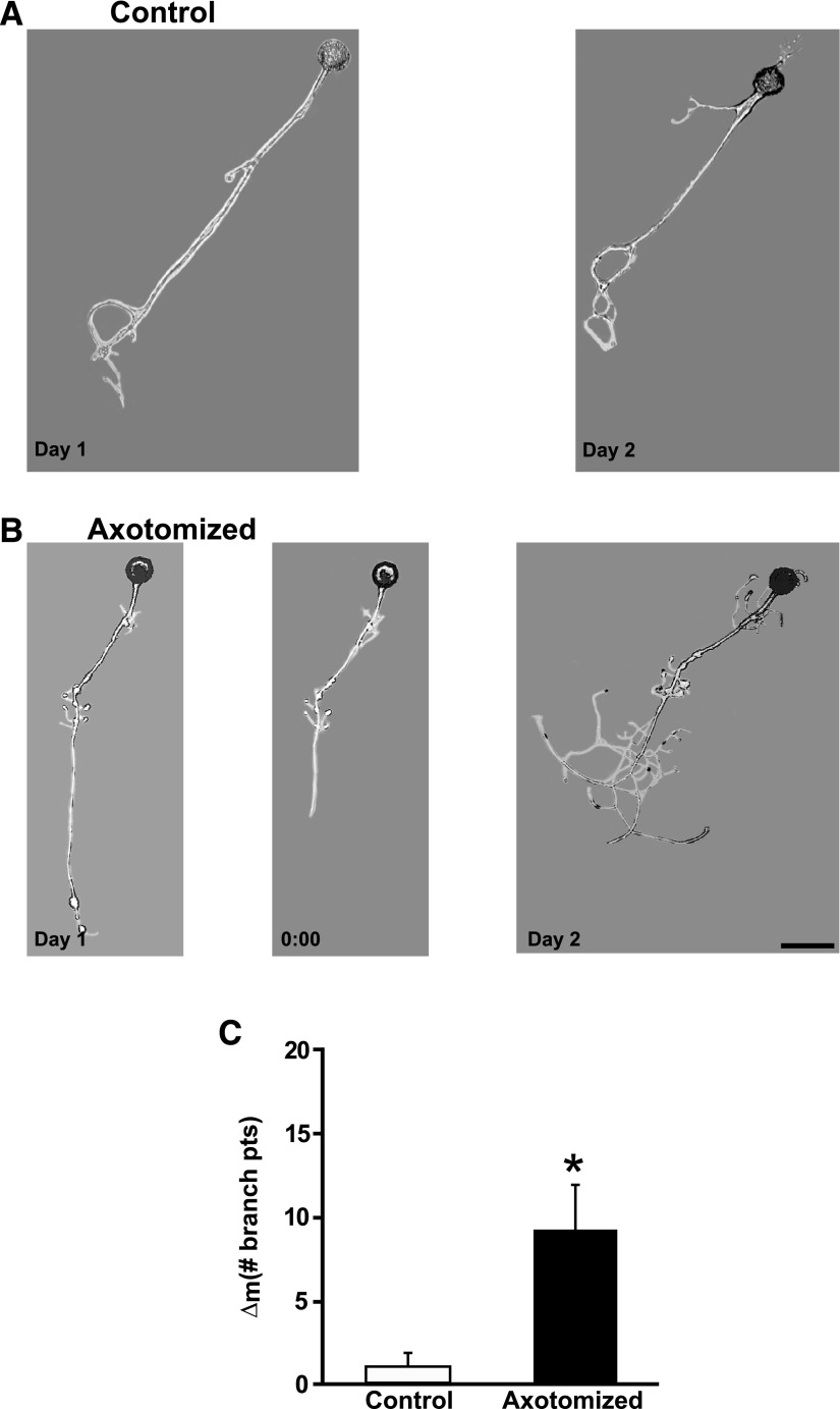
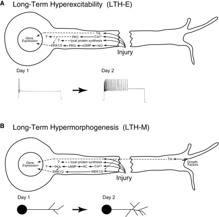
To facilitate an understanding of injury-induced changes within the nervous system, we used a single-cell, in vitro model of axonal injury. Sensory neurons were individually dissociated from the CNS of Aplysia and placed into cell culture. The major neurite of some neurons was then transected (axotomized neurons). Axotomy in hemolymph-containing culture medium produced long-term hyperexcitability (LTH-E) and enhanced neuritic sprouting (long-term hypermorphogenesis [LTH-M]). Axotomy in the absence of hemolymph induced LTH-E, but not LTH-M. Hemolymph-derived growth factors may activate tyrosine receptor kinase (Trk) receptors in sensory neurons. To examine this possibility, we treated uninjured (control) and axotomized sensory neurons with K252a, an inhibitor of Trk receptor activity. K252a depressed the excitability of both axotomized and control neurons. K252a also produced a distinct pattern of arborizing outgrowth of neurites in both axotomized and control neurons. Protein kinase C (PKC) is an intracellular signal downstream of Trk; accordingly, we tested the effects of bisindolylmaleimide I (Bis-I), a specific inhibitor of PKC, on the axotomy-induced cellular changes. Bis-I blocked LTH-E, but did not disrupt LTH-M. Finally, because Trk activates the extracellular signal regulated kinase pathway in Aplysia sensory neurons, we examined whether this pathway mediates the injury-induced changes. Sensory neurons were axotomized in the presence of U0126, an inhibitor of mitogen-activated/extracellular receptor-regulated kinase. U0126 blocked the LTH-M due to axotomy, but did not impair LTH-E. Therefore distinct cellular signaling pathways mediate the induction of LTH-E and LTH-M in the sensory neurons.


Figures

FIG. 1.
FIG. 1.
Axotomy in the absence of the hemolymph induces long-term hyperexcitability (LTH-E) of isolated sensory neurons in culture. A1: representative response of a Control neuron to a 2-s depolarizing current pulse (2 nA in this and subsequent figures) on Day 1 of the experiment. A2: response of the same Control neuron in A1 on Day 2. B1: representative response to depolarizing current pulse on Day 1 of an Axotomized neuron, prior to axotomy. B2: response of the same Axotomized neuron in B1 on Day 2. Notice the significant increase in repetitive firing (decreased accommodation) 24 h after axotomy. Calibration bars in this and subsequent figures: 10 mV, 125 ms. C: change in number of spikes from Day 1 to Day 2 in Control and Axotomized groups. The difference between the 2 groups was significant, as indicated by the asterisk. Graph in this and subsequent figures shows means ± SE.
FIG. 2.
FIG. 2.
Axotomy in the absence of hemolymph does not induce long-term hypermorphogenesis (LTH-M). A: photomicrographs of a Control neuron on Day 1 and the same neuron on Day 2. B: photomicrographs of an Axotomized neuron on Day 1, prior to axotomy (left), the same neuron immediately after axotomy on Day 1 (0:00 h, middle), and the neuron 24 h later on Day 2 (right). Scale bar for this and subsequent micrographs, 100 μm. C: change in the number of branch points from Day 1 to Day 2 for the Axotomized and Control groups. There was no significant difference between the 2 groups.
FIG. 3.
FIG. 3.
The tyrosine receptor kinase (Trk) receptor inhibitor K252a depresses excitability in both Control and Axotomized neurons. A1: response of a Control neuron treated with K252a to a depolarizing current pulse on Day 1. A2: response of the same Control neuron on Day 2. There was a decrease in the number of spikes elicited. B1: response of an Axotomized neuron treated with K252a to a depolarizing current pulse on Day 1. B2: response of the Axotomized neuron on Day 2 (24 h after axotomy). The neuron did not exhibit the hyperexcitability typically observed after axotomy. C: change in number of spikes from Day 1 to Day 2 in Control and Axotomized groups. Both groups exhibited decreased excitability; the difference between the groups was not significant.
FIG. 4.
FIG. 4.
K252a induces exaggerated neuritic outgrowth in both Control and Axotomized neurons. A: photomicrographs of a Control neuron on Day 1 and 24 h later. Note the enhanced outgrowth of the neurites. B: photomicrographs of an Axotomized neuron on Day 1, prior to axotomy (left), the same neuron immediately after axotomy on Day 1 (0:00 h, middle), and the neuron 24 h later on Day 2 (right). The outgrowth of the neurites in this neuron was also enhanced. C: change in the number of branch points from Day 1 to Day 2 for the Control and Axotomized groups. There was no significant difference in neurite outgrowth between the 2 groups.
FIG. 5.
FIG. 5.
Bisindolylmaleimide I (Bis-I), a protein kinase C (PKC) inhibitor inhibits LTH-E. A1: representative response of a Control neuron to a 2-s depolarizing current pulse on Day 1 of the experiment. A2, response of the same Control neuron in A1 on Day 2. B1: representative response to depolarizing current pulse on Day 1 of an Axotomized neuron, prior to axotomy. B2: response of the same Axotomized neuron in B1 on Day 2. C: change in number of spikes from Day 1 to Day 2 in Control and Axotomized groups. The difference between the 2 groups was not significant.
FIG. 6.
FIG. 6.
U0126, a mitogen-activated/extracellular receptor-regulated kinase (MEK) 1/2 inhibitor, does not block axotomy-induced LTH-E. A1 and A2: responses of a Control neuron on Day 1 and Day 2. There was a modest increase in the number of evoked spikes to the 2-s pulse on the second day. B1 and B2: responses of an Axotomized neuron on Day 1 and Day 2. This neuron exhibited a significant increase in its excitability on Day 2. C: change in number of evoked spikes from Day 1 to Day 2 in Control and Axotomized groups. The difference between the 2 groups was significant, as indicated by the asterisk.
FIG. 7.
FIG. 7.
U0126 blocks LTH-M due to acute axotomy. A: photomicrographs of a Control neuron on Day 1 and the same neuron on Day 2. The neuron exhibited a modest increase in the number of neuritic branches. B: photomicrographs of an Axotomized neuron on Day 1, prior to axotomy (left), the same neuron immediately after axotomy on Day 1 (0:00 h, middle), and the neuron 24 h later on Day 2 (right). Treatment with the MEK1/2 inhibitor blocked the increase in the number of branches. C: the change in the number of branch points from Day 1 to Day 2 for the Axotomized and Control groups. The difference between the Control and Axotomized groups was not significant.
FIG. 8.
FIG. 8.
Axotomy induces LTH-E of isolated sensory neurons in culture medium containing 0.1% dimethylsulfoxide (DMSO). A1 and A2: representative responses of a Control neuron treated with DMSO on Day 1 and Day 2. There was a modest increase in excitability in this cell over the 24-h period. B1 and B2: representative responses of an Axotomized neuron treated with DMSO. This neuron exhibited a very large increase in its excitability 24 h after axotomy. C: change in the number of evoked spikes from Day 1 to Day 2 in Control and Axotomized groups. The difference between the 2 groups was statistically significant, as indicated by the asterisk. Note that the results in DMSO resemble those that we have observed earlier in control culture medium without DMSO (Bedi and Glanzman 2001; Bedi et al. 1998).
FIG. 9.
FIG. 9.
Axotomy induces LTH-M of isolated sensory neurons in culture medium containing 0.1% DMSO. A: photomicrographs of a Control neuron on Day 1 and the same neuron on Day 2. The neuron exhibited a modest increase in outgrowth over the 24-h period. B: photomicrographs of an Axotomized neuron on Day 1, prior to axotomy (left), the same neuron immediately after axotomy on Day 1 (0:00 h, middle), and the neuron 24 h later on Day 2 (right). Axotomy produced a more dramatic increase in outgrowth than was observed in the Control neuron (A). C: the change in the number of branch points from Day 1 to Day 2 for the Axotomized and Control groups. The difference between the 2 groups was statistically significant, as indicated by the asterisk. This pattern of results was like that observed in earlier experiments using culture medium without DMSO (Bedi and Glanzman 2001; Bedi et al. 1998).
FIG. 10.
FIG. 10.
Signaling pathways that mediate injury-induced, long-term cellular changes in sensory neurons. A: model for LTH-E following axotomy. Axonal injury causes activation of nitric oxide (NO) synthase, thereby leading to elevation of NO within the axon; enhanced levels of NO, in turn, activate the cyclic guanosine monophosphate (cGMP)–dependent protein kinase G (cGMP–PKG) pathway (Lewin and Walters 1999). PKG activates extracellular signal regulated kinase (ERK)1/2, which can then translocate to the nucleus to regulate gene expression (Sung et al. 2004). In the case of LTH-E, ERK1/2 activation does not appear to result from MEK activity (Fig. 5). In addition, axotomy also leads to stimulation of a Trk-dependent pathway, which also appears to be required for LTH-E (Fig. 3). The injury-induced stimulation of the Trk-dependent pathway must be downstream of Trk receptors because growth factors are not required for LTH-E (Ambron et al. 1996; and Fig. 1). Trk receptors can also translocate to the nucleus and regulate gene expression (Riccio et al. 1997). Influx of Ca2+ after injury leads to activation of PKC (Fig. 5), which can also interact with ERK1/2 (Kawasaki et al. 2004). Ongoing activity in the Trk-dependent pathway is also required for noninjury-dependent excitability (Fig. 3). Local protein synthesis (Weragoda and Walters 2007; Weragoda et al. 2004) may well contribute to LTH-E as well, but we do not yet know which proteins are locally synthesized due to injury. B: model for long-term hypermorphogenesis (LTH-M) following axotomy. Injury results in an influx of Ca2+, which activates adenylyl cyclase (AC), leading to synthesis of cAMP and activation of protein kinase A (PKA), which is required for the induction of LTH-M (Bedi et al. 1998). PKA also modulates ERK1/2 (Sweatt 2004), which can then translocate to the nucleus and regulate gene expression. In addition to its activation by PKA, ERK1/2 is also likely to be activated by MEK1/2 after injury (Zhao et al. 2007); MEK1/2-dependent activation of ERK1/2 is critical for injury-induced LTH-M (Fig. 7). As well as MEK1/2 activity, injury-induced LTH-M depends on growth factors (Fig. 2). The effect of growth factors on the morphology of sensory neurons is likely to depend on Trk receptors, particularly nork (Hislop et al. 2004). Trk receptors may also stimulate activation of MEK1/2 (Chao 2003). In addition, local protein synthesis and its products may contribute to the injury-induced morphological changes. Note that under normal conditions axonal injury leads to both LTH-E and LTH-M. Also, both phenomena appear to depend on gene expression, possibly involving CREB activity (Dash et al. 1998), but we do not know the specific genes that are involved in each of these long-term, injury-induced changes. Obviously, the patterns of gene activation that produce LTH-E must differ somewhat from those that produce LTH-M. The dashed lines in A and B represent hypothetical paths for which there is no evidence at present in Aplysia.

Similar articles

Cited by

References

    1. Alberini CM, Ghirardi M, Metz R, Kandel ER. C/EBP is an immediate-early gene required for the consolidation of long-term facilitation in Aplysia. Cell 76: 1099–1114, 1994. - PubMed
    1. Ambron RT, Zhang XP, Gunstream JD, Povelones M, Walters ET. Intrinsic injury signals enhance growth, survival, and excitability of Aplysia neurons. J Neurosci 16: 7469–7477, 1996. - PMC - PubMed
    1. Angeles TS, Yang SX, Steffler C, Dionne CA. Kinetics of trkA tyrosine kinase activity and inhibition by K-252a. Arch Biochem Biophys 349: 267–274, 1998. - PubMed
    1. Bartsch D, Casadio A, Karl KA, Serodio P, Kandel ER. CREB1 encodes a nuclear activator, a repressor, and a cytoplasmic modulator that form a regulatory unit critical for long-term facilitation. Cell 95: 211–223, 1998. - PubMed
    1. Bartsch D, Ghirardi M, Skehel PA, Karl KA, Herder SP, Chen M, Bailey CH, Kandel ER. Aplysia CREB2 represses long-term facilitation: relief of repression converts transient facilitation into long-term functional and structural change. Cell 83: 979–992, 1995. - PubMed

Publication types

MeSH terms

Substances

LinkOut - more resources