. 2008 Oct 8;3(10):e3341.
doi: 10.1371/journal.pone.0003341.

Allele-specific RNA silencing of mutant ataxin-3 mediates neuroprotection in a rat model of Machado-Joseph disease

Affiliations

Allele-specific RNA silencing of mutant ataxin-3 mediates neuroprotection in a rat model of Machado-Joseph disease

Sandro Alves et al. PLoS One. .

Abstract

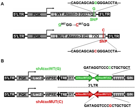
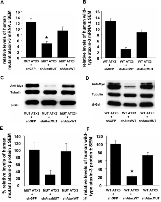
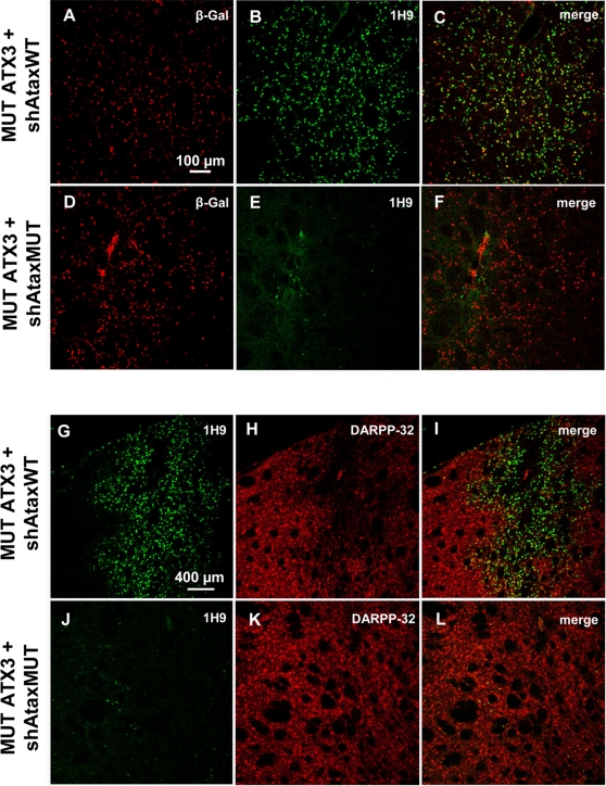
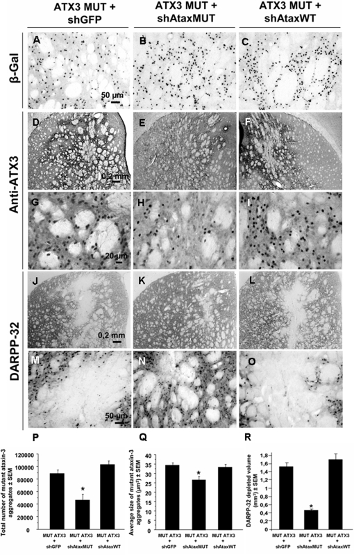
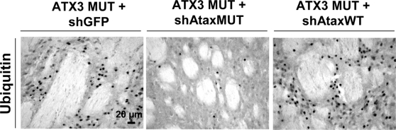
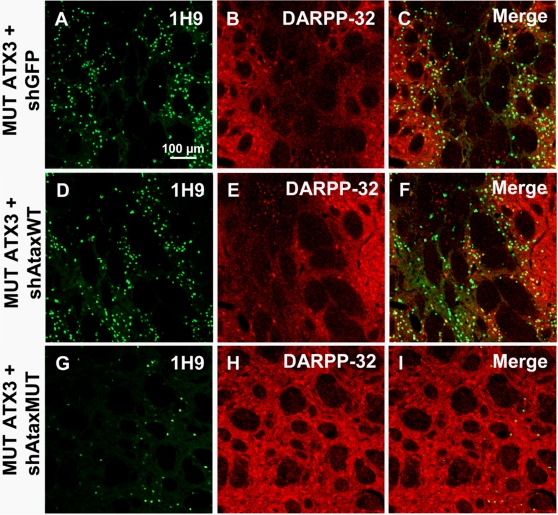
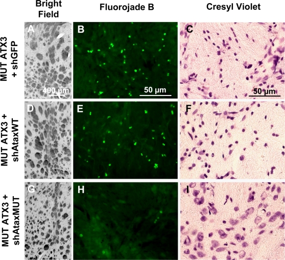
Recent studies have demonstrated that RNAi is a promising approach for treating autosomal dominant disorders. However, discrimination between wild-type and mutant transcripts is essential, to preserve wild-type expression and function. A single nucleotide polymorphism (SNP) is present in more than 70% of patients with Machado-Joseph disease (MJD). We investigated whether this SNP could be used to inactivate mutant ataxin-3 selectively. Lentiviral-mediated silencing of mutant human ataxin-3 was demonstrated in vitro and in a rat model of MJD in vivo. The allele-specific silencing of ataxin-3 significantly decreased the severity of the neuropathological abnormalities associated with MJD. These data demonstrate that RNAi has potential for use in MJD treatment and constitute the first proof-of-principle for allele-specific silencing in the central nervous system.


Conflict of interest statement

Competing Interests: The authors have declared that no competing interests exist.

Figures

Figure 1
Figure 1. The single nucleotide polymorphism strategy used for the specific elimination of mutant or wild-type human ataxin-3 (ATX3) by RNA interference.
A) Schematic representation of the lentiviral constructs encoding wild-type human ataxin-3 (27 CAG repeats) or mutant human ataxin-3 (72 CAG repeats) under control of the phosphoglycerate kinase-1 (PGK-1) promoter. Immediately after the last CAG repeat in the 3′ end, there is a linked single nucleotide polymorphism (SNP) (G 987GG→C 987GG) between wild-type and mutant human ataxin-3. B) Diagram of the shAtax vectors used to downregulate human ataxin-3: shRNA cassette under control of the H1 promoter (pol III) and a separate cassette containing the lacZ reporter gene under control of the PGK-1 promoter, making it possible to follow the expression of infected neurons. These shRNAs were designed to silence wild-type (shAtaxWT) or mutant human ataxin-3 (shAtaxMUT) selectively, making use of the (G 987GG→C 987GG) SNP.
Figure 2
Figure 2. shRNAs mediate the in vitro allele-specific suppression of mutant or wild-type human ataxin-3 by RNAi.
A–F) shAtaxMUT- or shAtaxWT-encoding plasmids selectively targeting mutant ataxin-3 and wild-type ataxin-3, respectively, resulted in much lower levels of these proteins than the mistargeted control (shGFP) or the non allele-specific shRNA. Quantitative real-time PCR analysis showing the silencing of human ATX3 mRNA in 293T cells co-expressing mutant human ataxin-3 (MUT ATX3) (A, top left) or wild-type human ataxin-3 (WT ATX3) (B, top right) and shAtaxWT, shAtaxMUT, or shGFP. Endogenous ß-actin mRNA was used as an internal control for the normalization and quantitative analysis of the ataxin-3 mRNA levels. Results are expressed as the mean elative mRNA level±SEM. C and D) Western-blot analysis of lysates of 293T cells co-transfected with the plasmid constructs encoding MUT ATX3 (C, middle left) or WT ATX3 (D, middle right) and the shAtax vectors (48 hours after calcium phosphate-mediated transfection; ratio ATX3/shRNA 1∶5). Tubulin staining is shown as a loading control. E and F) Optical densitometry was normalized according to the amount of tubulin loaded in the corresponding lane. A partition ratio was calculated and expressed as a percentage (bottom). All western blots and RT-PCRs shown are representative of three or four independent experiments. Statistical significance was evaluated using Fisher's test (*p<0.05).
Figure 3
Figure 3. shRNA-expressing plasmids mediate the allele-specific silencing of endogenous wild-type ataxin-3 in transiently transfected human 293T cells.
A) Western blot of human 293T cells transfected with different shRNA (shAtaxWT, shAtaxMUT and shGFP)-expressing plasmids (5 µg; 48 h post-transfection). The blot clearly indicates that shAtaxWT downregulates endogenous/wild-type human ATX3, whereas shAtaxMUT has no silencing effect. shGFP was used as a control vector and the protein tubulin was used as a loading control. This western blot is representative of three independent experiments.
Figure 4
Figure 4. Efficient allele-specific suppression of mutant human ataxin-3 in the rat brain at an early time point (3 weeks) mediates striatal neuroprotection.
A–L) Laser confocal microscopy showing the effects of recombinant lentiviral vectors encoding shAtaxMUT or shAtaxWT and MUT ATX3 in the rat brain striatum at an early time point (3 weeks). β-galactosidase, expressed from a separate PGK-lacZ cassette in the vectors, allows identification of infected neurons (A and D). shAtaxMUT specifically silences MUT ATX3, promoting the clearance of MUT ATX3-positive aggregates (E and J), whereas shAtaxWT has almost no effect on MUT ATX3 expression (B and G). A considerable loss of DARPP-32-immunoreactivity is observed in rat striatum co-infected with MUT ATX3 and shAtaxWT (H and the merged image I), whereas no DARPP-32 downregulation is observed in rat striatum co-infected with MUT ATX3 and shAtaxMUT (K and the merged image L), suggesting neuroprotection. The adult rats were co-injected bilaterally in the striatum with MUT ATX3 and the shAtaxWT or shAtaxMUT vectors (n = 2) and were killed three weeks later. All the pictures were taken around the injection site.
Figure 5
Figure 5. Allele-specific silencing of mutant human ataxin-3 in rat brain.
A) Laser confocal microscopy, showing neuronal transduction 2 months after injection in the rat striatum with recombinant lentiviral vectors encoding shAtaxMUT (n = 7), shAtaxWT (n = 8) or shGFP (n = 4) and mutant human ataxin-3 (MUT ATX3). The viral vectors also contained a separate PGK-LacZ cassette encoding β-galactosidase, to facilitate the detection of infected neurons (B, H, N and E, K, Q, high magnification). In adult rats expressing MUT ATX3 and shAtaxMUT (n = 7), the number of neurons containing MUT ATX3-positive aggregates was much smaller (M) and the high magnification merged image (R) indicates that the few cells positive for MUT ATX3 did not express the lacZ reporter gene present in the shAtaxMUT vector. These cells were therefore not transduced with the vectors encoding the silencing sequences. By contrast, in animals expressing MUT ATX3 and shGFP (n = 4) or the control shAtaxWT (n = 8) (A and G, respectively) high magnification merged images show many MUT ATX3-positive cells simultaneously expressing the lacZ reporter gene present in both shAtaxWT or shGFP (F and L, respectively). The figure shows representative images of immunohistochemical stainings that were reproducible among the different groups.
Figure 6
Figure 6. Allele-specific silencing of mutant human ataxin-3 mediates robust reduction of the number of ataxin-3 inclusions and preservation of DARPP-32 immunoreactivity in the rat striatum.
A–R) Co-overexpression of MUT ATX3 and various shRNAs (shAtaxWT, n = 8; shAtaxMUT, n = 7 and shGFP, n = 4) in the striatum of adult rats, 2 months post-injection. The vectors encoding the shRNA cassette and the lacZ reporter gene infect an extensive region of the rat striatum, as shown by β-galactosidase immunoreactivity (A, B and C). shAtaxMUT specifically downregulates MUT ATX3, promoting a significant decrease in the number of MUT ATX3-positive aggregates (E and H ), whereas shAtaxWT has almost no effect on MUT ATX3 expression (F and I), as shown by comparison with the results obtained with the mistargeted shGFP (D and G). A major loss of DARPP-32 immunoreactivity is observed in the striatum infected with MUT ATX3 and shAtaxWT (L and O) or shGFP (J and M), whereas minor DARPP-32 is observed in the striatum infected with shAtaxMUT (K and N), this downregulation being limited to the needle track area. P–R) Quantification of the effect of the different shRNAs on the absolute number (P) and mean size/surface* (Q) of MUT ATX3-positive cells (*p<0,05). R) Quantitative analysis of the DARPP-32-depleted region in the brains of rats in which the striatum was injected with MUT ATX3 and various shRNAs. The lesion volume in brains infected with shAtaxMUT and MUT ATX3 is much smaller than that in brains infected with shAtaxWT or shGFP, indicative of a neuroprotective effect conferred by the selective shAtaxMUT. Statistical significance was evaluated with Fisher's test. (* the mean size of the objects was estimated taking into account the pixels with a gray-scale level for intensity below the mean value, used as a threshold).
Figure 7
Figure 7. Reduction of ubiquitin-positive inclusions in the striatum of adult rats as result of mutant human ataxin-3 knock-down.
Animals infected with MUT ATX3 and the control shGFP (left; n = 4) or shAtaxWT (right, n = 8) show the accumulation of ubiquitin-positive inclusions, typical biomarkers of neuropathology, whereas no such accumulation is observed in animals co-infected with MUT ATX3 and the selective shAtaxMUT (middle, n = 7). The figure shows representative images of ubiquitin immunohistochemical stainings that were reproducible among the different groups.
Figure 8
Figure 8. Rescue of DARPP-32 immunoreactivity and reduced accumulation of ataxin-3 inclusions upon shAtaxMUT expression.
A–I) Laser confocal microscopy showing the expression of recombinant lentiviral vectors expressing MUT ATX3 and shGFP (n = 4), shAtaxWT (n = 8) or shMUT (n = 7) and its effects on DARPP-32 expression in the rat striatum 2 months post-injection. Slight DARPP-32 downregulation is observed in striatum infected with MUT ATX3 and the selective shAtaxMUT (H and the merged image I), whereas a significant loss of DARPP-32-immunoreactive neurons is observed in rat striatum co-infected with MUT ATX3 and shAtaxWT (E and the merged image F) or MUT ATX3 and the control shGFP (B and the merged image C). The figure shows representative images of immunohistochemical stainings that were reproducible among the different groups.
Figure 9
Figure 9. Allele-specific silencing of mutant human ataxin-3 prevents neurodegeneration in the adult rat striatum.
Coalescence of the internal capsule of the striatum is observed after co-infection with MUT ATX3 and shGFP (A, n = 4) or shAtaxWT (D, n = 8), at 2 months, on a bright-field photomicrograph, whereas no signs of striatal shrinkage are observed following co-infection with MUT ATX3 and the specific shAtaxMUT (G, n = 7) (left column). Neurodegeneration in rats co-infected with MUT ATX3 and shGFP (B) or shAtaxWT (E) is observed two months after injection on Fluorojade B-stained sections, but not in rats co-infected with MUT ATX3 and the selective shAtaxMUT (H) (middle column). Pycnotic nuclei are visible on cresyl violet-stained sections, suggesting cell injury and striatal degeneration after brain co-infection with MUT ATX3 and the control shGFP (C) or the non specific shAtaxWT (F), in adult rats at 2 months after injection. No such nuclei are observed on sections from rats co-infected with MUT ATX3 and the specific shAtaxMUT (I) (right column). All the pictures were taken around the injection site area and show representative immunohistochemical stainings that were reproducible among the different groups.

Similar articles

Cited by

References

    1. Sudarsky L, Coutinho P. Machado-Joseph disease. Clin Neurosci. 1995;3:17–22. - PubMed
    1. Gwinn-Hardy K, Singleton A, O'Suilleabhain P, Boss M, Nicholl D, et al. Spinocerebellar ataxia type 3 phenotypically resembling parkinson disease in a black family. Arch Neurol. 2001;58:296–299. - PubMed
    1. Wullner U, Reimold M, Abele M, Burk K, Minnerop M, et al. Dopamine transporter positron emission tomography in spinocerebellar ataxias type 1, 2, 3, and 6. Arch Neurol. 2005;62:1280–1285. - PubMed
    1. Alves S, Regulier E, Nascimento-Ferreira I, Hassig R, Dufour N, et al. Striatal and nigral pathology in a lentiviral rat model of Machado-Joseph disease. Hum Mol Genet 2008 - PubMed
    1. Ranum LP, Lundgren JK, Schut LJ, Ahrens MJ, Perlman S, et al. Spinocerebellar ataxia type 1 and Machado-Joseph disease: incidence of CAG expansions among adult-onset ataxia patients from 311 families with dominant, recessive, or sporadic ataxia. Am J Hum Genet. 1995;57:603–608. - PMC - PubMed

Publication types