. 2008 Dec 5;283(49):34305-14.
doi: 10.1074/jbc.M803925200. Epub 2008 Oct 7.

Transcriptional repression of the prosurvival endoplasmic reticulum chaperone GRP78/BIP by E2F1

Affiliations

Transcriptional repression of the prosurvival endoplasmic reticulum chaperone GRP78/BIP by E2F1

Tomás Racek et al. J Biol Chem. .

Abstract

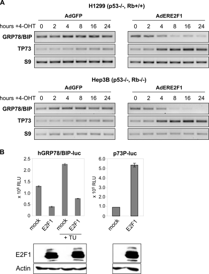
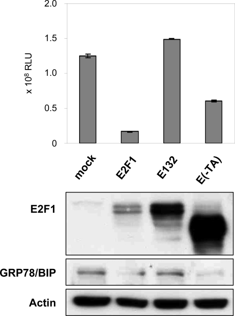
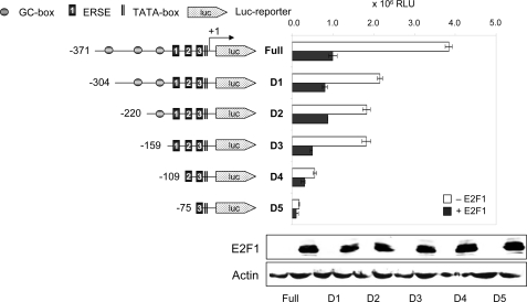
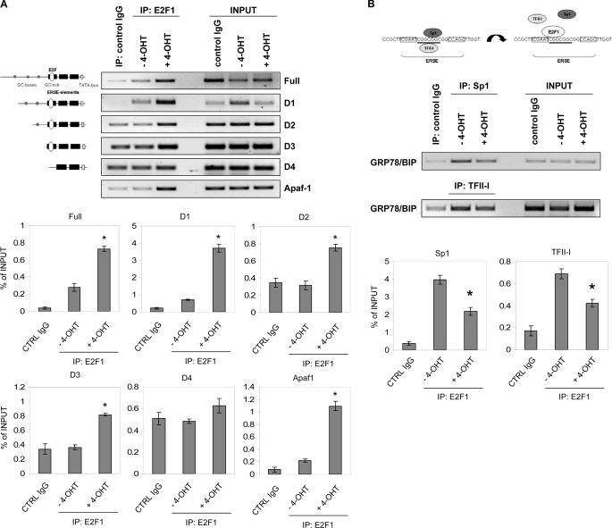
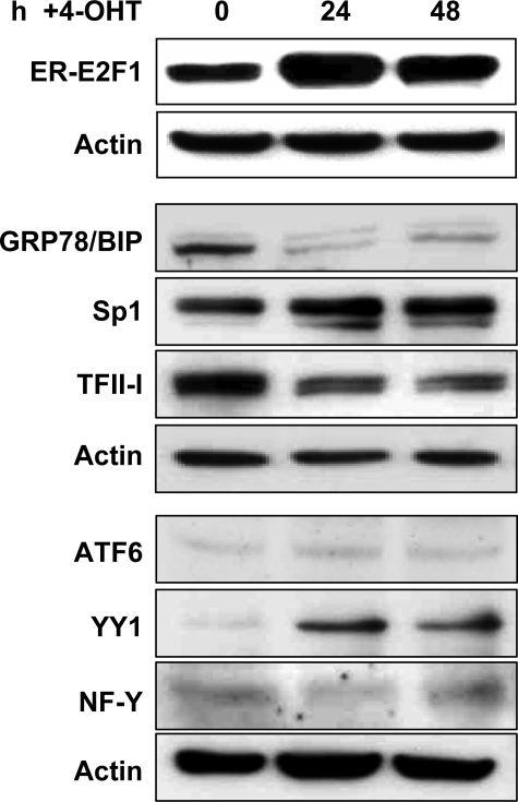
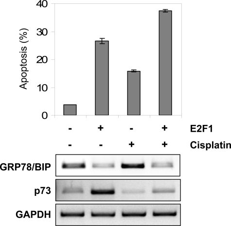
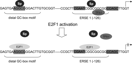
The endoplasmic reticulum chaperone GRP78/BIP plays a central role in the prosurvival machinery, and its enhanced expression has been implicated in drug resistance, carcinogenesis, and metastasis. E2F1, as part of an antitumor safeguard mechanism, promotes apoptosis regardless of functional p53. Using cells that are defective in p53, we show that E2F1 represses GRP78/BIP at the transcriptional level, and this requires its DNA binding domain. Analysis of human GRP78/BIP promoter reporter constructs revealed that the region between -371 and -109 of the proximal promoter contains major E2F1-responsive elements. Toward understanding the underlying mechanism of this regulation, we performed chromatin immunoprecipitation and gel shift assays, demonstrating that E2F1 directly binds to GC-rich regions in the distal GC-box and endoplasmic reticulum stress response element -126 by interfering with the binding of positive regulatory proteins Sp1 and TFII-I of the ER stress response element-binding factor complex. We further show that TFII-I, which is required for optimal stress induction of GRP78/BIP, is suppressed by E2F1 on the protein level. Finally, our studies suggest a molecular link between the inhibition of GRP78/BIP and E2F1-mediated chemosensitization of tumor cells, underscoring its relevance for cancer treatment. Together, the data provide a new mechanism for the incompletely understood tumor suppressor function of E2F1.


Figures

FIGURE 1.
FIGURE 1.
Repression of GRP78/BIP promoter activity by E2F1 is independent from p53 and Rb status. A, semiquantitative reverse transcription-PCR analysis of GRP78/BIP expression in p53-negative H1299 (Rb +/+) and Hep3B (Rb -/-) cells infected with Ad-vector expressing ER-E2F1. Infection by AdGFP was carried out as negative control. 16 h after infection, cells were grown in the presence of 4-OHT at the indicated times, followed by RNA isolation. p73 was used as positive control. Expression levels of RNA from the ribosomal S9 gene served as loading control. B, luciferase assay of p53-negative Hep3B cells co-transfected with 0.5 μg of GRP78/BIP promoter-luciferase reporter construct using the proximal core promoter (-371 to +2) and 0.5 μg of expression plasmid encoding E2F1 in the absence or presence of tunicamycin (TU; 0.5 μg/ml). 0.5 μg of the p73P-luc reporter construct was used as positive control. Luciferase activity (relative luciferase units; RLU) was measured 24 h after transfection. Error bars, S.D. of three independent measurements. E2F1 expression after transfection is shown in the bottom panel using actin as loading control.
FIGURE 2.
FIGURE 2.
E2F1-mediated GRP78/BIP inhibition requires the DNA binding and transactivation domain. Hep3B cells were co-transfected with 0.5 μg of GRP78/BIP reporter plasmid using the proximal core promoter region between -371 and +2 and 1 μg of expression plasmid encoding E2F1, DNA binding-defective mutant E132, E(-TA), lacking the transactivation domain, or pcDNA3.1 as mock control. Luciferase activity (RLU) was measured 36 h after transfection. Error bars, S.D. E2F1 and GRP78/BIP protein expression was verified by Western blotting. The blots were reprobed for α-actin as a loading control.
FIGURE 3.
FIGURE 3.
Identification of GRP78/BIP promoter elements responsible for transcriptional down-regulation by E2F1. The 5′-deletion mutants of the human GRP78/BIP promoter cloned upstream of the firefly luciferase gene are shown by nucleotide positions. Locations of the GC-boxes, ER stress elements (1-3), and TATA-box are indicated. The transcription start site of the GRP78/BIP gene is indicated by an arrow. 0.5 μg of the indicated reporter constructs either alone or together with 0.5 μg of E2F1 expression plasmid were transfected into H1299 cells. Luciferase activity (RLU) was measured 36 h after transfection. Data were obtained from three replicates. Error bars, one S.D. Protein expression levels in E2F1-transfected cells are indicated (bottom panel). Actin was used for equal loading.
FIGURE 4.
FIGURE 4.
E2F1 binds to the GRP78/BIP promoter in vivo by interfering with ER stress element-binding factors. A, serum-starved Saos-2 cells stably transfected with ER-E2F1 were grown in the presence or absence of 4-OHT for 24 h. Chromatin immunoprecipitation was performed using either a control IgG antibody or antibody against E2F1. PCR primers were designed to amplify the different GRP78/BIP promoter fragments spanning from -371 to +2 (full), -304 to +2 (D1), -220 to +2 (D2), -159 to +2 (D3), and -109 to +2 (D4). PCR primers for the Apaf-1 promoter were used as positive control. Input lane, 10% of total chromatin used in chromatin immunoprecipitation assay. B, chromatin immunoprecipitation was performed under the same conditions as in A using specific antibodies against Sp1 or TFII-I. PCR primers were used to amplify the GRP78/BIP promoter fragment between -220 and +2. Representative bands are indicated. Bar graphs show results from two independent experiments as relative software units cleared for background signals and normalized to input bands. Data represent the mean ± S.D. Significant differences in E2F1, Sp1, or TFII-I enrichment of chromatin in 4-OHT-treated versus untreated cells (A, p ≤ 0.005; B, p ≤ 0.01) are labeled with an asterisk (t test).
FIGURE 5.
FIGURE 5.
The GRP78/BIP promoter E2F1-like sequences in the distal GC-box and ERSE 1 element compete for nuclear binding with the specific Apaf-1 promoter E2F1 site. Biotin-labeled Apaf-1 E2F1 sequence (lanes 2-4) and GRP78/BIP Sp1 binding sequences -324 to -311 (distal GC-box; lanes 5-7) and -126 to -108 (GC-rich element of ERSE 1; lanes 8-10) were incubated with Saos-2ERE2F1 cell nuclear extracts and a 50-fold molar excess of cold double-stranded specific competitor for the Apaf-1 E2F1 site (lanes 3, 6, and 9) or an equimolar amount of mutant Apaf-1 E2F1 site competitor (lanes 4, 7, and 10). The arrow indicates a specific band competed by double-stranded cognate competitor. The faster migrating complexes B′ and C′ with the GC-box E2F site are not E2F1-specific. Lane 1, binding without nuclear extract.
FIGURE 6.
FIGURE 6.
E2F1 modulates ERSE-binding transcription factors on protein level. Serum-starved Saos-2 cells stably transfected with ER-E2F1 were grown in the presence of 4-OHT at the indicated times, followed by Western blot analysis. Equivalent amounts of total cell proteins were fractionated by SDS-PAGE and probed with antibodies specific for E2F1, GRP78/BIP, Sp1, TFII-I, ATF6, YY1, and NF-Y. Equal protein loading was confirmed by reprobing the stripped blots with anti-actin antibody. Detection was achieved by chemiluminescence.
FIGURE 7.
FIGURE 7.
Repression of GRP78/BIP by E2F1 accounts for E2F1-associated chemosensitization of cancer cells. Apoptosis induction after AdERE2F1 infection, 30 μm cisplatin, or combination treatment was analyzed in Hep3B cells by flow cytometry. The percentage of apoptotic cells (as determined by cells with sub-G1 DNA content) 24 h after treatment is as indicated. Semiquantitative reverse transcription-PCR analysis revealed differences in GRP78/BIP transcript levels for each treatment. p73 was used as a positive control. Data were normalized to glyceraldehyde-3-phosphate dehydrogenase (GAPDH) values.
FIGURE 8.
FIGURE 8.
Model of E2F1-inducible changes in transcription factor occupancy of the GRP78/BIP promoter. In stressed cells, Sp proteins are in contact with the distal GC-box and GC motifs of the ERSE-N9 region in the GRP78/BIP promoter. Upon E2F1 activation, E2F1 binds to its putative sites within the GC sequence motifs, resulting in the displacement of transcription factor binding to these regions, thereby acting as a transcriptional repressor.

Similar articles

Cited by

References

    1. Brown, J. M., and Giaccia, A. J. (1998) Cancer Res. 58 1408-1416 - PubMed
    1. Feldman, D. E., Chauhan, V., and Koong, A. C. (2005) Mol. Cancer Res. 3 597-605 - PubMed
    1. Ni, M., and Lee, A. S. (2007) FEBS Lett. 581 3641-3651 - PMC - PubMed
    1. Haze, K., Okada, T., Yoshida, H., Yanagi, H., Yura, T., Negishi, M., and Mori, K. (2001) Biochem. J. 355 19-28 - PMC - PubMed
    1. Wang, Y., Shen, J., Arenzana, N., Tirasophon, W., Kaufman, R. J., and Prywes, R. (2000) J. Biol. Chem. 275 27013-27020 - PubMed

Publication types

MeSH terms