. 2008 Dec 15;76(12):1694-704.
doi: 10.1016/j.bcp.2008.09.007. Epub 2008 Sep 17.

c-FLIP knockdown induces ligand-independent DR5-, FADD-, caspase-8-, and caspase-9-dependent apoptosis in breast cancer cells

Affiliations

c-FLIP knockdown induces ligand-independent DR5-, FADD-, caspase-8-, and caspase-9-dependent apoptosis in breast cancer cells

Travis W Day et al. Biochem Pharmacol. .

Abstract

Cellular-FLICE inhibitory protein (c-FLIP) is an inhibitor of apoptosis downstream of the death receptors Fas, DR4, and DR5, and is expressed as long (c-FLIP(L)) and short (c-FLIP(S)) splice forms. We found that the knockdown of c-FLIP using small interfering RNA (siRNA) triggered ligand-independent caspase-8- and -9-dependent spontaneous apoptosis and decreased the proliferation of MCF-7 breast cancer cells. Further analysis revealed that an apoptotic inhibitory complex (AIC) comprised of DR5, FADD, caspase-8, and c-FLIP(L) exists in MCF-7 cells, and the absence of c-FLIP(L) from this complex induces DR5- and FADD-mediated caspase-8 activation in the death inducing signaling complex (DISC). c-FLIP(S) was not detected in the AIC, and using splice form-specific siRNAs we showed that c-FLIP(L) but not c-FLIP(S) is required to prevent spontaneous death signaling in MCF-7 cells. These results clearly show that c-FLIP(L) prevents ligand-independent death signaling and provides direct support for studying c-FLIP as a relevant therapeutic target for breast cancers.


Figures
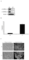
Fig.1
Fig.1. c-FLIP gene silencing triggers spontaneous apoptosis in MCF-7 cells
A, Immunoblot analysis of c-FLIPL, c-FLIPS, and β-actin in cell lysates following the treatment with 100 nM of non-targeting siRNA (SC) or c-FLIP-targeting siRNA (FT) for 48 h. B, Cells were transfected with 100 nM non-targeting siRNA (SC) or c-FLIP-specific siRNA (FT) for 48 h, and cells were harvested and apoptosis was determined by FACS analysis as described in the Materials and Methods section. Compensation was executed for each experiment using untreated cells stained with Annexin V and propidium iodide. Error bars show standard deviation from duplicate measurements. C, Cells were transfected with 100 nM non-targeting siRNA (SC) or c-FLIP-specific siRNA (FT) for 48 h, and Phase contrast (a and c) and fluorescence microscopy images (b and d) are shown after staining the DNA with Hoechst 33342. D, Cells were transfected with 100 nM non-targeting siRNA (SC) or c-FLIP-specific siRNA (FT) for 48 h, and the percentage of apoptotic cells was determined by counting fragmented nuclei stained with Hoechst 33342 among 200 cells. All values are representative of three independent experiments, and error bars show standard deviation from triplicate counts.
Fig. 2
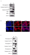
Fig. 2. c-FLIP knockdown induces the activation of caspases, Bid, and mitochondrial disintegration
A, Immunoblot analysis of caspase-8, BID, and β-actin in cell lysates following the transfection with non-targeting siRNA (SC) or c-FLIP-specific siRNA (FT) for 48 h. The asterisk (*) represents a non-specific protein that is recognized by the capase-8-specific antibody. B, Cells were transfected with non-targeting siRNA (SC) or c-FLIP-specific siRNA (FT) for 48 h, stained with 10 μM of Hoechst 33342 and 500 nM MitoTracker Red, and visualized by fluorescent microscopy. C, Immunoblot analysis of caspase-9, caspase-7, caspase-6, and β-actin in cell lysates following the transfection of MCF-7 with non-targeting siRNA (SC) or c-FLIP-specific siRNA (FT) for 48 h.
Fig. 3
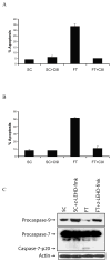
Fig. 3. c-FLIP knockdown triggers caspase-8- and caspase-9-dependent apoptosis
A, Cells were transfected with 100 nM non-targeting siRNA (SC) or c-FLIP-specific siRNA (FT) and treated with or without 20 μM of caspase-8 inhibitor (z-IETD-fmk) for 48 h, and apoptosis was determined by counting fragmented nuclei after staining with Hoecsht 33342 among 200 cells. All values are representative of three independent experiments, and error bars show standard deviation from triplicate counts. B, Cells were transfected with 100 nM non-targeting siRNA (SC) or c-FLIP-specific siRNA (FT) and incubated with or without 20 μM of caspase-9 inhibitor (z-LEHD-fmk) for 48 h, and apoptosis was determined by counting fragmented nuclei after staining with Hoecsht 33342 among 200 cells. All values are representative of three independent experiments, and error bars show standard deviation from triplicate counts. C, Immunoblot analysis of caspase-9, caspase-7, and β-actin in cell lysates after transfecting with 100 nM non-targeting siRNA (SC) or c-FLIP-specific siRNA (FT) and incubated with or without 20 μM of the caspase-9 inhibitor (z-LEHD-fmk) for 48 h.
Fig. 4
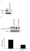
Fig. 4. Silencing of c-FLIP triggers FADD-dependent apoptosis
A, Immunoblot analysis of FADD and β-actin in cell lysates following the transfection with 100 nM of non-targeting siRNA (SC) or FADD-specific siRNA for 72 h. B, Immunoblot analysis of caspase-8 and β-actin in cell lysates treated with 100 nM FADD-specific siRNA for 24 h, and treated for an additional 48 h with 100 nM of non-targeting siRNA (SC) or c-FLIP-specific siRNA (FT). The asterisk (*) represents a non-specific protein that is recognized by the capase-8-specific antibody. C, The density of the caspase-8-p18 band in the c-FLIP-specific siRNA (FT) or FADD siRNA and FT siRNA treated cell lysates was quantified using the Image J 1.37c software (values +/− s.d.). D, Cells were treated with 100 nM FADD-specific siRNA for 24 h and treated for an additional 48 h with 100 nM non-targeting siRNA (SC) or c-FLIP-specific siRNA (FT), and apoptosis was determined by counting fragmented nuclei after staining with Hoecsht 33342 among 200 cells. All values are representative of three independent experiments, and error bars show standard deviation from triplicate counts.
Fig. 5
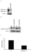
Fig. 5. The knockdown of c-FLIP triggers DR5-dependent apoptosis
A, Immunoblot analysis of DR5 and β-actin in cell lysates following the transfection with 100 nM of non-targeting siRNA (SC) or DR5-specific siRNA for 72 h. B, Immunoblot analysis of caspase-8 and β-actin in cell lysates treated with 100 nM DR5-specific siRNA for 24 h, and treated for an additional 48 h with 100 nM of non-targeting siRNA (SC) or c-FLIP-specific siRNA (FT). The asterisk (*) represents a non-specific protein that is recognized by the capase-8-specific antibody. C, The density of the caspase-8-p18 band in the c-FLIP-specific (FT) siRNA or DR5-specific siRNA and FT siRNA treated cell lysates was quantified using the Image J 1.37c software (values +/− s.d.). D, Cells were treated with 100 nM DR5-specific siRNA for 24 h and treated for an additional 48 h with 100 nM non-targeting siRNA (SC) or c-FLIP-specific siRNA (FT), and apoptosis was determined by counting fragmented nuclei after staining with Hoecsht 33342 among 200 cells. All values are representative of three independent experiments, and error bars show standard deviation from triplicate counts.
Fig. 6
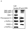
Fig. 6. DR5 binds to FADD, caspase-8, and c-FLIP
Input cell lysates and anti-DR5 immunoprecipitates were analyzed by Western blotting using DR5-, DR4-, caspase-8-, c-FLIP-, and FADD-specific antibodies. No proteins were immunoprecipitated using a control isotype-specific antibody.
Fig. 7
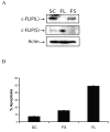
Fig. 7. c-FLIPL inhibits spontaneous apoptosis
A, Immunoblot analysis of c-FLIPL, c-FLIPS, and β-actin in cell lysates treated with 100 nM of non-targeting siRNA (SC), c-FLIPL-targeting siRNA (FL), or c-FLIPS-targeting siRNA (FS) for 48 h. B, Cells were treated with 100 nM of non-targeting siRNA (SC), c-FLIPL-targeting siRNA (FL), or c-FLIPS-targeting siRNA (FS) for 48 h, and apoptosis was quantified by counting fragmented nuclei after staining with Hoecsht 33342 among 200 cells. All values are representative of three independent experiments, and error bars show standard deviation from triplicate counts.
Fig. 8
Fig. 8. Knockdown of c-FLIP decreases the proliferation of MCF-7 cells
Cells were transfected with 100 nM of non-targeting siRNA (SC), c-FLIP-specific siRNA (FT), c-FLIPL-targeting siRNA (FL), or c-FLIPS-targeting siRNA (FS) for 48 h. The cells were harvested and 1,000 cells were transferred to 6-well plates containing complete media. Colonies were counted after incubation for 5 days. The values are representative of three independent experiments, and error bars show standard deviation from triplicate counts.
Fig. 9
Fig. 9. Proposed signaling pathway of c-FLIP siRNA-triggered spontaneous apoptosis in MCF-7 cells
In this signaling pathway, we propose that an apoptotic inhibitory complex (AIC) comprised of DR5, FADD, caspase-8, and c-FLIPL exists, and c-FLIPL inhibits caspase-8 activation. The knockdown of c-FLIPL using a c-FLIP-specific siRNA induces DISC activation by triggering DR5- and FADD-mediated activation of procaspase-8, forming active caspase-8. Active caspase-8 cleaves Bid to form tBid, leading to the release of cytochrome c from the mitochondria. Mitochondrial cytochrome c release activates procaspase-9 producing active caspase-9, which activates downstream caspases-6 and -7 thereby inducing apoptosis. The solid lines indicate protein binding partners and the brackets show protein complexes that comprise the AIC and DISC.

Similar articles

Cited by

References

    1. Cereghetti GM, Scorrano L. The many shapes of mitochondrial death. Oncogene. 2006;25:4717–24. - PubMed
    1. Debatin KM, Krammer PH. Death receptors in chemotherapy and cancer. Oncogene. 2004;23:2950–66. - PubMed
    1. Green D, Kroemer G. The central executioners of apoptosis: caspases or mitochondria? Trends Cell Biol. 1998;8:267–71. - PubMed
    1. Ashkenazi A, Dixit VM. Death receptors: signaling and modulation. Science. 1998;281:1305–8. - PubMed
    1. Sprick MR, Weigand MA, Rieser E, Rauch CT, Juo P, Blenis J, et al. FADD/MORT1 and caspase-8 are recruited to TRAIL receptors 1 and 2 and are essential for apoptosis mediated by TRAIL receptor 2. Immunity. 2000;12:599–609. - PubMed

Publication types

MeSH terms