. 2008 Oct 3;3(10):e3338.
doi: 10.1371/journal.pone.0003338.

Meiotic regulation of TPX2 protein levels governs cell cycle progression in mouse oocytes

Affiliations

Meiotic regulation of TPX2 protein levels governs cell cycle progression in mouse oocytes

Stéphane Brunet et al. PLoS One. .

Abstract

Formation of female gametes requires acentriolar spindle assembly during meiosis. Mitotic spindles organize from centrosomes and via local activation of the RanGTPase on chromosomes. Vertebrate oocytes present a RanGTP gradient centred on chromatin at all stages of meiotic maturation. However, this gradient is dispensable for assembly of the first meiotic spindle. To understand this meiosis I peculiarity, we studied TPX2, a Ran target, in mouse oocytes. Strikingly, TPX2 activity is controlled at the protein level through its accumulation from meiosis I to II. By RNAi depletion and live imaging, we show that TPX2 is required for spindle assembly via two distinct functions. It controls microtubule assembly and spindle pole integrity via the phosphorylation of TACC3, a regulator of MTOCs activity. We show that meiotic spindle formation in vivo depends on the regulation of at least a target of Ran, TPX2, rather than on the regulation of the RanGTP gradient itself.


Conflict of interest statement

Competing Interests: The authors have declared that no competing interests exist.

Figures

Figure 1
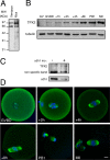
Figure 1. Characterization of TPX2 during mouse oocyte meiotic maturation.
A: Mouse oocytes (n = 200) and Xenopus metaphase II oocyte lysate (equivalent to 1/4 of oocyte) were immunoblotted using an anti-TPX2 antibody. The upper band corresponds to TPX2. The lower band is non-specific . B: TPX2 accumulates progressively during meiotic maturation. For each time point, 140 mouse oocytes were immunoblotted using anti-TPX2 (only the TPX2 specific band is presented) and anti-α-tubulin antibodies. C: Cdh1 controls TPX2 levels at GV (Prophase I). Western blot analysis of TPX2 and cdh1 in control oocytes and cdh1 morpholino injected oocytes (n = 100 for each group). D: Localization of TPX2 during meiotic maturation. Oocytes were double stained for TPX2 (green) and nucleic acids (blue) at the times indicated relative to GVBD. In meiosis I and II, TPX2 decorates the spindle microtubules. Scale bar is 10 µm.
Figure 2
Figure 2. TPX2 overexpresssion induces an arrest in meiosis I.
A: Immunoblot analysis of endogenous (TPX2) and exogenous (YFP-TPX2) protein levels in metaphase II oocytes (n = 72) expressing low levels of YFP-TPX2. B and C: Representative time-lapse videomicroscopy of oocytes expressing low (B; n = 78) or high (C; n = 29) levels of YFP-TPX2. Upper panels are the bright field images of the oocyte and the lower panels show the fluorescent YFP signal at the corresponding time points (germinal vesicle breakdown is 0 h). While the polar body extrusion (asterisks) was normal in (B), it was blocked in (C). In this case, multipolar microtubules structures were observed (see arrowheads).
Figure 3
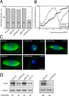
Figure 3. Meiotic maturation and spindle formation upon TPX2 depletion.
A: Percentages of polar body extrusion after siRNA treatment. Non-injected (non inj), control and TPX2 siRNA-injected oocytes maintained in prophase for 2 or 5 hours (siCo-2h, siCo-5h, siTPX2-2h and siTPX2-5h, respectively) were analyzed. The total number of oocytes for each condition is indicated in the bar. B: Semi-quantitative analysis of the Cyclin B1-GFP fluorescence during meiotic maturation (a.u.: arbitrary units). For each time point, the fluorescence was normalized against the maximum value reached for each oocyte during meiotic maturation. The curves represent the mean curves extracted from 10 oocytes. C: Representative spindle morphology upon TPX2 depletion. Non-injected, siCo-5h and siTPX2-2h injected oocytes at metaphase II. SiTPX2-5h injected oocytes arrested in meiosis I. Both non-injected and siCo-5h injected oocytes showed similar barrel-shaped bipolar spindles. However, siTPX-2h injected showed either small aster around the chromosomes (21%) or a mini spindles (79%). siTPX2-5h injected oocytes, on the other hand, displayed more severe perturbation in spindle assembly with only few microtubules present around the chromosomes in most of the cases (86%). Microtubules are in green and chromosomes are in blue. Scale bar is 10 µm. D: Immunoblot analysis of TPX2 upon siRNA depletion. Both siCo-2h and siCo-5h showed similar TPX2 levels as the non-injected oocytes. SiTPX2-2h depleted about half of the TPX2 protein while the siTPX2-5h almost completely depleted the protein. The number of oocytes in each sample is indicated below the immunoblots. α-tubulin was used as loading control.
Figure 4
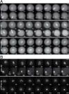
Figure 4. Microtubules and chromosomes behaviour upon TPX2 depletion.
A: Representative sequence of fluorescent images showing the microtubule organization in siCo-5h (top panel), siTPX2-2h (middle panel) and siTPX2-5h (bottom panel) injected oocytes expressing a GFP-tagged β-tubulin. Both siCo-5h and siTPX2-2h injected oocytes extruded the polar bodies. However, siTPX2-2h injected oocyte formed small spindle after the polar body extrusion (see panel 12 h). The siTPX2-5h depletion blocked polar body extrusion and the bipolar spindle formed started to collapse three to five hours after GVBD (panels 5 h–15 h). Times are relative to GVBD. B: Representative sequence of fluorescent images showing the morphology and movement of the chromosomes in siCo-5h (upper panel) or siTPX2-5h (bottom panel) injected oocytes expressing RFP-histone H2B. While the chromosomes progressively aligned onto the metaphase plate in the siCo-5h injected oocyte (panel 6 h), the chromosomes in the siTPX2-5h injected oocyte appeared to be stationary during the few hours after GVBD (panels 2 h–8 h). Times are relative to GVBD.
Figure 5
Figure 5. TPX2 activity relies on two distinct protein domains in the oocyte.
A: Percentage of polar body extrusion in different experimental conditions. Non-injected, siTPX2-5h, YFP-TPX2ΔN injected oocytes as well as oocytes co-injected with siTPX2-5h and YFP-TPX2 or YFPTPX2ΔN were analyzed. The total number of oocytes examined is indicated in each bar. B: Representative sequence of fluorescent images showing the microtubules organization in siTPX2-5h injected oocytes, which were co-injected either with YFP-TPX2 (upper panel) or YFP-TPX2ΔN (lower panel). Both YFP-TPX2 and YFP-TPX2ΔN rescued the spindle collapse. Asterisk indicates the polar body and the dotted line shows the outline of the oocyte. C: Representative fluorescent images of siTPX2-5h-injected oocytes co-injected with YFP-TPX2ΔN show spindle poles splitting (arrowheads). D: Representative sequence of fluorescent images showing the chromosomes siTPX2-5h injected oocytes expressing RFP-histone H2B alone (top panel), co-injected with either YFP-TPX2 (middle panel) or YFP-TPX2ΔN (bottom panel). The asterisk indicates the polar body and the dotted line shows the outline of the oocyte. Times are relative to the GVBD.
Figure 6
Figure 6. Characterization of TACC3 during mouse oocyte meiotic maturation.
A: Immunoblot analysis of TACC3 and phosphorylated TACC3 (P-TACC3) in GV and metaphase II (MII) oocytes. Oocytes (n = 150) were immunoblotted using an anti-TACC3 antibody (left) and an antibody raised against xTACC3 phosphorylated on Ser626 (right). B: Immunoblot analysis of TACC3 and P-TACC3 during meiotic maturation. Oocytes (n = 140) were immunoblotted at the indicated time points. C: Localization of P-TACC3 during meiotic maturation. Oocytes were triple stained for P-TACC3 (red), microtubules (green) and nucleic acids (blue). The P-TACC3 staining becomes restricted to the spindle poles during meiotic maturation. Scale bar is 10 µm.
Figure 7
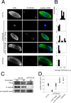
Figure 7. TACC3 is an indirect target of TPX2 in the oocyte.
A: Localization of P-TACC3 upon TPX2 depletion. Oocytes were triple labelled for P-TACC3 (red), microtubules (green) and nucleic acids (blue). P-TACC3 staining is absent in siTPX2-5h, siTPX2-5h co-injected with YFP-TPX2ΔN and siTACC3 oocytes. Scale bar is 10 µm. B: Semi-quantitative analysis of the distribution of the average P-TACC3 fluorescence intensity in individual oocytes. C: Western blot analysis of P-TACC3 and TPX2 levels upon TPX2 depletion. Both TPX2 and P-TACC3 proteins are significantly reduced after siTPX2-5h treatment. Numbers of oocytes used were indicated in each lane. The TPX2 non-specific band (lower band in Fig 1A) is used as a loading control. D: Spindle length measurements in the different experimental conditions. TPX2 depletion induces a dramatic reduction of the spindle size. The size is restored upon co-injection with YFP-TPX2. Large spindles, characteristic of meiosis I, did form in TPX2 depleted oocytes co-injected with YFP-TPX2ΔN as well as in TACC3 depleted oocytes. Each circle represents an individual measurement.
Figure 8
Figure 8. A model for TPX2 function in mouse oocytes.
TPX2 (red dots) accumulates during meiotic maturation. It induces microtubule assembly around the chromosomes. In parallel, it locally activates TACC3 (yellow dots) in the vicinity of MTOCs (green discs), which in turn stimulates spindle pole stability. Chromosomes appear in blue, microtubules in green and MTOCs as green discs.

Similar articles

Cited by

References

    1. Eichenlaub-Ritter U. Age-related non-disjunction, spindle formation and progression through maturation of mammalian oocytes. Progress in Clinical and Biological Research. 1989;318:259–269. - PubMed
    1. Hassold T, Hunt P. To err (meiotically) is human: the genesis of human aneuploidy. Nature Review Genetics. 2001;2:280–291. - PubMed
    1. Szöllösi D, Calarco P, Donahue RP. Absence of centrioles in the first and second meiotic spindles of mouse oocytes. Journal of Cell Science. 1972;11:521–541. - PubMed
    1. Schuh M, Ellenberg J. Self-organization of MTOCs replaces centrosome function during acentrosomal spindle assembly in live mouse oocytes. Cell. 2007;130:484–498. - PubMed
    1. Brunet S, Santa Maria A, Guillaud P, Dujardin D, Kubiak JZ, et al. Kinetochore fibers are not involved in the formation of the first meiotic spindle in mouse oocytes, but control the exit from the first meiotic M phase. J Cell Biol. 1999;146:1–11. - PMC - PubMed

Publication types

Substances