. 2008 Dec;28(24):7296-308.
doi: 10.1128/MCB.00662-08. Epub 2008 Sep 29.

Functional regulation of MyD88-activated interferon regulatory factor 5 by K63-linked polyubiquitination

Affiliations

Functional regulation of MyD88-activated interferon regulatory factor 5 by K63-linked polyubiquitination

Mumtaz Yaseen Balkhi et al. Mol Cell Biol. 2008 Dec.

Abstract

Interferon regulatory factor 5 (IRF-5) plays an important role in the innate antiviral and inflammatory response. Specific IRF-5 haplotypes are associated with dysregulated expression of type I interferons and predisposition to autoimmune disorders. IRF-5 is activated by Toll-like receptor 7 (TLR7) and TLR9 via the MyD88 pathway, where it interacts with both MyD88 and the E3 ubiquitin ligase, TRAF6. To understand the role of these interactions in the regulation of IRF-5, we examined the role of ubiquitination and showed that IRF-5 is subjected to TRAF6-mediated K63-linked ubiquitination, which is important for IRF-5 nuclear translocation and target gene regulation. We show that while the murine IRF-5 and human IRF-5 variant 4 (HuIRF-5v4) and HuIRF-5v5 are ubiquitinated, an IRF-5 bone marrow variant mutant containing an internal deletion of 288 nucleotides is not ubiquitinated. Lysine residues at positions 410 and 411 in a putative TRAF6 consensus binding domain of IRF-5 are the targets of K63-linked ubiquitination. Mutagenesis of these two lysines abolished IRF-5 ubiquitination, nuclear translocation, and the IFNA promoter-inducing activity but not the IRF-5-TRAF6 interaction. Finally, we show that IRAK1 associates with IRF-5 and that this interaction precedes and is required for IRF-5 ubiquitination and activation. Thus, our findings offer a new mechanistic insight into IRF-5 gene induction program through hitherto unknown processes of IRF-5 ubiquitination.


Figures
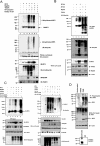
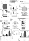
FIG. 1.
FIG. 1.
IRF-5 undergoes K63-linked polyubiquitination in a MyD88-dependent pathway. (A) 293T cells transfected with a total (4 μg) of His-tagged IRF-5 (lanes 2 to 6), MyD88 (lanes 4, 6, and 7), and TRAF6 (lanes 5, 6, and 7) together with HA-ubiquitin (lanes 3 to 7) IRF-5 were affinity purified from cell lysates, using Probond Ni2+-charged resin. Bound proteins were separated on an SDS gel, and IRF-5 was identified by immunoblotting (IB) with anti-IRF-5 (upper panel) and antiubiquitin antibody (lower panel) after stripping the same blot. Whole-cell lysate was also immunoblotted with tubulin, anti-MyD88, anti-TRAF6, and anti-HA antibodies. (B) 293T cells were transfected with IRF-5v4 or IRF-5v5 and ubiquitin together with MyD88 and TRAF6. At 24 h after transfection cells were lysed, and IRF-5v4 was immunoprecipitated with Flag antibodies. IRF-5v5 was affinity purified on Ni resin. Proteins were separated on an SDS gel and immunoblotted with anti-IRF-5 and antiubiquitin antibody (upper panels). The relative levels of IRF-5, β-actin, and TRAF6 in the whole-cell lysate determined by immunoblotting are shown. (C) 293T cells were transfected with His-tagged IRF-5 (lanes 2 to 6 in the left panel and lanes 2 to 8 in the right panel), MyD88 (lanes 4 to 6 in the left panel and lanes 4 to 6 in the right panel), TRAF6 (lanes 4 to 6 in the left panel and lanes 3, 8, and 9 in the right panel) together with HA-ubiquitin (lanes 3 and 4 in the left panel and lanes 3 and 4 in the right panel) or a K63R HA-ubiquitin mutant (lane 5 in the left panel and lanes 6 and 7 in the right panel) or K48R ubiquitin mutant (lane 6 in the left panel and lanes 7 to 8 in the right panel). His-tagged IRF-5 was affinity purified on Ni resin and blotted with anti-IRF-5 (left panel) or antiubiquitin (right panel) serum. The expression of IRF-5, MyD88, TRAF6, and K48 or K63 HA-ubiquitin detected in the input cell lysates was detected by immune blotting (lower panels). (D) Ubiquitination of the endogenous IRF-5. RAW264.7 cells were either treated with DMSO or stimulated with a 100 nM concentration of TLR7 agonist R848 for 8 h to engage TLR7-MyD88 pathway that leads to IRF-5 activation. As a positive control, cells were transfected with IRF-5 together with MyD88, TRAF6, and ubiquitin. Cells were lysed with radioimmunoprecipitation assay buffer, and ubiquitinated IRF-5 was immunopurified using antiubiquitin beads. The specifically bound proteins were subjected to SDS-PAGE and immunoblotted with IRF-5 antibodies (upper panel). The total cell lysate was probed with IRF-5 (lower panel). The cell lysate from unstimulated RAW264.7 cells was immunoprecipitated with IRF-5 and immunoblotted with IRF-5 to demonstrate the expression of an endogenous IRF-5 (lower panel). Immune blotting with ubiquitin antibodies did not detect any IRF-5 (data not shown). Ub, ubiquitin; IgG, immunoglobulin G.
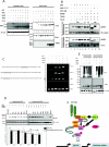
FIG. 2.
FIG. 2.
IRF-5 is polyubiquitinated by the E3 ligase TRAF6. (A) TRAF6−/− MEFs were cotransfected with IRF-5, MyD88, and HA-ubiquitin in the presence and absence of TRAF6. At 24 h posttransfection, cell lysates were immunoblotted with anti-IRF-5 (upper panel) or anti-HA epitope antibodies (middle panel). To verify TRAF6 expression, the blot was reprobed with anti-TRAF6 antibody (lower panel). Levels of β-actin show equal loading of protein. (B) Schematic structure of C-terminal deletions of IRF-5v4. DBD, DNA binding domain; IAD, interacting domain. Amino acid regions that are deleted in the IRF-5 mutants are indicated. Flag-tagged full-length IRF-5 or its deletion mutants were transfected to 293T cells together with MyD88, TRAF6, and ubiquitin. At 24 h posttransfection cells were lysed, and lysates were subjected to immunoprecipitation using anti-Flag-coupled beads followed by immunoblotting with anti IRF-5 antibody. IRF-5 and its deletion mutants were detected in cell lysates by immunoblotting with anti-Flag antibody. (C) A schematic illustration of point mutations at TRAF6 consensus recognition motif and a deletion in IRF-5-BMv. 293T cells were transfected with murine IRF-5 (positive control), IRF-5-BMv, and IRF-5 K410/K411R together with MyD88, TRAF6, and ubiquitin. His-purified IRF-5 was detected by immunoblotting with anti-IRF-5 (upper panel). IRF-5-BMv and IRF-5 mutant expression in cell lysates were detected by immunoblotting, and the relative levels of β-actin indicate equal protein loading. (D) Binding of TRAF6 to IRF-5 and its mutants. Cells were cotransfected with Flag-IRF-5 or its deletion mutants together with MyD88 and HA-ubiquitin expression plasmids (left). Twenty-four hours later the cells were lysed, and the lysates were precipitated with Flag antibodies; the presence of IRF-5 and TRAF6 in the precipitates was detected by Western blotting with IRF-5 or TRAF6 antibodies. The relative levels of transfected IRF-5, its mutants, and TRAF6 in the input lysates were detected by Western blotting. The IRF-5-positive band in the control sample (lane 1) was caused by a leaking well. Lysates from cells cotransfected with IRF-5, the IRF-5 K410/K411R mutant, and IRF-5-BMv with MyD888 and TRAF6 were immunoprecipitated with TRAF6 antibodies, and the levels of IRF-5 and TRAF6 in immunoprecipitated samples were detected by immunoblotting with IRF-5 or TRAF6 antibodies (right panels). The relative levels of transfected IRF-5, the IRF-5 K410/K411R mutant, IRF-5-BMv, and TRAF6 in the input lysates were detected by immunoblotting with respective antibodies. (E) The two C-terminal deletion mutants of IRF-5v5 containing the TRAF6 consensus recognition sequence PREKKL are shown schematically. The lysates of cells cotransfected with full-length IRF-5v5 or its mutants and MyD88 and TRAF6 were immunoprecipitated with Flag antibody, and IRF-5 was detected by immunoblotting with IRF-5 antibody (upper panel). The same blot was stripped of bound antibody and blotted with TRAF6 antibody (middle panel). The levels of expression of IRF-5 and TRAF6 in input lysates are shown in the lower panel. (F) Mutation in the TRAF6 consensus recognition motif of IRF-5 blocked activation of IFNA4 reporter. 293T cells were cotransfected with an IFNA4-luc reporter plasmid (10 ng) together with the indicated combination of MyD88 (10 ng), TRAF6 (10 ng), and ubiquitin (5 ng) and 10 ng each of IRF-5, IRF-5v5, IRF-5v4, and the IRF-5 K410/K411R mutant. Luciferase activity was measured 24 h after the transfections. Cells were also transfected with IRF-5, MyD88, and TRAF6 and together with TLR7 expression plasmid. Sixteen hours after transfections stimulated with 10 nM R848 for 8 h, luciferase activity was measured in cell lysates (right panel). (G) IFNA4-luciferase (Luc) reporter and IRF-5 plasmids were cotransfected into TRAF6−/− MEFs and wild-type MEFs together with MyD88, TRAF6, and ubiquitin as indicated. The levels of DNA were kept constant in all transfection experiments, and the data are expressed as means ± standard deviations of four replicates. Ub, ubiquitin; IP, immunoprecipitation; IB, immunoblotting; nt, nucleotides.
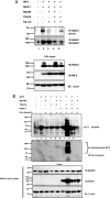
FIG. 3.
FIG. 3.
IRF-5 associates with IRAK1. (A) IRAK1−/− 293TI1A cells were transfected with IRAK1, His-tagged IRF-5, MyD88, TRAF6, and ubiquitin (Ub). Cell lysates were immunoprecipitated (IP) with anti-IRAK1 antibody and immunoblotted with either anti-His antibody or IRAK1 as indicated. The expression of transfected plasmids in cell lysates was detected by immunoblotting with respective antibodies (lower panel). (B) 293TI1A cells, transfected with His-tagged IRF-5 (lanes 2 to 8), MyD88 (lanes 3, 5, 6, 8, and 9), TRAF6 (lanes 4, 7, and 8), or IRAK1 (lanes 6 to 9) together with HA-ubiquitin (lanes 3 to 8). IRF-5 was His purified and analyzed on an SDS gel by immunoblotting (IB) with anti-IRF-5 antibody (upper panel) and subsequently with anti-HA antibodies (middle panel). The relative levels of MyD88 IRAK1 and β-actin in input cell lysates were detected by immunoblotting with the respective antibodies.
FIG. 4.
FIG. 4.
Ubiquitination of IRF-5 promotes nuclear transport, binding to type I IFN promoters and IRF-5 stabilization. (A). IRF-5 nuclear localization of IRF-5 in IRAK1−/− cells. 293TI1A cells were cotransfected with IRF-5, MyD88, TRAF6, and ubiquitin (Ub). Nuclear and cytoplasmic fractions were prepared 24 h after the transfections, and proteins were separated on SDS gels and immunoblotted (IB) with IRF-5 antibodies. To demonstrate equal loading, blots were stripped and immunoblotted with β-actin (cytoplasmic fraction), and the purity of the nuclear fraction was demonstrated by blotting with anti-RelA antibody. Immune blotting with anti-IRAK1 antibody was used to determine the levels of ectopic IRAK1. (B) 293T cells were transfected with IRF-5, the IRF-5 K410/K411R mutant, and the IRF-5Δ (with a deletion of residues 480 to 539) mutant, and cytoplasmic and nuclear proteins were resolved by SDS-PAGE and analyzed by Western blotting with anti-IRF-5, anti-Sp1 (nuclear fraction), and anti-α-tubulin (cytoplasmic fraction) antibody. (C) The association of IRF-5 with the promoters of the endogenous IFNA and IFNB genes was determine by ChIP assay in 293T cells transfected with IRF-5, MyD88, TRAF6, and ubiquitin as described in Materials and Methods. The sequences of the PCR-amplified promoter fragments containing the IRF-E binding site are shown. (D) Ubiquitination leads to stabilization of IRF-5. The left panel shows relative levels of IRF-5 in cells transiently expressing IRF-5, and the right panel shows IRF-5 levels in cells in which IRF-5 was coexpressed with MyD88, TRAF6, and ubiquitin. Ethanol control is a control sample cultured in the absence of CHX. Gels were scanned, and the ratios of IRF-5 to β-actin are shown as a function of time. (E) The relative levels of ubiquitinated proteins in cell lysates from 293T cells expressing IRF-5, MyD88, TRAF6, and ubiquitin and cultured with either DMSO (left panel) or 10 μM MG132 (right panel) for 6 h. The blots were reprobed with anti-IRF-5 (middle panel) and α-tubulin (lower panel) antibodies. (F). Proposed model of molecular mechanism leading to IRF-5 activation by a TLR7/9-activated, MyD88-dependent pathway.

Similar articles

Cited by

References

    1. Abbott, D. W., Y. Yang, J. E. Hutti, S. Madhavarapu, M. A. Kelliher, and L. C. Cantley. 2007. Coordinated regulation of Toll-like receptor and NOD2 signaling by K63-linked polyubiquitin chains. Mol. Cell. Biol. 276012-6025. - PMC - PubMed
    1. Akira, S., and K. Takeda. 2004. Toll-like receptor signalling. Nat. Rev. Immunol. 4499-511. - PubMed
    1. Akira, S., S. Uematsu, and O. Takeuchi. 2006. Pathogen recognition and innate immunity. Cell 124783-801. - PubMed
    1. Alexopoulou, L., A. C. Holt, R. Medzhitov, and R. A. Flavell. 2001. Recognition of double-stranded RNA and activation of NF-κB by Toll-like receptor 3. Nature 413732-738. - PubMed
    1. Barnes, B., B. Lubyova, and P. M. Pitha. 2002. On the role of IRF in host defense. J. Interferon Cytokine Res. 2259-71. - PubMed

Publication types

MeSH terms