. 2008 Dec;82(23):11837-50.
doi: 10.1128/JVI.01623-08. Epub 2008 Sep 24.

Human cytomegalovirus glycoproteins gB and gH/gL mediate epithelial cell-cell fusion when expressed either in cis or in trans

Affiliations

Human cytomegalovirus glycoproteins gB and gH/gL mediate epithelial cell-cell fusion when expressed either in cis or in trans

Adam L Vanarsdall et al. J Virol. 2008 Dec.

Abstract

Herpesviruses use a cascade of interactions with different cell surface molecules to gain entry into cells. In many cases, this involves binding to abundant glycosaminoglycans or integrins followed by interactions with more limited cell surface proteins, leading to fusion with cellular membranes. Human cytomegalovirus (HCMV) has the ability to infect a wide variety of human cell types in vivo. However, very little is known about which HCMV glycoproteins mediate entry into various cell types, including relevant epithelial and endothelial cells. For other herpesviruses, studies of cell-cell fusion induced by viral proteins have provided substantial information about late stages of entry. In this report, we describe the fusion of epithelial, endothelial, microglial, and fibroblast cells in which HCMV gB and gH/gL were expressed from nonreplicating adenovirus vectors. Fusion frequently involved the majority of cells, and gB and gH/gL were both necessary and sufficient for fusion, whereas no fusion occurred when either glycoprotein was omitted. Coexpression of UL128, UL130, and UL131 did not enhance fusion. We concluded that the HCMV core fusion machinery consists of gB and gH/gL. Coimmunoprecipitation indicated that HCMV gB and gH/gL can interact. Importantly, expression of gB and gH/gL in trans (gB-expressing cells mixed with other gH/gL-expressing cells) resulted in substantial fusion. We believe that this is the first description of a multicomponent viral fusion machine that can be split between cells. If gB and gH/gL must interact for fusion, then these molecules must reach across the space between apposing cells. Expression of gB and gH/gL in trans with different cell types revealed surface molecules that are required for fusion on HCMV-permissive cells but not on nonpermissive cells.


Figures
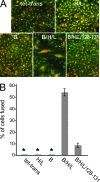
FIG. 1.
FIG. 1.
Cell-cell fusion of ARPE-19 human retinal epithelial cells by HCMV glycoproteins. (A) ARPE-19 cells were transduced with nonreplicating Ad vectors expressing the tet transactivator (tet-trans), gH and gL (H/L), gB (B), a combination of gB, gH, and gL (B/H/L), or a combination of gB, gH, and gL plus UL128, -130, and -131 (B/H/L/128-131) at 25 PFU/cell for each construct. Cell monolayers were transduced with the appropriate Ad vector for 12 h, trypsinized, mixed with nontransduced ARPE-19 cells, and incubated for 60 h to allow formation of syncytia. Cell monolayers were then fixed and fluorescently stained with propidium iodide and a rabbit polyclonal antibody to β-catenin, followed by a fluorescein isothiocyanate-conjugated donkey anti-rabbit antibody, to label cell nuclei and plasma membranes, respectively. (B) Quantification of cell-cell fusion from ARPE-19 cells expressing the tet transactivator (tet-trans), gH and gL (H/L), gB (B), a combination of gB, gH, and gL (B/H/L), or a combination of gB, gH, and gL plus UL128, -130, and -131 (B/H/L/128-131). Cell-cell fusion was quantified from micrographs by counting the number of nuclei involved in syncytium formation, dividing it by the total number of nuclei within the same field, and expressing it as the percentage of cells fused. Values represent the averages derived from three separate images, and the error bars indicate the standard deviations. Asterisks indicate that no fusion was observed.
FIG. 2.
FIG. 2.
Quantification of the extent of syncytium formation induced by HCMV glycoproteins gB, gH, and gL in ARPE-19 cells. ARPE-19 cells transduced with Ad vectors expressing gB, gH, and gL at 25 PFU/cell were mixed with nontransduced ARPE-19 cells and incubated for 60 h to induce syncytium formation. Images from fixed monolayers were analyzed to determine the number of independent nuclei within each syncytium. The x axis indicates the number of nuclei per syncytium, and the y axis represents the frequency of the total syncytia analyzed. Only syncytia containing at least five nuclei were included in the analysis. No syncytia were observed in non-glycoprotein-expressing ARPE-19 cells.
FIG. 3.
FIG. 3.
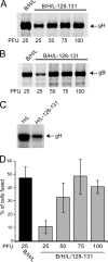
Analysis of dose responses and contribution of UL128-131 to cell-cell fusion of ARPE-19. (A and B) ARPE-19 cells were transduced with Ad vectors expressing HCMV glycoproteins at the indicated PFU/cell for 24 h and then radiolabeled with [35S]methionine-cysteine for 3 h. Proteins were immunoprecipitated with gB- or gH-specific MAbs 27-156 and 14-4b, respectively, and then analyzed by SDS-polyacrylamide gel electrophoresis (SDS-PAGE). (C) ARPE-19 cells were transduced with Ad vectors expressing either gH and gL or gH, gL, and UL128-131, using 25 PFU/cell, for 24 h and then surface labeled by lactoperoxidase-catalyzed iodination. gH was immunoprecipitated using anti-gH MAb 14-4b. Proteins were analyzed by SDS-PAGE. (D) Cell-cell fusion of ARPE-19 cells transduced with Ad vectors expressing gB, gH, and gL or gB, gH, gL, and UL128-131 at the doses indicated and incubated for 60 h. Cell fusion was quantified as described in the legend to Fig. 1. Values represent the averages derived from three separate images, and the error bars indicate the standard deviations.
FIG. 4.
FIG. 4.
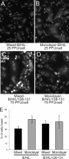
ARPE-19 cells fuse when gB and gH/gL are transduced into all of the cells in a monolayer. Two conditions for transduction of ARPE-19 cells were compared. In some instances, ARPE-19 cells were transduced with Ad vectors expressing gB, gH, and gL (A) or gB, gH, gL, and UL128-131 (C) and then mixed with untransduced ARPE-19 cells (mixed). In other cases, Ad vectors expressing gB, gH, and gL (B) or gB, gH, gL, and UL128-131 (D) were used to directly transduce monolayers of ARPE-19 cells (monolayers). Transductions of gB, gH, and gL were performed at 25 PFU/cell. To attain similar levels of expression, transductions involving gB, gH, gL, and UL128-131 involved higher doses (75 PFU/cell). (E) Quantification of cell-cell fusion under the conditions in panels A to D by the methods described in the legend to Fig. 1. Values represent the averages derived from three separate images, and the error bars indicate the standard deviations.
FIG. 5.
FIG. 5.
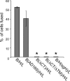
Analysis of cell-cell fusion of ARPE-19 cells transduced with mutant forms of gB and gH. Cell-cell fusion of ARPE-19 cells transduced with Ad vectors expressing gB, gH, and gL derived from HCMV strain TR was analyzed. Cell-cell fusion assays were also performed with Ad vectors expressing gB from AD169, AD169 gB lacking the CT domain (ΔCT), or AD169 gB lacking both the CT and TM domains (ΔCTΔTM). Fusion assays were also performed with an Ad vector expressing a mutant gH (sgH) that lacked both the CT and TM domains. Cell-cell fusion was quantified from images as described in the legend to Fig. 1. Values represent the averages derived from three separate images, and the error bars indicate the standard deviations. Asterisks indicate that no fusion was observed.
FIG. 6.
FIG. 6.
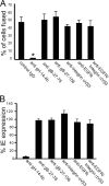
Effects of gH-, gB-, EGFR-, and integrin-specific antibodies on infectivity of HCMV and cell-cell fusion. (A) ARPE-19 cells were transduced with Ad vectors expressing gB, gH, and gL (25 PFU/cell) for 12 h and then mixed with nontransduced cells. Six hours after cell mixing, the medium was replaced with culture medium containing anti-gH MAb 14-4b at 10 μg/ml and anti-gB MAbs 27-156 and 27-78, anti-EGFR MAb LA1, anti-integrin αVβ3 MAb 23C6, or purified control mouse IgG at 50 μg/ml, and the cells were then incubated for 54 h. Cell-cell fusion was observed in triplicate wells and quantified as described in the legend to Fig. 1. Asterisks indicate that no fusion was observed. (B) For antibody inhibition of virus entry, ARPE-19 cells were incubated in culture medium containing EGFR MAb, integrin αVβ3 MAb, or purified mouse IgG at 50 μg/ml prior to infection with HCMV. Alternatively, HCMV virus inoculum diluted to 3 PFU/cell was incubated with MAb 14-4b, 27-156, or 27-78 or with purified mouse IgG at 50 μg/ml prior to being added to ARPE-19 cells. After 24 h, the cells were fixed and stained for HCMV IE protein IE-86. The number of cells expressing HCMV IE protein was normalized to that observed with the control mouse IgG.
FIG. 7.
FIG. 7.
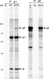
Coimmunoprecipitation of HCMV gB and gH. ARPE-19 cells were transduced with Ad vectors expressing gB alone (B), gH and gL (H/L), or a combination of gB, gH, and gL (B/H/L), using 25 PFU/cell, for 24 h and then labeled with [35S]methionine-cysteine for 3 h. Cell extracts were made with 0.5% NP-40 lysis buffer and subjected to immunoprecipitation with anti-gH (14-4b) or anti-gB (27-156) MAb, and proteins were analyzed by SDS-PAGE under reducing conditions. The molecular mass marker, in kilodaltons, is indicated on the left.
FIG. 8.
FIG. 8.
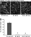
HCMV gB and gH/gL induce cell-cell fusion when expressed in ARPE-19 cells in trans. (A) Cell-cell fusion after ARPE-19 cells expressing gB were mixed with ARPE-19 cells expressing gH and gL (B→H/L). Control assays were performed after mixing ARPE-19 cells expressing either gB or gH and gL alone (B→B and H/L→H/L, respectively). Cell monolayers were fixed and fluorescently stained with propidium iodide to label cell nuclei. (B) Quantification of cell-cell fusion mediated by gB and gH/gL in trans under the conditions described in the legend to Fig. 1. Values represent the averages derived from three separate images, and the error bars indicate the standard deviations. Asterisks indicate that no fusion was observed.
FIG. 9.
FIG. 9.
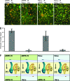
Analysis of cell-cell fusion mediated by gB and gH/gL in trans, using different cell types. (A) ARPE-19 cells expressing gB were mixed with ARPE-19 cells expressing gH and gL. (B) ARPE-19 cells expressing gB were mixed with HeLa cells expressing gH and gL. (C) HeLa cells expressing gB were mixed with ARPE-19 cells expressing gH and gL. (D) HeLa cells expressing gB were mixed with HeLa cells expressing gH and gL. The cells were fixed and stained with propidium iodide and β-catenin MAb (a cell surface marker) after 60 h. (E) Quantification of cell-cell fusion in the samples described above. Values represent the averages derived from three separate images, and the error bars indicate the standard deviations. (F) Cartoon depicting the orientation of gB and gH/gL in ARPE-19 and HeLa cells. Arrows and + or − signs indicate whether or not fusion was observed.

Similar articles

Cited by

References

    1. Adler, B., L. Scrivano, Z. Ruzcics, B. Rupp, C. Sinzger, and U. Koszinowski. 2006. Role of human cytomegalovirus UL131A in cell type-specific virus entry and release. J. Gen. Virol. 872451-2460. - PubMed
    1. Alford, C. A., and W. J. Britt. 1990. Cytomegalovirus, p. 1981-2010. In B. N. Fields and D. M. Knipe (ed.), Fields virology. Raven Press Ltd., New York, NY.
    1. Atanasiu, D., J. C. Whitbeck, T. M. Cairns, B. Reilly, G. H. Cohen, and R. J. Eisenberg. 2007. Bimolecular complementation reveals that glycoproteins gB and gH/gL of herpes simplex virus interact with each other during cell fusion. Proc. Natl. Acad. Sci. USA 10418718-18723. - PMC - PubMed
    1. Avitabile, E., C. Forghieri, and G. Campadelli-Fiume. 2007. Complexes between herpes simplex virus glycoproteins gD, gB, and gH detected in cells by complementation of split enhanced green fluorescent protein. J. Virol. 8111532-11537. - PMC - PubMed
    1. Bender, F. C., J. C. Whitbeck, H. Lou, G. H. Cohen, and R. J. Eisenberg. 2005. Herpes simplex virus glycoprotein B binds to cell surfaces independently of heparan sulfate and blocks virus entry. J. Virol. 7911588-11597. - PMC - PubMed

Publication types

Substances

LinkOut - more resources