. 2008 Nov 28;283(48):33611-9.
doi: 10.1074/jbc.M804840200. Epub 2008 Sep 22.

Regulation of dynactin through the differential expression of p150Glued isoforms

Affiliations

Regulation of dynactin through the differential expression of p150Glued isoforms

Ram Dixit et al. J Biol Chem. .

Abstract

Cytoplasmic dynein and dynactin interact to drive microtubule-based transport in the cell. The p150Glued subunit of dynactin binds to dynein, and directly to microtubules. We have identified alternatively spliced isoforms of p150Glued that are expressed in a tissue-specific manner and which differ significantly in their affinity for microtubules. Live cell assays indicate that these alternatively spliced isoforms also differ significantly in their microtubule plus end-tracking activity, suggesting a mechanism by which the cell may regulate the dynamic localization of dynactin. To test the function of the microtubule-binding domain of p150Glued, we used RNAi to deplete the endogenous polypeptide from HeLa cells, followed by rescue with constructs encoding either the full-length polypeptide or an isoform lacking the microtubule-binding domain. Both constructs fully rescued defects in Golgi morphology induced by depletion of p150Glued, indicating that an independent microtubule-binding site in dynactin may not be required for dynactin-mediated trafficking in some mammalian cell types. In neurons, however, a mutation within the microtubule-binding domain of p150Glued results in motor neuron disease; here we investigate the effects of four other mutations in highly conserved domains of the polypeptide (M571T, R785W, R1101K, and T1249I) associated in genetic studies with Amyotrophic Lateral Sclerosis. Both biochemical and cellular assays reveal that these amino acid substitutions do not result in functional differences, suggesting that these sequence changes are either allelic variants or contributory risk factors rather than causative for motor neuron disease. Together, these studies provide further insight into the regulation of dynein-dynactin function in the cell.


Figures

FIGURE 1.
FIGURE 1.
Alternative splicing results in the tissue-specific expression of multiple isoforms of p150Glued. A, full-length isoform of the dynactin subunit p150Glued has a N-terminal CAP-Gly domain (red), followed by a basic domain that binds more weakly to microtubules (green), and two coiled-coil domains that mediate homodimerization (blue). These coiled-coil domains also mediate binding to the DIC and to the Arp1 subunit of dynactin. The previously characterized p135 isoform is specifically expressed in neurons and lacks both of the N-terminal microtubule-binding domains. The newly characterized Δ5, Δ5,6, and Δ5,6,7 isoforms lack much of the basic domain that forms a secondary site of association with the microtubule; basic residues in the amino acid sequences of exons 4 through 8 are highlighted in green. B, isoform expression was analyzed by RT-PCR of mRNA isolated from adult human tissues including brain, kidney, and skeletal muscle using primers spanning exons 3-8, and compared with PCR products from cDNAs corresponding to Δ5,6,7, Δ5,6, and full-length p150Glued. The identity of the bands observed by gel electrophoresis was confirmed by direct sequencing of the PCR products. A minor upper band visible in the PCR reaction from human brain mRNA was determined to be unrelated by sequence analysis.
FIGURE 2.
FIGURE 2.
Alternatively spliced isoforms of p150Glued differ significantly in their binding affinity for microtubules. Microtubule binding affinities for the p150-FL and p150-Δ5,6,7 isoforms were measured in co-sedimentation assays with increasing concentrations of polymerized tubulin, and indicate that the basic domain encoded by exons 5, 6, and 7 contributes significantly to the overall affinity of p150Glued for the microtubule. Fitting the data to the equation [y = (Bmax×x)/(Kd+x)] by nonlinear regression yields a Kd of 0.40±0.07 μm for p150-FL; an estimated Kd of 5.3 ± 1.5 μm was obtained for p150-Δ5,6,7.
FIGURE 3.
FIGURE 3.
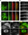
Regulation of tip tracking by differential expression of p150Glued isoforms. A, HeLa cells transfected with GFP-tagged p150Glued-FL (top) or GFP-tagged p150-Δ5,6,7 (bottom) were stained with antibodies to p150Glued (UP502, green) and tubulin (red). Full-length p150Glued demonstrates lattice binding of microtubules, but p150-Δ5,6,7 localizes predominantly to microtubule tips. B, still images from a time series observing GFP-tagged p150-FL (top) or p150-Δ5,6,7 (bottom). Arrowheads point to GFP- p150-Δ5,6,7 labeling of dynamic plus ends. Frames are displayed in 5-s intervals. The full-length isoform localizes along the length of the microtubule lattice, remaining associated with the microtubule during both growth and catastrophe. In contrast, the p150-Δ5,6,7 isoform demonstrates dynamic tip-tracking behavior with growing microtubule tips. See also supplemental Movies S1 and S2. C, HeLa cells were transfected with GFP-tagged p150-FL (top) or GFP-tagged p150-Δ5,6,7 (bottom). Cells were fixed 24 h after transfection and stained with antibodies to p150Glued (UP502, green) and TGN46 (Golgi, red). Overexpression of either isoform causes Golgi disruption. The extent of Golgi disruption is dependent on expression levels; compare the relatively intact Golgi (single arrowhead) seen in a cell expressing low levels of the GFP-p150 with the dispersed Golgi (double arrowheads) in the cell expressing higher levels of GFP-p150. Scale bars, 5 μm.
FIGURE 4.
FIGURE 4.
The microtubule-binding domain of p150Glued is not required to maintain Golgi organization in mammalian cells. A, images of HeLa-M cells transiently transfected with either the p150-FL or p135 isoforms of dynactin. The accompanying bar graph shows the percentage of transfected cells containing either normal, moderately dispersed, or severely dispersed Golgi determined from ∼100 cells from three independent experiments for each construct. B, endogenous p150Glued was depleted from HeLa-M cells by RNAi-induced knockdown; cells were rescued by transfection with either the p150-FL or p135 isoforms of dynactin. Immunofluorescence was carried out using antibodies to p150Glued (UP502) and to TGN46 to visualize the Golgi. T, transfected cell; U, untransfected cell. Scale bar, 20 μm. The accompanying bar graph shows the percentage of transfected cells containing either normal, moderately dispersed, or severely dispersed Golgi determined from ∼100 cells from three independent experiments for each construct.
FIGURE 5.
FIGURE 5.
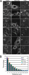
Expression of wild-type and mutant p150Glued in COS7 cells. A, COS7 cells transected with either wild-type or M571T, R785W, R1101K, and T1249I Myc-tagged constructs of p150Glued were analyzed by immunofluorescence using antibodies to tubulin, the Myc tag, and TGN46 (Golgi). Note the extensive Golgi disruption in transfected cells. Scale bar, 20 μm. B, histogram showing the quantification of Golgi disruption by wild-type and mutant p150Glued proteins. The distribution of the distance of individual Golgi fragments from the nuclear surface was measured in COS7 cells expressing either wild-type or the indicated mutant p150Glued proteins. Data from ∼20 cells with measurements of >1000 individual Golgi fragments for each construct are shown.
FIGURE 6.
FIGURE 6.
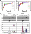
Comparison of microtubule and DIC binding of wild-type and mutant p150Glued. A, microtubule binding was measured in co-sedimentation assays at 0, 5, and 25 μm tubulin for in vitro translated wild-type or mutant p150Glued proteins. Data are shown as the bound fraction as a function of tubulin concentration, and are the mean ± S.D. obtained from three independent experiments. No significant differences were observed among the constructs. B, DIC binding assays were performed at increasing concentrations of p150Glued. Wild-type or mutant constructs were expressed using in vitro translation and binding to DIC-Sepharose beads was measured in pull-down assays. Data points are the mean ± S.D. obtained from three independent experiments; data were fit to the equation [y = (Bmax × x)/(Kd + x)] by nonlinear regression; no significant differences among the constructs were observed. C, sucrose density gradients of dynein-dynactin complex. Cell lysates from COS7 cells transiently expressing either wild-type or mutant p150Glued proteins (the R785W mutant is shown as a representative example) were fractionated through a 5-25% linear sucrose density gradient and the fractions subjected to immunoblot analysis using antibodies against Myc, DIC, and p50. The graphs show the relative protein amounts in each fraction as mean ± S.D. obtained from three independent experiments.
FIGURE 7.
FIGURE 7.
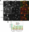
Comparison of wild-type and mutant p150Glued expressed in HeLa cells following depletion of endogenous p150Glued by RNAi. A, images of HeLa-M cells transiently expressing either the wild-type or mutant p150Glued proteins (only M571T and R785W images are shown) following RNAi-induced knockdown of the endogenous p150Glued protein. Cells were fixed 24 h after plasmid transfection and immunofluorescence staining carried out using monoclonal antibodies against p150Glued and TGN46 (Golgi). T, transfected cell; U, untransfected cell. Scale bar, 20 μm. B, bar graph shows the average percentage ± S.D. of cells containing either normal, moderately dispersed, or severely dispersed Golgi determined from >300 cells from three independent experiments.

Similar articles

Cited by

References

    1. Harada, A., Takei, Y., Kanai, Y., Tanaka, Y., Nonaka, S., and Hirokawa, N. (1998) J. Cell Biol. 141 51-59 - PMC - PubMed
    1. Lai, C., Lin, X., Chandran, J., Shim, H., Yang, W. J., and Cai, H. (2007) J. Neurosci. 27 13982-13990 - PMC - PubMed
    1. Hafezparast, M., Klocke, R., Ruhrberg, C., Marquardt, A., Ahmad-Annuar, A., Bowen, S., Lalli, G., Witherden, A. S., Hummerich, H., Nicholson, S., Morgan, P. J., Oozageer, R., Priestley, J. V., Averill, S., King, V. R., Ball, S., Peters, J., Toda, T., Yamamoto, A., Hiraoka, Y., Augustin, M., Korthaus, D., Wattler, S., Wabnitz, P., Dickneite, C., Lampel, S., Boehme, F., Peraus, G., Popp, A., Rudelius, M., Schlegel, J., Fuchs, H., Hrabe de Angelis, M., Schiavo, G., Shima, D. T., Russ, A. P., Stumm, G., Martin, J. E., and Fisher, E. M. (2003) Science 300 808-812 - PubMed
    1. Chen, X. J., Levedakou, E. N., Millen, K. J., Wollmann, R. L., Soliven, B., and Popko, B. (2007) J. Neurosci. 27 14515-14524 - PMC - PubMed
    1. Puls, I., Jonnakuty, C., LaMonte, B. H., Holzbaur, E. L., Tokito, M., Mann, E., Floeter, M. K., Bidus, K., Drayna, D., Oh, S. J., Brown, R. H., Jr., Ludlow, C. L., and Fischbeck, K. H. (2003) Nat. Genet. 33 455-456 - PubMed

Publication types

MeSH terms

LinkOut - more resources