. 2009 May;101(5):273-85.
doi: 10.1042/BC20080109.

Geminin is partially localized to the centrosome and plays a role in proper centrosome duplication

Affiliations

Geminin is partially localized to the centrosome and plays a role in proper centrosome duplication

Fei Lu et al. Biol Cell. 2009 May.

Abstract

Background information: Centrosome duplication normally parallels with DNA replication and is responsible for correct segregation of replicated DNA into the daughter cells. Although geminin interacts with Cdt1 to prevent loading of MCMs (minichromosome maintenance proteins) on to the replication origins, inactivation of geminin nevertheless causes centrosome over-duplication in addition to the re-replication of the genome, suggesting that geminin may play a role in centrosome duplication. However, the exact mechanism by which loss of geminin affects centrosomal duplication remains unclear and the possible direct interaction of geminin with centrosomal-localized proteins is still unidentified.

Results: We report in the present study that geminin is physically localized to the centrosome. This unexpected geminin localization is cell-cycle dependent and mediated by the actin-related protein, Arp1, one subunit of the dynein-dynactin complex. Disruption of the integrity of the dynein-dynactin complex by overexpression of dynamitin/p50, a well-characterized inhibitor of dynactin, reduces the centrosomal localization of both geminin and Arp1. Enrichment of geminin on centrosomes was enhanced when cellular ATP production was suppressed in the ATP-inhibitor assay, whereas the accumulation of geminin on the centrosome was disrupted by depolymerization of the microtubules using nocodazole. We further demonstrate that the coiled-coil motif of geminin is required for its centrosomal localization and the interaction of geminin with Arp1. Depletion of geminin by siRNA (small interfering RNA) in MDA-MB-231 cells led to centrosome over-duplication. Conversely, overexpression of geminin inhibits centrosome over-duplication induced by HU in S-phase-arrested cells, and the coiled-coil-motif-mediated centrosomal localization of geminin is required for its inhibition of centrosome over-duplication. Centrosomal localization of geminin is conserved among mammalian cells and geminin might perform as an inhibitor of centrosome duplication.

Conclusions: The results of the present study demonstrate that a fraction of geminin is localized on the centrosome, and the centrosomal localization of geminin is Arp1-mediated and dynein-dynactin-dependent. The coiled-coil motif of geminin is required for its targeting to the centrosome and inhibition of centrosome duplication. Thus the centrosomal localization of geminin might perform an important role in regulation of proper centrosome duplication.


Figures

Figure 1
Figure 1. Geminin localizes on centrosomes
(A) A fraction of GFP–geminin localized to ‘aster-like’ structures in a live cell. HeLa cells were transfected with pEGFPC2-geminin vector using a standard calcium phosphate transfection protocol. The culture dish was placed on to a heated sample stage within a heated chamber (37°C) at 5% CO2. GFP–geminin-expressing living cells were imaged by a Zeiss200M fluorescence microscope (Axiovert 200M) with the plan APO 63′/1.35 objective. GFP–geminin is expressed and partially localized to ‘aster-like’ structures in interphase cells as well as spindle fibres in cells undergoing mitosis as indicated with arrowheads (red). Scale bar=10 μm. (B) Centrosomal localization of both exogenous GFP–geminin and endogenous geminin. GFP–geminin-expressing HeLa cells were fixed with ice-cold methanol and immunostained with a γ-tubulin antibody. After incubation with a TRITC-conjugated secondary antibody, cells were mounted with mowiol containing DAPI for staining of DNA. Staining for γ-tubulin indicated the presence of centrosomes. Endogenous geminin was examined by immunostaining with an anti-geminin antibody. A clear centrosome co-staining of γ-tubulin and geminin (indicated with arrowheads) was achieved. Scale bar=10 μm. (C) The endogenous geminin from different cell lines was probed using the same antibody against human geminin as used in Western blot analysis, suggesting that the anti-geminin antibody specifically recognized geminin in these cells. α-Tubulin was used as a loading control. The molecular mass in kDa is indicated on the left-hand side of the gel. 293, HEK (human embryonic kidney)-293 cells; 231, MDA-MB-231 cells; 3T3, NIH 3T3 cells. (D) Co-localization of geminin and Arp1 on centrosomes in MDA-MB-231 cells and MEF cells. MDA-MB-231 and MEF cells were fixed with ice-cold methanol and co-immunostained using antibodies against geminin and γ-tubulin (γ-tub). The similar centrosomal localization of both geminin and γ-tubulin (indicated with arrowheads, red) was observed in these cell lines as in (B). Scale bar=10 μm. Gem, geminin.
Figure 2
Figure 2. Cell-cycle-dependent centrosomal localization of geminin
(A) The centrosomal localization of geminin is accompanied with the progression of the cell cycle. Synchronized HeLa cells were fixed in ice-cold methanol and subjected to immunofluorescence staining using anti-geminin and anti-γ-tubulin antibodies. Geminin (Gem) locates on to centrosomes from late G1- to M-phase, and disassociates from centrosomes in early G1-phase (indicated with arrowheads). Scale bar=10 μm. (B) Centrosomes purified from synchronized HeLa cells were analysed by Western blotting. The absence of geminin (Gem) in purified early G1-phase centrosomes correlated with the results in (A), that geminin released from centrosomes in early G1 phase. γ-tub, γ-tubulin. (C) Synchronized HeLa cells in (A) and (B) were analysed by FACS. HeLa cells were arrested at late G1-phase by double-thymidine treatment. Cells were then released into fresh medium for 4, 10 and 16 h to enter S-, G2- and early G1-phase. M-phase cells were obtained by 24 h thymidine treatment followed by 12 h nocodazole treatment.
Figure 3
Figure 3. Arp1 as a potential centrosomal protein co-localized with geminin
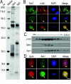
(A) Lysate from HeLa cells expressing exogenous GFP–geminin was immunoprecipitated (IP) using an antibody against geminin or rabbit IgG (rIgG) as a control. The proteins co-immunoprecipitated with geminin were separated on SDS/PAGE (10% gel) and stained with Coomassie Blue R-250. The protein bands were subjected to MS analysis. The bands indicated with a circle are (from the bottom to the top): dynactin 1 (p150Glued), Cdt1, GFP–geminin and Arp1 respectively. The molecular mass in kDa is indicated on the left-hand side of the gel. (B) Arp1 co-stained with γ-tubulin (γ-tub) in HeLa cells. HeLa cells were fixed and subjected to immunofluorescence staining using antibodies against Arp1 and γ-tubulin. The results showed that a fraction of Arp1 is located on centrosomes and Arp1 was well co-localized with γ-tubulin (indicated with arrowheads). Scale bar=10 μm. (C) Components of purified centrosomes from HeLa cells were examined for the co-existence of geminin (Gem) and Arp1. Fractions of centrosomes isolated from a discontinuous sucrose gradient were separated using SDS/PAGE (10% gel) and immunoblotted using antibodies against geminin and Arp1. γ-Tubulin (γ-tub) was immunoblotted as a positive control for centrosome purification. (D) Co-localization of endogenous geminin (Gem) and Arp1 was viewed by immunofluorescence staining using antibodies against geminin and Arp1 in HeLa cells fixed with ice-cold methanol. Both geminin and Arp1 were apparently co-localized in the centrosome in interphase and spindle poles in mitosis (indicated with arrowheads). Scale bar=10 μm.
Figure 4
Figure 4. Geminin directly interacts with Arp1
(A) Geminin and Arp1 interacted with each other. In the upper panels, Arp1 was co-immunoprecipitated with geminin. Lysates of HeLa cells were immunoprecipitated (IP) with antibodies against geminin or rabbit IgG (rIgG) as a control. The co-existence of Arp1 and geminin was examined by immunoblotting analysis. Cdt1 was immunoblotted as a positive control for binding proteins of geminin. In the bottom panels, geminin was co-immunoprecipitated with Arp1. Lysates from GFP–Arp1-overexpressing HeLa cells were tested for immunosedimentation of geminin by Arp1 using an antibody against Arp1 or rIgG as a control. (B) In vitro pulldown assay for testing the association between Arp1 and GST–geminin. Purified GST–geminin was coupled to glutathione–Sepharose 4B beads and incubated with the lysate of HeLa cells for 2 h. The proteins on the beads were separated on SDS/PAGE (10% gel) and transferred on to a nitrocellulose membrane and stained with Fast Green (upper panel). The nitrocellulose membrane was then immunoprobed for Arp1 with a specific antibody. Cdt1 was immunoblotted as a positive control of geminin-associated proteins (bottom panels). Gem, geminin.
Figure 5
Figure 5. Disruption of the dynein–dynactin complex by overexpressed exogenous dynamitin/p50 impairs both geminin and Arp1 centrosomal localization
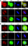
GFP–dynamitin/p50 was introduced into HeLa cells using Lipofectamine™ 2000. At 24 h after transfection, the cells were fixed with ice-cold methanol and examined by immunofluorescence staining using antibodies against geminin and Arp1. Transfection of GFP was taken as a negative control. In GFP-expressing control HeLa cells, the centrosomal/spindle pole localization of both geminin and Arp1 was not affected, as indicated in the upper panels of (A) and (B). Similar results were obtained in low-level dynamitin/p50-expressing cells, in which both geminin and Arp1 were normally localized on centrosome/spindle poles [middle panels in (A) and (B)]. High expression of dynamitin/p50 in HeLa cells did impair centrosomal localization of both geminin and Arp1, as indicated in the bottom panels of (A) and (B). Scale bar=10 μm.
Figure 6
Figure 6. Dynein–dynactin-mediated centrosomal targeting of geminin is dependent on microtubules
(A) Geminin accumulation on centrosomes depends on microtubules. Before the cells were fixed, nocodazole (20 μM) was added to the medium and cells were further incubated for 30 min. Co-immunofluorescence staining of geminin (Gem; green) and α-tubulin (red) was performed to examine altered localization of endogenous geminin when microtubules were depolymerized by nocodazole. As indicated with the arrowheads, centrosomal enrichment of geminin was inhibited after nocodazole treatment. Scale bar=10 μm. (B) Geminin accumulated on centrosomes after cellular ATP reduction. An ATP-inhibitor assay was performed as described in the Material and methods section. HeLa cells were transfected with GFP–geminin (GFP-Gem) and treated with either saline/glucose or saline/glucose plus 5 mM Az and 1 mM DOG and processed for microscopy. As indicated with arrowheads, after incubation for 30 min in Az/DOG, the accumulation of GFP–geminin on centrosomes was enhanced. When microtubules were depolymerized with 20 μM nocodazole, centrosomal enrichment of geminin was inhibited even in the presence of Az/DOG. (C) Live-cell analysis of geminin on centrosomes after Az/DOG treatment. Cells were imaged at 0, 15 and 30 min after Az/DOG was added to the medium. As indicated with arrowheads, GFP–geminin gradually accumulated on centrosomes. For microtubule-depolymerization assays, 20 μM nocodazole was added to the medium 30 min before Az/DOG treatment. The centrosomal localization of GFP–geminin was disrupted in the present of Az/DOG. Scale bar=10 μm. (D) Co-immunofluorescence staining of geminin (Gem) and γ-tubulin (red) showed geminin enriched on centrosomes after cellular ATP induction. The altered localization of endogenous geminin (right-hand panels) was consistent with that of GFP–geminin (GFP-Gem; left-hand panels) under the same treatment. Scale bar=10 μm.
Figure 7
Figure 7. The coiled-coil motif is required for geminin centrosomal localization and interaction with Arp1
(A) Schematic diagrams of geminin and its truncated mutants. (B) The coiled-coil motif is required for geminin to interact with Arp1. For in vitro pulldown assays, four mutants of GST–geminin were purified and coupled on to glutathione–Sepharose 4B beads and incubated with the lysate of HeLa cells for 2 h. The proteins on the beads were separated on SDS/PAGE (10% gel) and subjected to Western blot analysis using antibodies against Arp1. Cdt1 was immunoprobed as a positive control. Coomassie Blue staining of the proteins represents the protein loading in the pulldown assay (upper panel). The molecular mass in kDa is indicated on the left-hand side of the gel. (C) The coiled-coil motif is required for centrosomal localization of geminin. HeLa cells were transfected with geminin or its mutants in the form of GFP-fusion proteins, and subjected to an ATP-inhibitor assay as described in the Materials and methods section. Only mutant 94–160 which contains the coiled-coil motif of geminin presents centrosomal enrichment in a similar manner to wild-type geminin. γ-Tubulin (γ-tub; red) was immunostained as a marker of centrosomes. Scale bar=10 μm.
Figure 8
Figure 8. Geminin regulates centrosome duplication in MDA-MB-231 cells
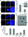
(A) siRNA of geminin resulted in centrosome over-duplication in MDA-MB-231 cells. Cells were transfected with siRNA targeting geminin (Gem siRNA) or non-specific siRNA as a control. Immunofluorescence staining of γ-tubulin (red) showed that the centrosomes were over-duplicated in geminin siRNA cells. Scale bar=10 μm. The cells with supernumerary centrosomes were counted and are shown in (B). The efficiency of geminin-knockdown was detected by Western blot analysis and is shown on the top right panel in (B). (C) Centrosomes were over-duplicated after being treated with HU in MDA-MB-231 cells. MDA-MB-231 cells were treated with/without 4 mM HU for 48 h and then immunofluorescence staining of γ-tubulin was performed to observe centrosomes. Scale bar=10 μm. (D and E) The coiled-coil motif is required for geminin to inhibit centrosome over-duplication in HU-arrested MDA-MB-231 cells. MDA-MB-231 cells were treated with 4 mM HU for 12 h and then transfected with geminin and its mutants as a GFP-fusion protein and cultured for another 48 h in the presence of HU to maintain the cells in S-phase. Cells were then either processed for immunofluorescence staining of γ-tubulin to observe centrosomes or Western blotted using anti-geminin or anti-GFP antibodies to exam the level of GFP-fusion proteins of geminin and its mutants. Note that over-duplication of centrosomes induced by HU was inhibited by overexpression of mutant 94–160, which contains the coiled-coil motif, as well as wild-type geminin. These results indicated that the coiled-coil motif is required for geminin to inhibit centrosome over-duplication. Supernumerary centrosomes were counted (E) and error bars represent 1 S.D.

Similar articles

Cited by

References

    1. Bell S.P., Dutta A. DNA replication in eukaryotic cells. Annu. Rev. Biochem. 2002;71:333–374. - PubMed
    1. Benjamin J.M., Torke S.J., Demeler B., McGarry T.J. Geminin has dimerization, Cdt1-binding, and destruction domains that are required for biological activity. J. Biol. Chem. 2004;279:45957–45968. - PubMed
    1. Chong J.P., Mahbubani H.M., Khoo C.Y., Blow J.J. Purification of an MCM-containing complex as a component of the DNA replication licensing system. Nature. 1995;375:418–421. - PubMed
    1. Clark S.W., Meyer D.I. Centractin is an actin homologue associated with the centrosome. Nature. 1992;359:246–250. - PubMed
    1. D'Assoro A.B., Busby R., Suino K., Delva E., Almodovar-Mercado G.J., Johnson H., Folk C., Farrugia D.J., Vasile V., Stivala F. Genotoxic stress leads to centrosome amplification in breast cancer cell lines that have an inactive G1/S cell cycle checkpoint. Oncogene. 2004;23:4068–4075. - PubMed

Publication types