. 2008 Nov;28(22):6889-902.
doi: 10.1128/MCB.01192-08. Epub 2008 Sep 15.

Transforming growth factor beta-induced Smad1/5 phosphorylation in epithelial cells is mediated by novel receptor complexes and is essential for anchorage-independent growth

Affiliations

Transforming growth factor beta-induced Smad1/5 phosphorylation in epithelial cells is mediated by novel receptor complexes and is essential for anchorage-independent growth

Amanda C Daly et al. Mol Cell Biol. 2008 Nov.

Abstract

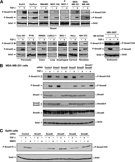
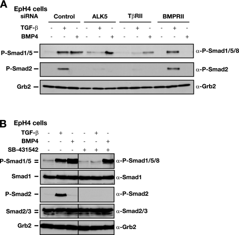
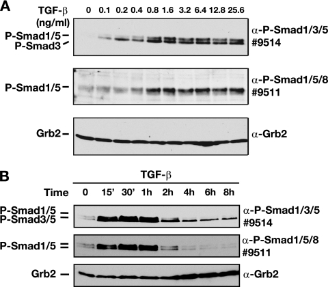
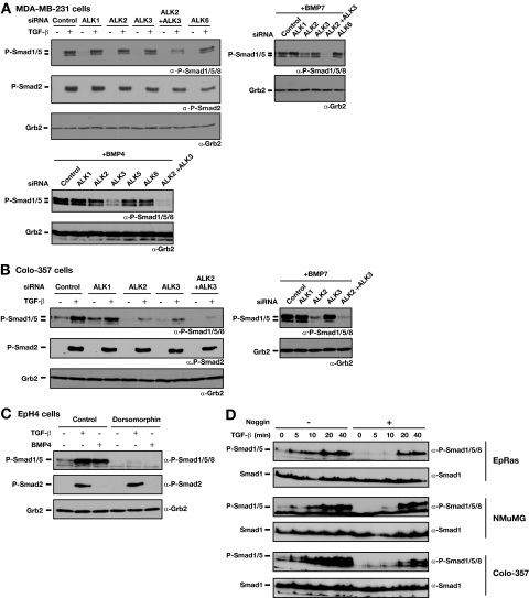
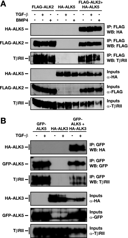
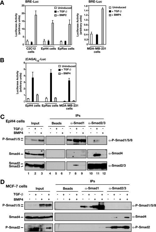
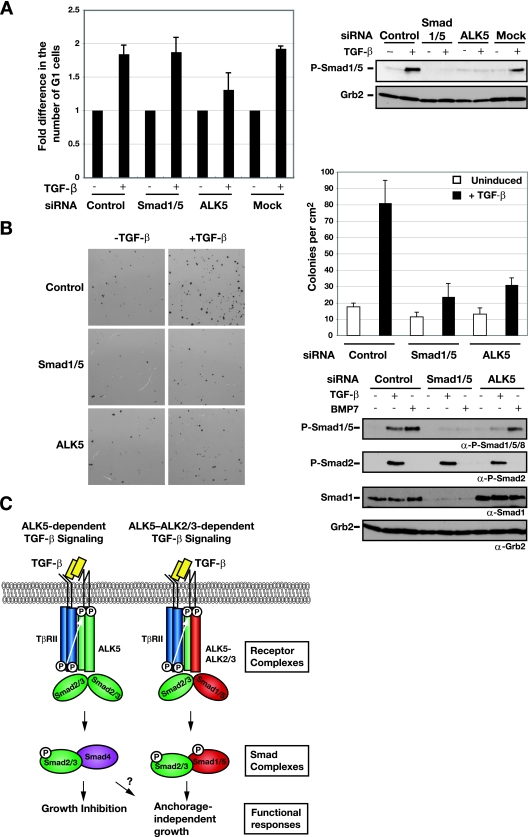
Transforming growth factor beta (TGF-beta) signals predominantly through a receptor complex comprising ALK5 and TbetaRII to activate receptor-regulated Smads (R-Smads) Smad2 and Smad3. In endothelial cells, however, TGF-beta can additionally activate Smad1 and Smad5. Here, we report that TGF-beta also strongly induces phosphorylation of Smad1/5 in many different normal epithelial cells, epithelium-derived tumor cells, and fibroblasts. We demonstrate that TbetaRII and ALK5, as well as ALK2 and/or ALK3, are required for TGF-beta-induced Smad1/5 phosphorylation. We show that the simultaneous activation of the R-Smads Smad2/3 and Smad1/5 by TGF-beta results in the formation of mixed R-Smad complexes, containing, for example, phosphorylated Smad1 and Smad2. The prevalence of these mixed R-Smad complexes explains why TGF-beta-induced Smad1/5 phosphorylation does not result in transcriptional activation via bone morphogenetic protein (BMP)-responsive elements, which bind activated Smad1/5-Smad4 complexes that are induced by BMP stimulation. Thus, TGF-beta induces two parallel pathways: one signaling via Smad2-Smad4 or Smad3-Smad4 complexes and the other signaling via mixed R-Smad complexes. Finally, we assess the function of the novel arm of TGF-beta signaling and show that TGF-beta-induced Smad1/5 activation is not required for the growth-inhibitory effects of TGF-beta but is specifically required for TGF-beta-induced anchorage-independent growth.


Figures

FIG. 1.
FIG. 1.
TGF-β induces phosphorylation of Smad1 and Smad5 in epithelial cells. α, anti. (A) A panel of cell lines including normal and cancerous breast cell lines and various human cancer cell lines and fibroblasts were tested for the ability of TGF-β to induce phosphorylation of Smad1/Smad5. Cells were either left untreated (−) or stimulated with 2 ng/ml TGF-β1 (+) for 1 h as indicated. An asterisk indicates the cell line in which TGF-β did not induce Smad1/5 phosphorylation. The lower right panel shows results for HEK-293T cells that were not treated with SB-431542 or treated with 10 μM SB-431542 overnight. The untreated cells were treated with or without TGF-β for 1 h. Of the cells treated with SB-431542, those to be induced with TGF-β were washed twice with phosphate-buffered saline and then stimulated with TGF-β for 1 h. Whole-cell extracts were analyzed by Western blotting using antibodies against phosphorylated Smad1/5/8 (P-Smad1/5/8), P-Smad2, and Grb2 as a loading control. (B) Smad1 and Smad5 are phosphorylated in response to TGF-β in MDA-MB-231 cells. MDA-MB-231 cells were transfected with siRNA SMARTpools against the R-Smads or a control siRNA oligonucleotide as indicated. At 72 h posttransfection, cells were left untreated (−) or treated with TGF-β1 for 45 min (+), and whole-cell extracts were prepared and analyzed by Western blotting using antibodies against Smad1, Smad2/3, Smad5, P-Smad1/5/8, P-Smad2, and Grb2. Note that P-Smad1/5 is a doublet. The siRNA knockdown results suggest that the upper band is a mixture of Smad1 and Smad5 and the lower band is predominantly an isoform of Smad5. (C) EpH4 cells were transfected with siRNA SMARTpools against the R-Smads or a control siRNA oligonucleotide as indicated. Cells were either left uninduced or treated with TGF-β1 (2 ng/ml) or BMP2 (200 ng/ml) for 45 min as indicated. Cell lysates were analyzed as described for panel A.
FIG. 2.
FIG. 2.
ALK5 and TβRII are required for the activation of Smad1/5 by TGF-β. α, anti. (A) Depletion of ALK5 and TβRII by siRNA silencing abolishes TGF-β-induced Smad1/5 phosphorylation. EpH4 cells were transfected with siRNA SMARTpools against TβRII, BMPRII, an siRNA duplex against ALK5, or a control siRNA oligonucleotide. After 72 h, cells were either left uninduced or stimulated with TGF-β1 or BMP4 for 45 min as indicated. Whole-cell extracts were analyzed by Western blotting using antibodies against phosphorylated Smad1/5/8 (P-Smad1/5/8), P-Smad2, and Grb2 as a control for protein loading. (B) ALK5 kinase activity is required for TGF-β-induced Smad1/5 phosphorylation. EpH4 cells were either left untreated or treated with the ALK5 inhibitor SB-431542 (10 μM) 15 min prior to stimulation with TGF-β or BMP4 for 45 min. Cell lysates were analyzed by Western blotting using antibodies against P-Smad1/5/8, Smad1, P-Smad2, Smad2/3, and Grb2 as a loading control.
FIG. 3.
FIG. 3.
Differential regulation of TGF-β-induced phosphorylated Smad1/5 (P-Smad1/5) and P-Smad2/3. α, anti. (A) High doses of ligand are required for TGF-β-induced Smad1/5 phosphorylation. EpH4 cells were stimulated with increasing concentrations of TGF-β1 for 1 h as indicated. Whole-cell extracts were analyzed by Western blotting using antibodies against P-Smad1/5/8 (antibody 9511), P-Smad1/3/5 (antibody 9514), and Grb2 as a loading control. Note that the Cell Signaling Technology antibody 9514 recognizes phosphorylated Smad3 in addition to phosphorylated Smad1/5. See Fig. S2C in the supplemental material for data on which the assignment of the different bands to the different Smads is based. (B) TGF-β-induced Smad1/5 phosphorylation is transient and disappears between 2 and 4 h after stimulation, whereas Smad3 phosphorylation is readily detectable for at least 8 h. EpH4 cells were stimulated with TGF-β1 (2 ng/ml) for different time periods at 37°C before lysis. Cell lysates were fractionated by SDS-polyacrylamide gel electrophoresis and analyzed by Western blotting with antibodies against P-Smad1/3/5, P-Smad1/5/8, and Grb2.
FIG. 4.
FIG. 4.
ALK2 and ALK3 are required for TGF-β-induced phosphorylation of Smad1/5. α, anti. (A) Effect of knockdown of ALK2 and ALK3 on Smad1/5 phosphorylation in MDA-MB-231 cells. Cells were transfected with siRNA SMARTpools against the ALKs for 72 h. Cells were either left uninduced or stimulated with 2 ng/ml TGF-β1 (upper-left panel), 200 ng/ml BMP7 (upper-right panel), or 20 ng/ml BMP4 (lower panel) for 45 min. Whole-cell extracts were analyzed by Western blotting using antibodies against phosphorylated Smad1/5/8 (P-Smad1/5/8), P-Smad2, and Grb2 as a loading control. (B) Colo-357 cells were transfected with the indicated siRNA SMARTpools for 72 h. Cells were either left uninduced or stimulated with 2 ng/ml TGF-β1 (left panel) or 200 ng/ml BMP7 (right panel) for 45 min. Whole-cell extracts were analyzed by Western blotting using antibodies against P-Smad1/5/8, P-Smad2, and Grb2 as a loading control. (C) Inhibition of ALK2, ALK3, and ALK6 activity abolishes phosphorylation of Smad1/5 by TGF-β. EpH4 cells were incubated with dorsomorphin (10 μM), a selective inhibitor of ALK2, ALK3, and ALK6, for 1 h prior to stimulation with TGF-β1 or BMP4 for 45 min. Cell lysates were analyzed by Western blotting as described above. (D) Phosphorylation of Smad1/5 in response to TGF-β is direct and independent of BMP signaling. EpRas, NMuMG, and Colo-357 cells were treated with or without 300 ng/ml Noggin for 2 h and then treated with TGF-β for the times indicated. Whole-cell extracts were analyzed by Western blotting using antibodies against phospho-Smad1/5/8 and Smad1.
FIG. 5.
FIG. 5.
ALK2 and ALK3 form a heteromeric complex with ALK5. α, anti. (A and B) Interaction of ALK2 or ALK3 with ALK5 was assayed by IP with overexpressed tagged ALKs and Western blot (WB) analysis. NIH 3T3 cells were transfected with either FLAG-tagged ALK2 (A) or HA-ALK3 (B) and/or HA-ALK5 (A) or GFP-ALK5 (B) together with TβRII. After 48 h, cells were treated with TGF-β1 or BMP4 for 1 h, as indicated. Whole-cell extracts were analyzed by Western blotting using antibodies against HA, FLAG, GFP, or TβRII either directly (input) or after IP with anti-FLAG beads (A) or anti-GFP antibody and protein G beads (B).
FIG. 6.
FIG. 6.
TGF-β-induced phospho-Smad1/5 (P-Smad1/5) fails to activate transcription from a BRE because it forms mixed R-Smad complexes with Smad2/3. α, anti. (A and B) Luciferase reporter assays with EpH4, EpRas, MDA-MB-231, and C2C12 cells. Cells were transfected with either BRE-Luc (A) or (CAGA)12-Luc (B) reporter and induced with TGF-β1 or BMP4 or BMP2 for 8 h as indicated. Luciferase activity was assayed and normalized. The data are the means and standard deviations for three independent experiments. (C) Smad1 forms complexes with Smad2 and Smad3 after stimulation with TGF-β and with Smad4 after stimulation with BMP4. Interaction of Smad1/5 with Smad2/3 was assayed by IP with anti-Smad antibodies and Western blot analysis. EpH4 cells were either left untreated or stimulated with TGF-β1 (2 ng/ml) or BMP4 (20 ng/ml) for 45 min before lysis. Whole-cell extracts were prepared, and equal amounts of protein were immunoprecipitated with antibodies against Smad1 or Smad2/3 or with beads alone. The IP reactions were analyzed by Western blotting with antibodies against Smad2/3, Smad4, and P-Smad1/5/8. As controls, 20% inputs are also shown. (D) Mixed R-Smad complexes are not formed by dual phosphorylation of Smad1/5 and Smad2/3 after costimulation with TGF-β and BMP4. Interactions of Smad1/5 with Smad2, Smad3, and Smad4 were assayed by IP with anti-Smad antibodies and Western blot analysis. MCF-7 cells were either untreated or stimulated with TGF-β1 (2 ng/ml) and/or BMP4 (20 ng/ml) for 45 min before lysis. Whole-cell extracts were prepared, and equal amounts of protein were immunoprecipitated with antibodies against Smad1 and Smad2/3. The IP reactions were analyzed by Western blotting with antibodies against Smad4, P-Smad2, and P-Smad1/5/8. As controls, inputs are also shown on the left of the panel.
FIG. 7.
FIG. 7.
Knockdown of Smad1/5 has no effect on TGF-β-induced growth arrest, but Smad1/5 is required for anchorage-independent growth in soft agar. (A) Knockdown of Smad1/5 has no effect on TGF-β-induced growth arrest. EpH4 cells were transfected with siRNA SMARTpools against Smad1/5, ALK5 siRNA duplexes, or a control siRNA oligonucleotide. After 48 h, cells were either left uninduced (−) or stimulated with TGF-β1 (+) for 20 h. Samples were collected for FACS analysis to determine the percentages of cells in the G1, S, and G2/M phases. The percentage of cells in G1 was normalized to the percentage of cells in G1 under unstimulated conditions (no TGF-β). The values shown are the averages from three independent experiments. For the control siRNA samples, the average percentage of cells in G1 in the absence of TGF-β was 24.5%, and that in the presence of TGF-β was 44.8%. Samples were also analyzed to confirm depletion of TGF-β-induced phosphorylation of Smad1/5. After 72 h incubation, cells were induced with TGF-β1 for 45 min and cell lysates were analyzed by Western blotting using antibodies against phosphorylated Smad1/5 (P-Smad1/5) and Grb2 (right panel). (B) Activation of Smad1/5 in combination with Smad2/3 by TGF-β is required for the growth of EpRas cells in soft agar in response to TGF-β. EpRas cells were transfected with siRNA SMARTpools against Smad1/5, an ALK5 siRNA duplex, or a control siRNA oligonucleotide, as indicated. After 48 h, the cells were assayed for their ability to grow in soft agar in the absence or presence of 2 ng/ml TGF-β as described in Materials and Methods. After 12 days, the number of colonies was assessed by staining with thiazolyl blue tetrazolium bromide (left panel). Each field is equal to 1 cm2. The means and standard deviations (error bars) for three replicate wells from a representative experiment are shown (upper-right panel). Confirmation of depletion of TGF-β-induced phosphorylation of Smad1/5 Western blot analysis as described for panel A is also shown (lower-right panel). α, anti. (C) Schematic illustration of the signaling events downstream of TGF-β stimulation in epithelial cells. Different sets of receptor complexes are formed to phosphorylate the R-Smads, which then form complexes in a variety of combinations. The complexes accumulate in the nucleus, where they are involved in transcriptional activation and repression of a plethora of gene promoters. See the text for a discussion. For simplicity, Smad complexes are portrayed as dimers.

Similar articles

Cited by

References

    1. Bardwell, V. J., and R. Treisman. 1994. The POZ domain: a conserved protein-protein interaction motif. Genes Dev. 81664-1677. - PubMed
    1. Bharathy, S., W. Xie, J. M. Yingling, and M. Reiss. 2008. Cancer-associated transforming growth factor β type II receptor gene mutant causes activation of bone morphogenic protein-Smads and invasive phenotype. Cancer Res. 681656-1666. - PMC - PubMed
    1. Byfield, S. D., and A. B. Roberts. 2004. Lateral signaling enhances TGF-β response complexity. Trends Cell Biol. 14107-111. - PubMed
    1. Chen, X., M. J. Rubock, and M. Whitman. 1996. A transcriptional partner for MAD proteins in TGF-β signalling. Nature 383691-696. - PubMed
    1. Chen, Y. G., A. Hata, R. S. Lo, D. Wotton, Y. Shi, N. Pavletich, and J. Massagué. 1998. Determinants of specificity in TGF-β signal transduction. Genes Dev. 122144-2152. - PMC - PubMed

Publication types

MeSH terms

Substances

LinkOut - more resources