. 2008 Sep 15;68(18):7638-49.
doi: 10.1158/0008-5472.CAN-07-6614.

Targeting V600EB-Raf and Akt3 using nanoliposomal-small interfering RNA inhibits cutaneous melanocytic lesion development

Affiliations

Targeting V600EB-Raf and Akt3 using nanoliposomal-small interfering RNA inhibits cutaneous melanocytic lesion development

Melissa A Tran et al. Cancer Res. .

Abstract

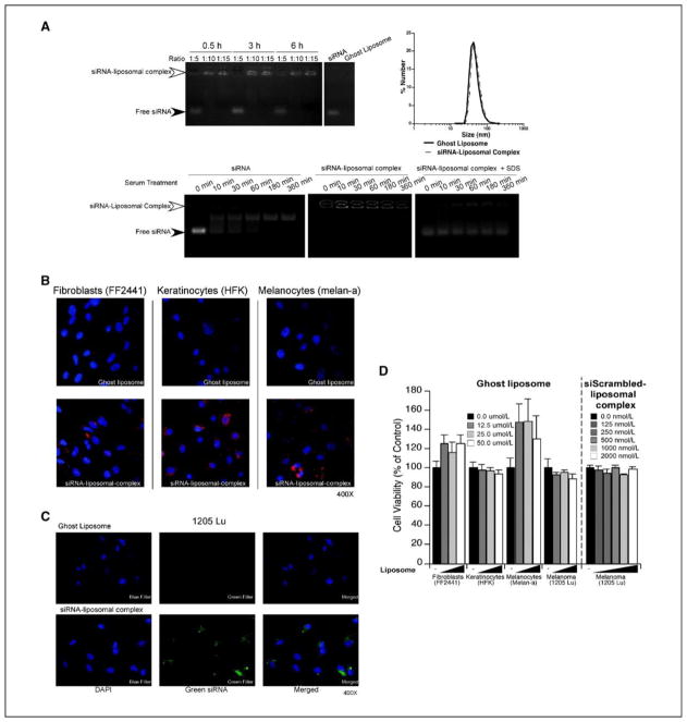
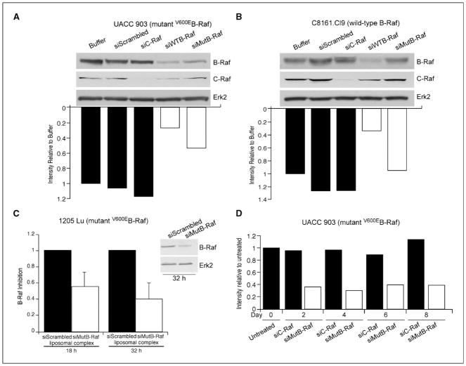
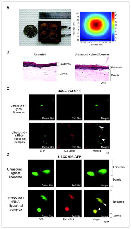
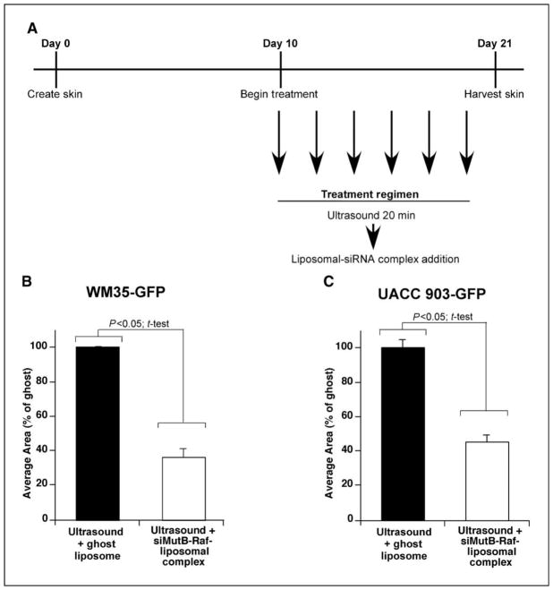
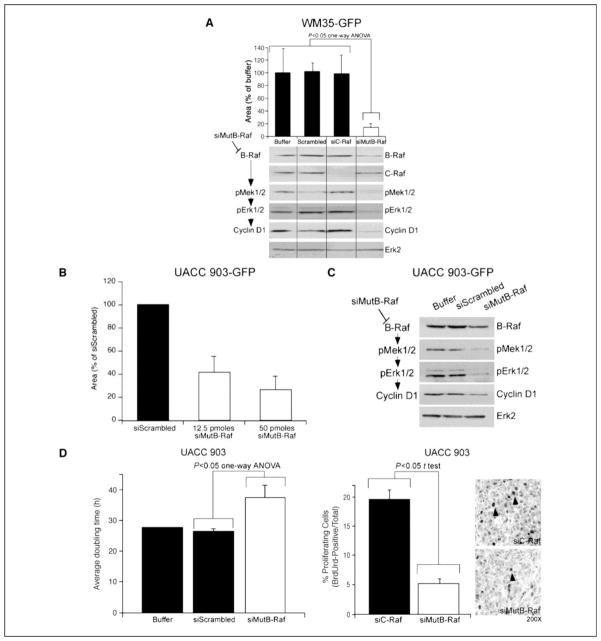
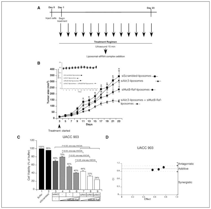
Most events promoting early melanoma development are yet to be identified, but deregulation of the B-Raf and Akt3 signaling cascades is an important regulator of this process. Approximately 90% of normal moles and approximately 60% of early invasive cutaneous melanomas contain a T1799A B-Raf mutation ((V600E)B-Raf), leading to 10 times higher enzyme activity and constitutive activation of the mitogen-activated protein kinase pathway. Furthermore, approximately 70% of melanomas have elevated Akt3 signaling due to increased gene copy number and PTEN loss. Therefore, targeting (V600E)B-Raf and Akt3 signaling is necessary to prevent or treat cutaneous melanocytic lesions. Agents specifically targeting these proteins are needed, having fewer side effects than those inhibiting both normal and mutant B-Raf protein or targeting all three Akt isoforms. In this study, a unique nanoliposomal-ultrasound-mediated approach has been developed for delivering small interfering RNA (siRNA) specifically targeting (V600E)B-Raf and Akt3 into melanocytic tumors present in skin to retard melanoma development. Novel cationic nanoliposomes stably encapsulate siRNA targeting (V600E)B-Raf or Akt3, providing protection from degradation and facilitating entry into melanoma cells to decrease expression of these proteins. Low-frequency ultrasound using a lightweight four-cymbal transducer array enables penetration of nanoliposomal-siRNA complex throughout the epidermal and dermal layers of laboratory-generated or animal skin. Nanoliposomal-mediated siRNA targeting of (V600E)B-Raf and Akt3 led to a cooperatively acting approximately 65% decrease in early or invasive cutaneous melanoma compared with inhibition of each singly with negligible associated systemic toxicity. Thus, cationic nanoliposomes loaded with siRNA targeting (V600E)B-Raf and Akt3 provide an effective approach for targeted inhibition of early or invasive cutaneous melanomas.


Figures

Figure 1
Figure 1. Characterization of cationic siRNA-liposome complexes
A. Loading of fluorescently tagged siRNA into nanoliposomes. Fluorescently tagged siRNA was complexed with cationic nanoliposomes at ratios of 1:5, 1:10, or 1:15, for 0.5, 3, or 6 h and run on a 2% agarose gel to determine loading efficiency. Maximal loading was reached at a 1:10 ratio following a 0.5-h incubation (upper left panel). siRNA at a 1:10 ratio with nanoliposomes were sized using dynamic light scattering and similar size ranges observed for ghost or nanoliposomes loaded with siRNA (upper right panel). siRNA protection by cationic nanoliposomes was measured by complexing fluorescent siRNA with nanoliposomes overnight followed by exposure to serum for 10, 30, 60, 180, or 360 m. Free fluorescent siRNA alone was used as a control (lower left & middle panels). Release of siRNA in nanoliposomes was accomplished by collapsing serum treated siRNA-nanoliposomal complexes with SDS to release free siRNA, which was then run on an agarose gel (lower right panel). B. siRNA-nanoliposomal complex is taken up into normal cells. Uptake of siRNA-nanoliposomal complex into normal cells was measured by adding fluorescently tagged siRNA-nanoliposomal complex (200 nM) to fibroblasts, keratinocytes, and melanocytes for 3 h followed by fixation and imaging using fluorescence microscopy (magnification; 400X). Ghost nanoliposomes lacking fluorescent siRNA were used as a control. C. siRNA-nanoliposomal complex is taken up into the cytoplasm of cells and is not merely surface bound. siRNA-nanoliposomal complex localization in cells following treatment with siRNA-nanoliposomal complexes (100 nM) was ascertained by exposing 1205 Lu melanoma cells for 3 h after which cells were trypsinized to remove surface bound complex and replated overnight onto coverslips. Cells were fixed and imaged using fluorescent microscopy (magnification; 400X). D. Ghost liposome or siRNA-nanoliposomal complex exerted negligible toxicity on normal or cancer cells. Cellular toxicity of ghost liposome and siRNA-nanoliposomal complex was measured by adding nanoliposomes (12.5, 25, and 50 μM) to fibroblasts, keratinocytes, melanocytes, or melanoma cells for 24 h followed by MTS assay analysis. Untreated cells (−) served as controls for comparison. Results are mean±SE
Figure 2
Figure 2. siMutB-Raf decreases protein expression of V600EB-Raf
A., B. siRNA can be designed to decrease expression of V600EB-Raf but not normal protein expression. To verify specificity of siMutB-Raf for decreasing expression of mutant but not wild-type protein, melanoma cells containing mutant (UACC 903) (A.) or normal (C8161.Cl9) (B.) B-Raf protein were nucleofected with buffer, siScrambled, siC-Raf, siWTB-Raf, or siMutB-Raf siRNA, protein lysates harvested 48 hours later, and analyzed by Western blot analysis for B-Raf and C-Raf knockdown. Erk2 served as a loading control. C. siMutB-Raf-nanoliposomal complex decreased expression of V600EB-Raf protein in cells. 1205 Lu cells were exposed to siMutB-Raf or siScrambled-nanoliposomal complex, protein lysates harvested at 18 and 32 hours and analyzed by Western blot analysis. Erk2 served as a control for protein loading. Densitometry results are mean±SE. D. Duration of V600EB-Raf protein knockdown following exposure to siRNA targeting mutant protein is beyond 8-d. Cells were nucleofected with C-Raf or V600EB-Raf siRNA, replated in culture dishes and protein harvested 2, 4, 6 and 8 d later to measure duration of protein knockdown. Untreated cells or cells nucleofected with siRNA targeting C-Raf served as controls.
Figure 3
Figure 3. Ultrasound treatment permeabilizes skin enabling melanocytic lesions to take up siRNA-nanoliposomal complexes
A. Ultrasound assembly. A lightweight, low-profile ultrasound array was constructed using four cymbal transducers, which were connected in parallel and encased in polymer. The temporal peak intensity was determined in a spatial plane 1 mm from the face of the transducer for exposure conditions. B. Ultrasound treatment does not damage skin. Laboratory generated skin was ultrasound treated followed by addition of ghost liposome, skin sectioned and H&E stained. No changes cellular structure or in skin morphology was observed compared to untreated control skin (magnification; 200X). C., D. Following ultrasound treatment of skin, siRNA-nanoliposomal complex is taken up by melanocytic lesions in the epidermis and at the epidermal-dermal junction. Following 20 m ultrasound treatment, fluorescent siRNA-nanoliposomal complex or ghost nanoliposomes were applied topically onto reconstructed skin. 1-h later, skin was fixed and analyzed using fluorescent microscopy looking down at the skin (Magnification; 4X) (C.), or by cross sections (magnification; 400X) (D.). Fluorescence (red) indicating presence of siRNA-nanoliposomal complex was evident in melanocytic lesions in both epidermis and at epidermal-dermal junction (arrows).
Figure 4
Figure 4. Ultrasound treatment followed by topical application of siMutB-Raf-nanoliposomal complex inhibits melanocytic lesion development in reconstructed skin
A. Schematic showing treatment regime. Beginning on d 10 and on alternate days thereafter up to d 20, reconstructed skin was treated with ultrasound for 20 m followed by topical administration of siMutB-Raf-nanoliposomal complex (100 pmoles) or ghost nanoliposomes. B., C. Ultrasound followed by addition of siMutB-Raf-nanoliposomal complex decreases melanocytic lesion development in skin. Reconstructed skin containing UACC 903-GFP or WM35-GFP cells were treated with ultrasound for 20 m followed by topical administration of siMutB-Raf-nanoliposomal complex on alternated days from d 10–20. Skin was harvested on d 21, and average area occupied by GFP-tagged tumors calculated for each group. Ultrasound treatments followed by exposure to ghost nanoliposomes served as a control. Results are mean±SE.
Figure 5
Figure 5. siMutB-Raf inhibits melanocytic lesion growth in reconstructed skin
A. Targeting melanocytic lesions using siRNA against V600EB-Raf decreases melanocytic lesion development in laboratory-generated skin. Effectiveness of siRNA targeting V600EB-Raf for decreasing cutaneous tumor development was established by nucleofecting GFP-tagged WM35 cells with buffer, scrambled siRNA or siRNA targeting C-Raf, or V600EB-Raf (100 pmoles). Cells were then seeded into laboratory-generated skin at time of creation and 10 d later, average area occupied by green melanocytic lesions quantified. A statistically significant reduction in green fluorescent lesions was observed following siMutB-Raf treatment (p<0.05; One-Way ANOVA) (upper panel). Results are mean±SE. Protein lysates harvested from cells were analyzed by Western blot for B-Raf, C-Raf, p-Mek1/2, pErk1/2, and Cyclin D1 protein expression. Erk2 served as a control for protein loading (lower panel). B. siRNA-mediated inhibition of V600EB-Raf protein expression in GFP-tagged UACC 903 cells decreases lesion formation in skin reconstrtucts. UACC 903-GFP cells were nucleofected with siScrambled or siB-Raf (12.5 or 50 pmoles) and cells seeded into laboratory generated skin at time of creation. Reconstructed skin was analyzed using fluorescence microscopy 10 d later and area occupied by developing GFP lesions quantified. Results are mean±SE. C. Inhibition of V600EB-Raf decreased MAP kinase signaling in UACC 903-GFP cells. UACC 903-GFP cells were nucleofected with buffer, siScrambled or siB-Raf (50 pmoles) and harvested at 48 h for Western blot analysis. Westerns were probed with B-Raf, p-Mek1/2, p-Erk1/2, and cyclin D1 to show decreased MAP kinase pathway signaling. Erk2 served as a loading control. D. Mechanistically, siRNA-mediated targeting of V600EB-Raf protein decreased the proliferative capacity of cells. Cultured UACC 903 melanoma cells treated with siMutB-Raf had an increased doubling time indicating cells were proliferating at a slower rate (left panel). Quantifying proliferating cells showed a 2–3 fold decrease following siMutB-Raf treatment of tumor cells in size and time matched tumor controls treated with siRNA to C-Raf (middle and right panels). Results are mean±SE.
Figure 6
Figure 6. Ultrasound treatment followed by topical application of siMutB-Raf-nanoliposomal complex alone or in combination with siAkt3-nanoliposomal complex inhibits melanocytic lesion development in animal skin
A. Schematic showing treatment regime. Ultrasound treatment followed by topical application of siMutB-Raf-nanoliposomal complexes started the day after injection of melanoma cells and continued on alternate days to day 23. During the procedure, anesthetized mice were treated with ultrasound at the injection site for 15 m followed by topical application of siMutB-Raf-nanoliposomal complex. B. Ultrasound treatment followed by topical application of siAkt3-liposomal complex + siMutB-Raf-liposomal complex decreased melanoma development in animal skin. UACC 903-GFP cells (1×10⁶), were injected subcutaneously into nude mice and after 24 h, tumors forming at injection sites were treated on alternate days with ultrasound for 15 m followed by topical administration of siMutB-Raf-liposomal complex, siAkt3-liposomal complex, or siAkt3-liposomal complex + siMutB-Raf liposomal complex. Tumors were measured on alternate days beginning on d-3. Control mice were ultrasound treated followed by addition of siScrambled-liposomal complex. Ghost liposomes were added to single treatments so that mice were treated with equivalent amounts of liposomal vehicle. Statistically significant differences between control and siMutB-Raf-liposomal complex + siAkt3-liposomal complex treated tumors were observed beginning on day 11 (Two-Way ANOVA; p<0.05). Results are mean±SE. Ultrasound treatment followed by topical application of siAkt3-liposomal complex + siMutB-Raf-liposomal complex does not cause significant change in animal body weight. Animal weights were measured on alternate days beginning on d 1 to determine whether any weight-related toxicity occurred. No significant weight loss was observed between control and experimental groups (Two-Way ANOVA; p>0.05) (B. inset). Results are mean±SE. C. siMutB-Raf and siAkt3 cooperate to reduce anchorage independent growth in cell culture. UACC 903 cells were nucleofected with siAkt3 (200 pmoles) and siMutB-Raf (1.5, 3, 6, or 12 pmoles) in combination and compared to single siRNAs to siAkt3 (200 pmoles), siMutB-Raf (1.5, 3, 6, or 12 pmoles), siScrambled, or Buffer only for the ability to inhibit anchorage independent growth. D. siAkt3 and siMutB-Raf act additively to inhibit cell viability. Calculation of the CI index for the combination of siAkt3 and siMutB-Raf showed additive inhibition of cell viability with CI values between 0.94 and 1.10.

Similar articles

Cited by

References

    1. Satyamoorthy K, Herlyn M. Cellular and molecular biology of human melanoma. Cancer Biol Ther. 2002;1:14–7. - PubMed
    1. Brose MS, Volpe P, Feldman M, et al. BRAF and RAS mutations in human lung cancer and melanoma. Cancer Res. 2002;62:6997–7000. - PubMed
    1. Davies H, Bignell GR, Cox C, et al. Mutations of the BRAF gene in human cancer. Nature. 2002;417:949–54. - PubMed
    1. Yazdi AS, Palmedo G, Flaig MJ, et al. Mutations of the BRAF gene in benign and malignant melanocytic lesions. J Invest Dermatol. 2003;121:1160–2. - PubMed
    1. Miller CJ, Cheung M, Sharma A, et al. Method of mutation analysis may contribute to discrepancies in reports of (V599E)BRAF mutation frequencies in melanocytic neoplasms. J Invest Dermatol. 2004;123:990–2. - PubMed

Publication types

MeSH terms