. 2008 Nov;82(22):11383-97.
doi: 10.1128/JVI.01293-08. Epub 2008 Sep 10.

Internal deletions of IE2 86 and loss of the late IE2 60 and IE2 40 proteins encoded by human cytomegalovirus affect the levels of UL84 protein but not the amount of UL84 mRNA or the loading and distribution of the mRNA on polysomes

Affiliations

Internal deletions of IE2 86 and loss of the late IE2 60 and IE2 40 proteins encoded by human cytomegalovirus affect the levels of UL84 protein but not the amount of UL84 mRNA or the loading and distribution of the mRNA on polysomes

Rebecca L Sanders et al. J Virol. 2008 Nov.

Abstract

The major immediate-early (IE) region of human cytomegalovirus encodes two IE proteins, IE1 72 and IE2 86, that are translated from alternatively spliced transcripts that differ in their 3' ends. Two other proteins that correspond to the C-terminal region of IE2 86, IE2 60 and IE2 40, are expressed at late times. In this study, we used IE2 mutant viruses to examine the mechanism by which IE2 86, IE2 60, and IE2 40 affect the expression of a viral DNA replication factor, UL84. Deletion of amino acids (aa) 136 to 290 of IE2 86 results in a significant decrease in UL84 protein during the infection. This loss of UL84 is both proteasome and calpain independent, and the stability of the protein in the context of infection with the mutant remains unaffected. The RNA for UL84 is expressed to normal levels in the mutant virus-infected cells, as are the RNAs for two other proteins encoded by this region, UL85 and UL86. Moreover, nuclear-to-cytoplasmic transport and the distribution of the UL84 mRNA on polysomes are unaffected. A region between aa 290 and 369 of IE2 86 contributes to the UL84-IE2 86 interaction in vivo and in vitro. IE2 86, IE2 60, and IE2 40 are each able to interact with UL84 in the mutant-infected cells, suggesting that these interactions may be important for the roles of UL84 and the IE2 proteins. Thus, these data have defined the contribution of IE2 86, IE2 60, and IE2 40 to the efficient expression of UL84 throughout the infection.


Figures

FIG. 1.
FIG. 1.
Schematic representation of the UL122-123 transcripts and deletions created within the IE2 86 coding regions of mutant viruses. (A) UL122-123 transcripts and representation of the MIE genes, IE1 72 and IE2 86, in the wild-type HCMV coding region. Exons 1, 2, 3, and 4 specify IE1 72 mRNA, while exons 1, 2, 3, and 5 specify IE2 86 mRNA. Translation begins at exon 2 for both IE1 72 and IE2 86, while translation of IE2 60 and IE2 40 begins at aa 170 and 242, respectively. (B) Schematic of the mutant viruses used in these analyses to identify the dysregulation of UL84, as well as the important binding domain within IE2 86 for UL84. The WT, IE2 86ΔSX (ΔSX), and a revertant form of the IE2 86ΔSX (REV) are represented in the schematic. Other deletion mutants created, including Δ88-290, Δ88-135, IE2 Δ40, and M170L, are also shown. Diagrams are not to scale.
FIG. 2.
FIG. 2.
Analysis of UL84, UL85, UL86, and UL83 RNA and protein at late times postinfection. (A) G0-synchronized HFFs were infected at an MOI of 5 PFU/cell with the WT-EGFP (WT), IE2 86ΔSX-EGFP (SX), or Rev-EGFP (Rev) viruses or were mock infected (M) and harvested at the indicated times postinfection (12 to 96 h). The mock lane was added to indicate that the UL84 antibody was specific for the viral protein. An equal amount of protein from the cell lysates was analyzed by Western blot analyses. β-Actin was used as a loading control. (B) Analysis of the UL84, UL85, and UL86 transcripts in the IE2 86ΔSX-EGFP infection. HFFs were infected or mock infected (M) at an MOI of 5 PFU/cell with the WT-EGFP, IE2 86ΔSX-EGFP, or Rev-EGFP viruses and then harvested at either 24 or 96 h p.i. mRNA was oligo(dT) selected, resolved by agarose gel electrophoresis, and then transferred to a nitrocellulose membrane. 32P-labeled probes were synthesized that recognize UL84, UL85, and UL86 or β-actin. Following hybridization of the probe, membranes were exposed to film for autoradiography. The three transcripts occurring at 2.1, 3.0, and 9.0 kb encode the UL84, UL85, and UL86 RNA transcripts, respectively. Cellular β-actin mRNA served as a loading control. A representative example of three experiments is shown. (C) The same mRNA as for panel B was analyzed by Northern blotting for expression of the UL83 mRNA transcript at 96 h p.i., when UL83 is most abundant. Both the WT-EGFP and IE2 86ΔSX-EGFP virus-infected cells are shown. The UL84 and UL83 protein from this infection is also shown for comparison at 96 h p.i. for the two viruses. (D) Western blot assay results for the UL84, UL85, and UL86 proteins at 72, 96, and 120 h p.i. are shown for the WT-EGFP and IE2 86ΔSX-EGFP viruses. Actin served as a loading control.
FIG. 3.
FIG. 3.
Nuclear-cytoplasmic distribution of UL84 RNA in WT and IE2 86ΔSX virus infection. At 24 h p.i., cells were harvested and the nuclear and cytoplasmic fractions were separated by centrifugation. RNA was prepared from each fraction and quantitated by quantitative real-time RT-PCR. The percent nuclear versus cytoplasmic was calculated for each sample set. The nuclear fraction (Nuc) and cytoplasmic fraction (Cyto) are shown for both the WT and IE2 86ΔSX (ΔSX) virus-infected cells. Each fraction was normalized to a cellular housekeeping gene (G6PD) to account for the amount of RNA in each reaction mixture. Averages of three experiments are shown, with error bars representing the margins of error between experiments.
FIG. 4.
FIG. 4.
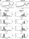
Polysome distribution of UL84, IE2 86, UL44, and G6PD RNAs in the WT and IE2 86ΔSX (ΔSX) infections. (A) Absorbance measured during fractionation from the heaviest to the lightest fractions. Fractions were pooled into six separate samples (fractions 1 to 6) and are represented in the figure. Absorbance (A254) was measured throughout the collection. The polysomal fractions are represented in fractions 1, 2, and 3. The monosomal fractions are represented in fractions 4 and 5. Fraction 6 represents the free RNP RNA. (B) The distribution of the RNA and quantification of each fraction is shown for UL84, IE2 86, UL44, and G6PD. Samples were either treated with EDTA (+EDTA) or mock treated (-EDTA). Quantified RNA was measured by real-time RT-PCR and then normalized to the total amount of RNA collected in that fraction. Relative amounts of RNA are shown compared to the total WT RNA present in fraction 6 for each gene, which was set to a value of 1.
FIG. 5.
FIG. 5.
Analysis of proteasome- and calpain-dependent degradation in IE2 86ΔSX- and WT-infected cells and assessment of the stability of UL84 and IE2 86 proteins in these infections. (A) HFFs were infected at an MOI of 5 PFU/cell with either the WT-EGFP or IE2 86ΔSX-EGFP (SX). Lactacystin (10 μM; Lac, +), or DMSO for mock treatment (-), was added at either 18 or 90 h p.i., and the cells were incubated for 6 h. Cells were harvested at 24 and 96 h p.i. and then analyzed for UL84 protein expression by Western blot analyses. (B) HFFs were infected as above and then treated with calpeptin (50 μM; Cal, +) for 6 h or mock treated with DMSO (-). Cells were harvested and then lysates were analyzed by Western blotting for UL84 protein expression. (C) Cells were infected at an MOI of 1 PFU/cell with the WT or IE2 86ΔSX (SX) and at 36 h p.i. were either harvested to assess protein concentration before drug treatment or were treated with cycloheximide (100 μg/ml; CHX, +) or DMSO (−) as a control for 6 h. Following treatment, cells were harvested at 42 h p.i. to analyze the loss of UL84 and IE2 86 during that time. Two exposures of the IE2 86 blot are shown in order to visualize the small amount of IE2 86 in the IE2 86ΔSX infection (*). β-Actin was analyzed in all of the above experiments in order to assess protein loading.
FIG. 6.
FIG. 6.
UL84 interacts with IE2 60 and IE2 40 without the contribution of IE2 86. (A) Schematic representation of the experiment, indicating that the post-CH16.0 IP product (step 1) is the same sample as that used for the pre-IP in the mIE2 86 IPs (step 2). The corresponding post and pre samples are denoted with a * in all subsequent parts of the figure. (B) IPs were carried out exactly as described for panel A, and cells were harvested 24 to 96 h p.i. The pre- (lanes 1 to 3) and post- (lanes 7 to 9) IP samples equal approximately 10% of the IP (lanes 4 to 6). (C) The post-binding supernatant (in panel A, this corresponds to Post*, lanes 7 to 9) was subjected to a subsequent IP with an antibody that recognizes IE2 86, IE2 60, and IE2 40 (mIE2 86) to determine if UL84 interacts with IE2 60 and IE2 40 in combination, without the presence of IE2 86. UL84 was analyzed again by Western blotting (lanes 1 to 9). PRE* indicates that this corresponds to the POST* supernatant of the first IP in panel B. The second set of IPs (mIE2 86 IPs) were also analyzed by Western blotting for the presence IE2 86, IE2 60, and IE2 40 using the mIE2 86 antibody (lanes 10 to 18). Only the samples at 72 and 96 h p.i. of the Western blot assay with the mIE2 86 antibody are shown, as IE2 86, IE2 60, and IE2 40 were most abundant at these times. A representative of two experiments is shown.
FIG. 7.
FIG. 7.
Sequence and characterization of the M170L mutant. (A) A portion of the IE2 86 coding region is shown. The mutated initiator methionine (M170, originally ATG) for IE2 60 was changed to a leucine (CTC) to create this mutant virus (M170L). (B) Expression of IE2 86, IE2 60, and IE2 40 in the M170L mutant compared to the WT and the M170L revertant (M170L R) at 24 to 96 h p.i. in HFFs infected at an MOI of 3 PFU/cell. A mock-infected sample (M) is also shown. Western blot analyses were used to examine expression of UL84 as well. Cellular β-actin was used as a control for protein loading.
FIG. 8.
FIG. 8.
IE2 60 and IE2 40 interact individually with UL84 in the absence of IE2 86. As in Fig. 6, sequential IPs were performed, with the exception of the mutant viruses used, which were the IE2 Δ40 and M170L mutants, and the time points examined (only 96 h p.i. is shown here). At 96 h p.i., cells were harvested and the first IP (lanes 2, 5, and 8) was carried out using the CH16.0 antibody. Western blot analyses were performed to assess the interactions occurring and expression patterns of UL84, IE2 86, IE2 60, and IE2 40. The supernatants following IP (POST*, lanes 3, 6, and 9) were used for the next immunoprecipitation (mIE2 86 IPs, lanes 10 to 18). POST* (lanes 3, 6, and 9) and PRE* (lanes 10, 13, and 16) indicate that the post-binding supernatant for the first IP was used for the pre-binding sample for the second IP. The pre and post samples represent approximately 10% of the IP. The interaction of UL84 and IE2 86 were assessed in lanes 2, 5, and 8, while the interactions between UL84, IE2 60, and IE2 40 were assessed in lanes 11, 14, and 17. Lanes 14 and 17 represent the individual interactions with UL84 and IE2 60 or IE2 40, respectively. The arrow indicates the sequential nature of the IPs from the initial CH16.0 IPs to the mIE2 86 IPs.
FIG. 9.
FIG. 9.
Identification of a C-terminal domain involved in the interaction between IE2 86 and UL84. (A) HFFs were infected at an MOI of 3 PFU/cell with the WT, Δ88-135, or Δ88-290 viruses and harvested at either 72 or 96 h p.i. Lysates were prepared, a small aliquot was removed (PRE), and the remainder was incubated with beads conjugated with the CH16.0 antibody (which recognizes IE1 72 and IE2 86). Following incubation, an aliquot of the supernatant was removed (POST). The beads were then washed and IP products were eluted from the beads (IP). The PRE and POST lanes represent 10% of the protein loaded in the IP lanes. UL84 was analyzed by Western blotting as described in Materials and Methods. (B) Schematic of the IE2 86 coding region and all of the mutated forms assayed. Amino acid deletions: SX, Δ136-290; MN, Δ85-369; MX, Δ85-290; TM293, Δ290-579; XN, Δ290-369; MX364, Δ85-290 and Δ369-579; STOP, Δ85-579. (C) UL84 and IE2 86 binding was assessed as described in Materials and Methods. Eluted protein was analyzed by agarose gel electrophoresis, and the amount of UL84 bound was analyzed by autoradiography. The input was 10% of bound reaction mixtures. All mutants are described above. GST alone was used as a negative control for nonspecific binding. A representative of four experiments is shown.

Similar articles

Cited by

References

    1. Ahn, J. H., C. J. Chiou, and G. S. Hayward. 1998. Evaluation and mapping of the DNA binding and oligomerization domains of the IE2 regulatory protein of human cytomegalovirus using yeast one and two hybrid interaction assays. Gene 21025-36. - PubMed
    1. Ahn, J. H., Y. Xu, W. J. Jang, M. J. Matunis, and G. S. Hayward. 2001. Evaluation of interactions of human cytomegalovirus immediate-early IE2 regulatory protein with small ubiquitin-like modifiers and their conjugation enzyme Ubc9. J. Virol. 753859-3872. - PMC - PubMed
    1. Andreoni, K. A., X. Wang, S. M. Huang, and E. S. Huang. 2002. Human cytomegalovirus hyperimmune globulin not only neutralizes HCMV infectivity, but also inhibits HCMV-induced intracellular NF-κB, Sp1, and PI3-K signaling pathways. J. Med. Virol. 6733-40. - PubMed
    1. Barrasa, M. I., N. Harel, Y. Yu, and J. C. Alwine. 2003. Strain variations in single amino acids of the 86-kilodalton human cytomegalovirus major immediate-early protein (IE2) affect its functional and biochemical properties: implications of dynamic protein conformation. J. Virol. 774760-4772. - PMC - PubMed
    1. Barrasa, M. I., N. Y. Harel, and J. C. Alwine. 2005. The phosphorylation status of the serine-rich region of the human cytomegalovirus 86-kilodalton major immediate-early protein IE2/IEP86 affects temporal viral gene expression. J. Virol. 791428-1437. - PMC - PubMed

Publication types

LinkOut - more resources