. 2008 Oct;118(10):3440-52.
doi: 10.1172/JCI34721.

TNF-alpha and TLR agonists increase susceptibility to HIV-1 transmission by human Langerhans cells ex vivo

Affiliations

TNF-alpha and TLR agonists increase susceptibility to HIV-1 transmission by human Langerhans cells ex vivo

Marein A W P de Jong et al. J Clin Invest. 2008 Oct.

Abstract

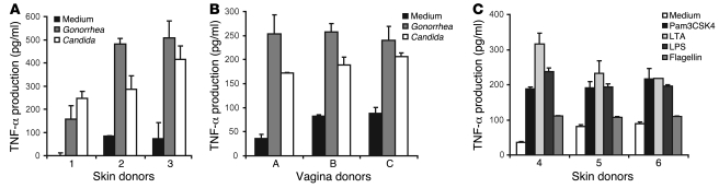
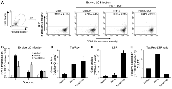
Genital coinfections increase an individual's risk of becoming infected with HIV-1 by sexual contact. Several mechanisms have been proposed to explain this, such as the presence of ulceration and bleeding caused by the coinfecting pathogen. Here we demonstrate that Langerhans cells (LCs) are involved in the increased susceptibility to HIV-1 in the presence of genital coinfections. Although LCs are a target for HIV-1 infection in genital tissues, we found that immature LCs did not efficiently mediate HIV-1 transmission in an ex vivo human skin explant model. However, the inflammatory stimuli TNF-alpha and Pam3CysSerLys4 (Pam3CSK4), the ligand for the TLR1/TLR2 heterodimer, strongly increased HIV-1 transmission by LCs through distinct mechanisms. TNF-alpha enhanced transmission by increasing HIV-1 replication in LCs, whereas Pam3CSK4 acted by increasing LC capture of HIV-1 and subsequent trans-infection of T cells. Genital infections such as Candida albicans and Neisseria gonorrhea not only triggered TLRs but also induced TNF-alpha production in vaginal and skin explants. Thus, during coinfection, LCs could be directly activated by pathogenic structures and indirectly activated by inflammatory factors, thereby increasing the risk of acquiring HIV-1. Our data demonstrate a decisive role for LCs in HIV-1 transmission during genital coinfections and suggest antiinflammatory therapies as potential strategies to prevent HIV-1 transmission.


Figures

Figure 1
Figure 1. An ex vivo skin model to investigate the effect of inflammation on HIV-1 transmission is shown.
(AC) Epidermal sheets were floated on medium in a 24-well plate and where indicated stimulated with different stimuli. After 6 hours the sheets were inoculated with HIV-1–eGFP. After 3 days, the epidermal sheets were removed and CCR5+ Jurkat T cells were added for an additional 7 days. Filled dendritic forms indicate LCs; open dendritic forms indicate GFP+-infected LCs; filled circles indicate CCR5+ Jurkat T cells; open circles indicate GFP+-infected T cells. (B) The migrated epidermal cells (day 3) were stained with antibodies against CD1a, Langerin, CD86, and CD3 and analyzed by flow cytometry to determine their phenotype and HIV-1 infection. Gates are based on isotype control (Supplemental Figure 1). R1 is gated on the larger cells in the population. Open histograms represent isotype-control; filled histogram specific antibody staining; the mean ± SD of the specific staining is depicted in the upper-right corner. The percentage of GFP+, CD3+, and CD1a+ cells ± SD is depicted in the upper-right corner of the dot plots. (C) Samples of the cocultures (day 5, 7, and 10) that were not stimulated with any stimulus were analyzed for GFP expression by flow cytometry. The percentage of GFP+ cells ± SD is depicted in the upper-right corner. As control for HIV-1 infection, the same concentration of HIV-1–eGFP was added to wells without epidermal sheets and processed similarly as the other conditions.
Figure 2
Figure 2. Different pathogens and pathogenic ligands induce TNF-α production in skin and vaginal biopsies.
Skin or vagina epithelial biopsies were treated with Candida albicans, Neisseria gonorrhea (A and B), Pam3CSK4, LTA, LPS, or flagellin (C). After 24 hours, the supernatant was collected, and TNF-α production was measured by ELISA. Error bars represent the mean ± SD of duplicates. For each panel, the different donors were measured in 2 independent experiments.
Figure 3
Figure 3. TNF-α and Pam3CSK4 enhance HIV-1 transmission ex vivo.
(AD) Epidermal sheets were stimulated with TNF-α, Pam3CSK4, LTA, LPS, or flagellin for 6 hours, and ex vivo transmission was determined as in Figure 1. (A) Cocultures of donors number 1–4 (day 7) were analyzed for GFP expression by flow cytometry. HIV-1 transmission is depicted as percentage of CCR5+ Jurkat T cells positive for GFP expression. Error bars represent the mean ± SD of duplicates. (B) Results obtained with donors number 1–8 are depicted, and the values were analyzed for statistical differences by ANOVA (**P < 0.01, ***P < 0.001 versus the no treatment condition). (C) Before addition of the CCR5+ Jurkat T cells the migrated cells were extensively washed. At day 7, supernatant was collected, and viral content was monitored by p24 ELISA. A representative experiment of 2 is depicted. (D) To analyze donor differences, 4 different skin donors were incubated with TNF-α, LTA, LPS, or flagellin at 2 different days, and ex vivo transmission was determined as described in Figure 1. The samples were measured for 13 days. Error bars represent the mean ± SD of duplicates.
Figure 4
Figure 4. TNF-α and Pam3CSK4 enhance HIV-1 transmission ex vivo.
(A) Epidermal single-cell suspensions were stimulated with TNF-α and Pam3CSK4, and after 2 hours, the cells were inoculated with HIV-1–eGFP. After 2 hours the cells were washed and CCR5+ Jurkat T cells were added for 7 days and analyzed for GFP expression by flow cytometry. A representative experiment out of 2 donors is depicted. (B) Epidermal sheets were stimulated with TNF-α, Pam3CSK4, LTA, LPS, or flagellin for 6 hours, and ex vivo transmission was determined as in Figures 1 and 3. A part of the migrated cells was extensively washed before addition of the CCR5+ Jurkat T cells (right panel). HIV-1 transmission is depicted as percentage of CCR5+ Jurkat T cells positive for GFP expression. Error bars represent the mean ± SD of duplicates. A representative experiment out of 2 donors is depicted. (C) Epidermal sheets were stimulated with TNF-α and Pam3CSK4 before incubation with HIV-1–eGFP. After 3 days, the migrated cells were washed and LCs were isolated by CD1a-selection using magnetic beads. The CD1a+ and CD1a fraction were added to CCR5+ Jurkat T cells. The cocultures were monitored by flow cytometry at different days. Error bars represent the mean ± SD of duplicates. A representative experiment out of 2 is shown.
Figure 5
Figure 5. TNF-α and Pam3CSK4 enhance HIV-1 infection.
(A and B) Epidermal sheets were stimulated with TNF-α or Pam3CSK4. After 6 hours, the sheets were inoculated with HIV-1–eGFP. After 3 days, the epidermal sheets were removed, the migrated cells were harvested, stained for CD86, and subsequently analyzed for GFP expression by flow cytometry. (A) Infection is depicted in dot plots. The percentage of GFP+ cells ± SD is depicted in the upper-right corner. (B) HIV-1 infection is depicted as percentage of cells positive for GFP expression. Error bars represent the mean ± SD of duplicates. The 3 depicted donors have been measured in 2 independent experiments. (CE) Epidermal single-cells suspensions were stimulated with TNF-α or Pam3CSK4 for 30 minutes and inoculated with NL4.3-BaL. After 6 hours, the cells were washed and analyzed by quantitative real-time PCR analysis for HIV-1 replication by Tat/Rev transcripts and viral uptake by full-length viral RNAs (LTR). The Ct values were normalized for cellular GAPDH, and relative mRNA expression of HIV-1–inoculated medium control samples was set at 1. (C) Transcription per viral copy in cell was determined by the ratio of Tat/Rev mRNA and full-length viral RNA. A representative experiment out of 4 donors is depicted. Error bars represent the mean ± SD of duplicates.
Figure 6
Figure 6. Pam3CSK4 enhances trans-infection of HIV-1.
(A and B) Epidermal sheets or (CE) epidermal single-cells suspensions were stimulated with TNF-α or Pam3CSK4 (E) after replication-competent HIV-1–eGFP to show where the virus is used. After 6 hours the sheets/cells were inoculated with replication-competent HIV-1–eGFP or single-cycle HIV-1–eGFP (HIV-1 NL4.3-eGFPΔ envelope pseudotyped with the BaL envelope). After 3 days, the epidermal sheets were removed or the cell suspensions were washed and CCR5+ Jurkat T cells were added. At day 5, the cocultures were analyzed for GFP expression by flow cytometry. (A) The response to different virus doses is depicted. (B and D) The mean of the responses of different donors are depicted, the values were analyzed by ANOVA for statistical differences. **P < 0.01. Error bars represent the mean ± SD of duplicates.
Figure 7
Figure 7. Pam3CSK4 increases HIV-1 capture and primarily transmits cell-bound HIV-1.
(A and B) Emigrant LCs were stimulated with Pam3CSK4 for 1 hour and inoculated with NL4.3 BaL. After 2 hours, cells were extensively washed. The cells were fixed, permeabilized, and stained for the LC-marker CD1a and HIV-1 p24. The cells were counterstained with isotype-specific Alexa antibodies (red, HIV-1 p24; green, CD1a). Single HIV-1 particles are indicated with arrows. Original magnification, ×630. A representative experiment out of 2 is depicted. (C and D) Epidermal single-cells suspensions were stimulated with Pam3CSK4. After 6 hours, cells were inoculated with single-cycle HIV-1. After 2 hours, the cell suspensions were washed, and subsequently cells were treated with trypsin at 37°C to remove cell-bound HIV-1 or a PBS control (C), or with HIV-1 neutralizing antibody b12 at 4°C to neutralize cell-bound, but not internalized, HIV-1 and an isotype control (D). Cells were washed and CCR5+ Jurkat T cells were added. At day 5, the cocultures were analyzed for GFP expression by flow cytometry. A representative experiment out of 3 is depicted. Errors bars represent the SD of duplicates. (E) Epidermal single-cells suspension was stimulated with TNF-α or Pam3CSK4 before being inoculated with different concentrations of HIV-1–eGFP. After 2 hours, the cell suspensions were washed, and CCR5+ Jurkat T cells were added. HIV-1 transmission was followed by flow cytometry. A representative experiment out of 2 donors is depicted. Error bars represent the mean ± SD of duplicates. TCID, tissue-culture infectious dose.

Similar articles

Cited by

References

    1. Joint United Nations Programme on HIV/AIDS and World Health Organisation 2007. 2007 AIDS epidemic update. http://www.unaids.org .
    1. Royce R.A., Sena A., Cates W., Jr., Cohen M.S. Sexual transmission of HIV. N. Engl. J. Med. 1997;336:1072–1078. doi: 10.1056/NEJM199704103361507. - DOI - PubMed
    1. Ito A., et al. Monoclonal antibody (5F3) defining renal cell carcinoma-associated antigen disialosyl globopentaosylceramide (V3NeuAcIV6NeuAcGb5), and distribution pattern of the antigen in tumor and normal tissues. Glycoconj. J. 2001;18:475–485. doi: 10.1023/A:1016281002344. - DOI - PubMed
    1. Naarding M.A., et al. Lewis X component in human milk binds DC-SIGN and inhibits HIV-1 transfer to CD4+ T lymphocytes. J. Clin. Invest. 2005;115:3256–3264. doi: 10.1172/JCI25105. - DOI - PMC - PubMed
    1. Munch J., et al. Semen-derived amyloid fibrils drastically enhance HIV infection. Cell. 2007;131:1059–1071. doi: 10.1016/j.cell.2007.10.014. - DOI - PubMed

Publication types

MeSH terms