

What does the anatomical organization of the entorhinal cortex tell us?
- PMID: 18769556
- PMCID: PMC2526269
- DOI: 10.1155/2008/381243
What does the anatomical organization of the entorhinal cortex tell us?
Abstract
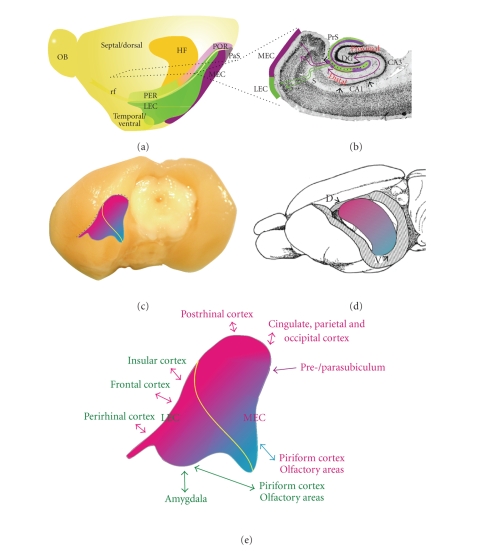
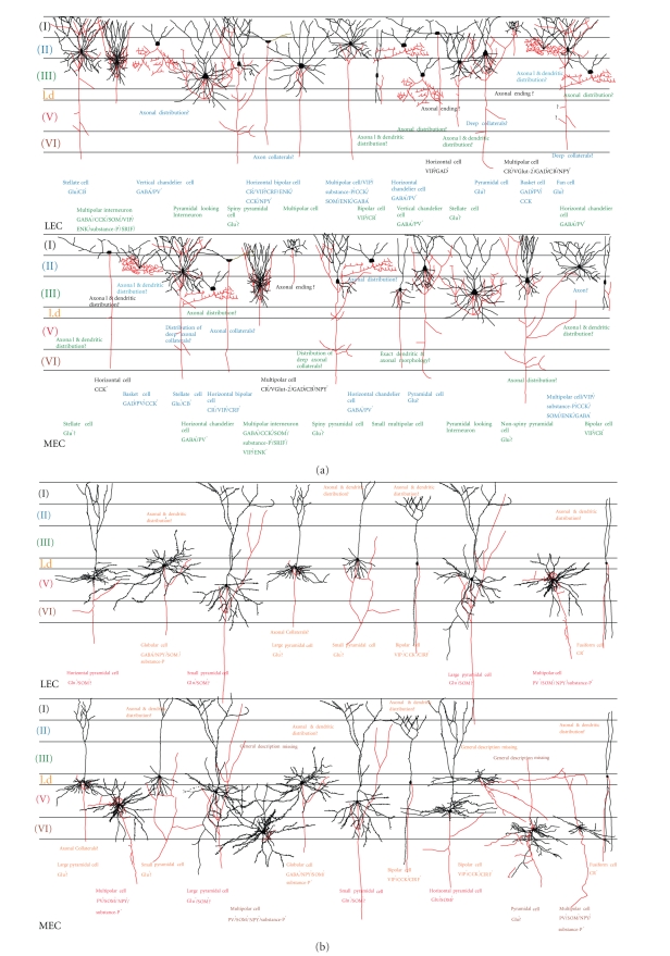
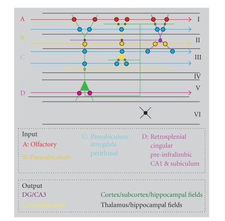
The entorhinal cortex is commonly perceived as a major input and output structure of the hippocampal formation, entertaining the role of the nodal point of cortico-hippocampal circuits. Superficial layers receive convergent cortical information, which is relayed to structures in the hippocampus, and hippocampal output reaches deep layers of entorhinal cortex, that project back to the cortex. The finding of the grid cells in all layers and reports on interactions between deep and superficial layers indicate that this rather simplistic perception may be at fault. Therefore, an integrative approach on the entorhinal cortex, that takes into account recent additions to our knowledge database on entorhinal connectivity, is timely. We argue that layers in entorhinal cortex show different functional characteristics most likely not on the basis of strikingly different inputs or outputs, but much more likely on the basis of differences in intrinsic organization, combined with very specific sets of inputs. Here, we aim to summarize recent anatomical data supporting the notion that the traditional description of the entorhinal cortex as a layered input-output structure for the hippocampal formation does not give the deserved credit to what this structure might be contributing to the overall functions of cortico-hippocampal networks.
Figures
Similar articles
-
Anatomical organization of the parahippocampal-hippocampal network.Ann N Y Acad Sci. 2000 Jun;911:1-24. doi: 10.1111/j.1749-6632.2000.tb06716.x. Ann N Y Acad Sci. 2000. PMID: 10911864 Review.
-
Processing of Hippocampal Network Activity in the Receiver Network of the Medial Entorhinal Cortex Layer V.J Neurosci. 2020 Oct 28;40(44):8413-8425. doi: 10.1523/JNEUROSCI.0586-20.2020. Epub 2020 Sep 25. J Neurosci. 2020. PMID: 32978288 Free PMC article.
-
Intrinsic Projections of Layer Vb Neurons to Layers Va, III, and II in the Lateral and Medial Entorhinal Cortex of the Rat.Cell Rep. 2018 Jul 3;24(1):107-116. doi: 10.1016/j.celrep.2018.06.014. Cell Rep. 2018. PMID: 29972772
-
Neurons and networks in the entorhinal cortex: A reappraisal of the lateral and medial entorhinal subdivisions mediating parallel cortical pathways.Hippocampus. 2019 Dec;29(12):1238-1254. doi: 10.1002/hipo.23145. Epub 2019 Aug 13. Hippocampus. 2019. PMID: 31408260 Review.
-
Entorhinal cortex of the monkey: IV. Topographical and laminar organization of cortical afferents.J Comp Neurol. 2008 Aug 20;509(6):608-41. doi: 10.1002/cne.21753. J Comp Neurol. 2008. PMID: 18551518 Free PMC article.
Cited by
-
Propagation of tau pathology in a model of early Alzheimer's disease.Neuron. 2012 Feb 23;73(4):685-97. doi: 10.1016/j.neuron.2011.11.033. Neuron. 2012. PMID: 22365544 Free PMC article.
-
G9a/GLP histone lysine dimethyltransferase complex activity in the hippocampus and the entorhinal cortex is required for gene activation and silencing during memory consolidation.J Neurosci. 2012 Apr 18;32(16):5440-53. doi: 10.1523/JNEUROSCI.0147-12.2012. J Neurosci. 2012. PMID: 22514307 Free PMC article.
-
Storage fidelity for sequence memory in the hippocampal circuit.PLoS One. 2018 Oct 4;13(10):e0204685. doi: 10.1371/journal.pone.0204685. eCollection 2018. PLoS One. 2018. PMID: 30286147 Free PMC article.
-
Aging-Related Alterations to Persistent Firing in the Lateral Entorhinal Cortex Contribute to Deficits in Temporal Associative Memory.Front Aging Neurosci. 2022 Mar 11;14:838513. doi: 10.3389/fnagi.2022.838513. eCollection 2022. Front Aging Neurosci. 2022. PMID: 35360205 Free PMC article. Review.
-
cFOS as a biomarker of activity maturation in the hippocampal formation.Front Neurosci. 2023 Jul 13;17:929461. doi: 10.3389/fnins.2023.929461. eCollection 2023. Front Neurosci. 2023. PMID: 37521697 Free PMC article.
References
-
- Cajal SRY. Sobre un ganglio especial de la corteza esfeno-occipital. Trabajos del Laboratorio de Investigaciones Biológicas de la Universidad de Madrid. 1902;1:189–206.
-
- Witter MP, Groenewegen HJ, Lopes da Silva FH, Lohman AH. Functional organization of the extrinsic and intrinsic circuitry of the parahippocampal region. Progress in Neurobiology. 1989;33(3):161–253. - PubMed
-
- Naber PA, Caballero-Bleda M, Jorritsma-Byham B, Witter MP. Parallel input to the hippocampal memory system through peri- and postrhinal cortices. NeuroReport. 1997;8(11):2617–2621. - PubMed
-
- Witter MP, Wouterlood FG, Naber PA, van Haeften T. Anatomical organization of the parahippocampal-hippocampal network. Annals of the New York Academy of Sciences. 2000;911(1):1–24. - PubMed
Publication types
MeSH terms
LinkOut - more resources
Full Text Sources
