Review
. 2008 Aug:224:44-57.
doi: 10.1111/j.1600-065X.2008.00663.x.

Regulation of hematopoietic cell function by inhibitory immunoglobulin G receptors and their inositol lipid phosphatase effectors

Affiliations
Review

Regulation of hematopoietic cell function by inhibitory immunoglobulin G receptors and their inositol lipid phosphatase effectors

Carol T Cady et al. Immunol Rev. 2008 Aug.

Abstract

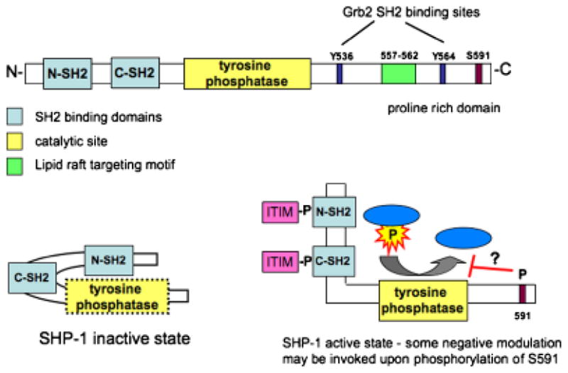
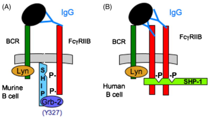
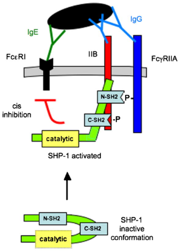
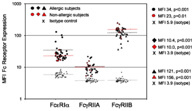
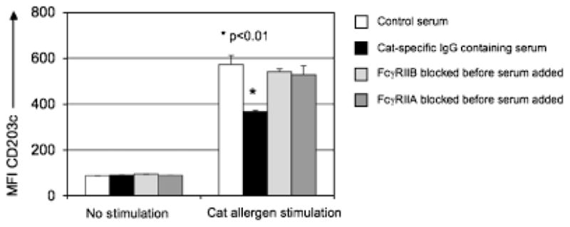
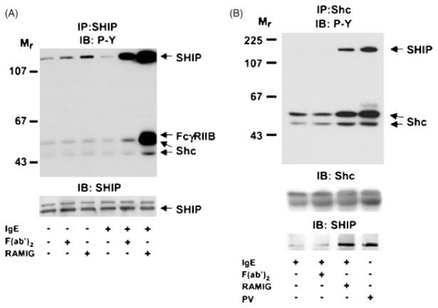
Numerous autoimmune and inflammatory disorders stem from the dysregulation of hematopoietic cell activation. The activity of inositol lipid and protein tyrosine phosphatases, and the receptors that recruit them, is critical for prevention of these disorders. Balanced signaling by inhibitory and activating receptors is now recognized to be an important factor in tuning cell function and inflammatory potential. In this review, we provide an overview of current knowledge of membrane proximal events in signaling by inhibitory/regulatory receptors focusing on structural and functional characteristics of receptors and their effectors Src homology 2 (SH2) domain-containing tyrosine phosphatase 1 and SH2 domain-containing inositol 5-phosphatase-1. We review use of new strategies to identify novel regulatory receptors and effectors. Finally, we discuss complementary actions of paired inhibitory and activating receptors, using Fc gammaRIIA and Fc gammaRIIB regulation human basophil activation as a prototype.


Figures

Fig. 1
Fig. 1. Structure of SHIP-1
The multiple functional domains of SHIP-1 include an N-terminal SH2 domain, which has been shown to interact with FcγRIIB. Immediately C-terminal of the catalytic domain is a newly recognized C2 allosteric activation domain. When bound by PtdIns3,4P2, this region was found to increase the enzyme activity of SHIP-1. The C-terminal domain NPXY motifs (917 and 1017) interact with phosphotyrosine binding domain (PTB) of Shc, p62dok, and DAB-1. The proline-rich PXXP motifs have been shown to interact with Grb2.
Fig. 2
Fig. 2. Proposed progressive activation of SHIP-1 resulting in formation of anMPSC
(A) Lyn is associated with a portion of resting BCR. (B) Co-aggregation of the BCR with FcγRIIB by antigen/IgG complexes results in phosphorylation of the ITIM consensus sequence Y(319)SSL. (C) The pITIM binds the SHIP-1 SH2 domain, and the Y327 terminal phosphotyrosine recruits Grb2 to form a bidentate interaction between SHIP1/Grb-2/FcγRIIB. (D) Lyn phosphorylates the tyrosine in the NPXY motif, creating a docking site for the adapter molecule p62dok (E). (F) Lyn phosphorylates the ITIM on Dok-1 (Y361DEP), creating a ‘preferred’ SH2 binding site for the N-terminal SH2 domain of SHIP-1. (G) This interaction results in release of SHIP-1 from FcγRIIB, and results in (H), an mobile PtdIns3,4,5P3 scavenger complex (MPSC), which is now free to migrate to remote membrane sites where it inhibits lipid hydrolysis.
Fig. 3
Fig. 3. MPSC interaction with the plasma membrane
The Dok-1 PH domain docks to PtdIns3,4,5P3 stabilizing SHIP-1/Dok-1 interaction with the plasma membrane. Recent work has shown the C2 domain of SHIP-1 interacts with PtdIns3,4P2, resulting in allosteric activation of SHIP-1. The conversion of PtdIns3,4,5P3 to PtdIns3,4P2 prevents further activation of Tec family kinases. In addition, PtdIns3,4P2 is now available to interact with TAPP1, the outcome of which remains uncertain.
Fig. 4
Fig. 4. Co-aggregation of FcγRIIB with FcεR1 inhibits LTB4 production and T-cell migration
Murine bone marrow-derived mast cells from wildtype mice were sensitized with IgE, washed and stimulated with either rabbit F(ab′)2 anti-mouse immunoglobulin antibodies, resulting in aggregation of FcεR1 alone, or with intact antibodies, resulting in co-aggregation of FcεR1 with FcγRIIB. Co-aggregation of IgE with FcγRIIB resulted in inhibition of migration (A) LTB4 production (B).
Fig. 5
Fig. 5. SHIP-1 is required for FcγRIIB-mediated inhibition of FcεRI-induced LTB4 production
Bone marrow-derived mast cells from wildtype and SHIP-1 knockout mice were sensitized with IgE, washed, and stimulated with either rabbit F(ab′)2 anti-mouse immunoglobulin antibodies resulting in cross-linking of FcεR1, or with intact antibodies, resulting in co-aggregation of FcεR1 with FcγRIIB. In the absence of SHIP-1, co-aggregation of IgE with FcγRIIB did not inhibit migration (A) or LTB4 production (B).
Fig. 6
Fig. 6. SHP-1 structure
(A) SHP-1 is composed of two SH2 domains, which are proposed to have distinct specificities, followed by the catalytic site. The C-terminal tail contains Grb2 SH2 binding sites (Y536 and Y564), a newly identified lipid raft targeting motif, and a C-terminal serine, which when phosphorylated is thought to inhibit SHP-1 function. (B) In a resting state, the N-terminal SH2 domain interacts with the tyrosine phosphatase domain, preventing enzyme activity. Interaction of the two SH2 domains with pITIMs on adjacent proteins results in exposure and resulting activation of the catalytic domain.
Fig. 7
Fig. 7. Proposed SHIP-1 versus SHP-1 binding
(A) In a murine B cell, tripartate interaction of FcγRIIB/Grb-2/SHIP-1 follows FcR and BCR co-aggregation. (B) In the human B cell, Y327 in the C-terminal tail of FcγRIIB is absent, possibly allowing for SHP-1 interaction. Because SHP-1 has two SH2 domains, higher order cross-linking of FcγRIIB may be required for this interaction to occur.
Fig. 8
Fig. 8. Proposed SHP-1 binding in cell expressing both FcγRIIA and FcγRIIB
SHP-1 has two SH2 domains with distinct binding capabilities: one may bind the pITIM on FcγRIIB, the other the N-terminal pITAM of FcγRIIA (106). This interaction results in exposure and activation of the catalytic domain, which dephosphorylate tyrosines in cis.
Fig. 9
Fig. 9. Analysis of FcR expression by human peripheral blood basophils
Human peripheral blood was collected in sodium heparin tubes. Cells were fixed, and then stained with the basophil-specific marker, CD203c antibodies against FcεR1α (polyclonal rabbit from Serotec), FcγRIIA (IV.3), or FcγRIIB (2B6) were used to determine surface expression of the respective Fc receptors (108). Both antibodies have been altered so that their Fc regions no longer bind FcγR (both were kind gifts from Macrogenics). Subjects were divided into those denying any allergy symptoms (open symbols), or those who reported suffering from allergies (closed symbols). FcεR1 expression was significantly correlated with serum IgE levels, P < 0.01 (data not shown).
Fig. 10
Fig. 10. FcγRIIA and FcγRIIB are both required for immune complex-mediated inhibition of FcεRI signaling
Human peripheral blood was collected from a cat allergen-sensitized individual in a sodium heparin tube. Next, the cells were incubated with blocking antibodies against either FcγRIIA or FcγRIIB (figure legend). Serum from a subject on immunotherapy containing high levels of cat allergen-specific IgG was added (20% w/v) to the whole blood samples for 3 h before a 10 min stimulation with cat hair extract (normalized to a Fel d1 concentration of 0.01 μg/ml). Activation was stopped by placing the cells on ice. After stimulation, red blood cells were lysed, and cells were stained with pan-leukocyte marker anti-CD45, basophil-specific marker anti-CD203c, and anti-IgE. CD203c was also used as a marker of basophil activation (109). CD203c expression was lower when serum containing cat allergen-specific IgG was added before stimulation with cat allergen, when compared with control serum (no cat allergen-specific IgG), P < 0.01. When FcγRIIA or FcγRIIB was blocked before addition of the serum, the inhibitory effect was reversed.
Fig. 11
Fig. 11. Tyrosine phosphorylation and association of SHIP-1 and Shc following FcγRIIB co-aggregation with FcεRI
RBL-mFcγRIIB cells were cultured with or without sensitizing IgE and stimulated with rabbit anti-mouse IgG F(ab′)2, rabbit anti-mouse intact IgG, or pervanadate (PV) for 4 min as indicated. Lysates of total cellular proteins were prepared and proteins were immunoprecipitated with anti-SHIP-1 (A) or anti-Shc (B) antibodies. Immune complexes were resolved by SDS-PAGE, transferred to membranes, and analyzed by immunoblotting with anti-phosphotyrosine antibodies. Membranes were stripped and reprobed with anti-SHIP-1 or anti-Shc antibodies as indicated. Published with permission from Ott et al. (104).

Similar articles

Cited by

References

    1. Takai T. Fc receptors and their role in immune regulation and autoimmunity. J Clin Immunol. 2005;25:1–18. - PubMed
    1. Malbec O, Daeron M. The mast cell IgG receptors and their roles in tissue inflammation. Immunol Rev. 2007;217:206–221. - PubMed
    1. Ujike A, et al. Modulation of immunoglobulin (Ig)E-mediated systemic anaphylaxis by low-affinity Fc receptors for IgG. J Exp Med. 1999;189:1573–1579. - PMC - PubMed
    1. Huber M, Helgason CD, Damen JE, Liu L, Humphries RK, Krystal G. The Src homology 2-containing inositol phosphatase (SHIP) is the gatekeeper of mast cell degranulation. Proc Natl Acad Sci USA. 1998;95:11330–11335. - PMC - PubMed
    1. Helgason CD, et al. A dual role for Src homology 2 domain-containing inositol-5-phosphatase (SHIP) in immunity: aberrant development and enhanced function of b lymphocytes in ship −/− mice. J Exp Med. 2000;191:781–794. - PMC - PubMed

MeSH terms

Substances