. 2008 Nov;82(21):10444-54.
doi: 10.1128/JVI.00833-08. Epub 2008 Aug 13.

Binding STAT2 by the acidic domain of human cytomegalovirus IE1 promotes viral growth and is negatively regulated by SUMO

Affiliations

Binding STAT2 by the acidic domain of human cytomegalovirus IE1 promotes viral growth and is negatively regulated by SUMO

Yong Ho Huh et al. J Virol. 2008 Nov.

Abstract

The human cytomegalovirus (HCMV) 72-kDa immediate-early 1 (IE1) protein is thought to modulate cellular antiviral functions impacting on promyelocytic leukemia (PML) nuclear bodies and signal transducer and activator of transcription (STAT) signaling. IE1 consists of four distinct regions: an amino-terminal region required for nuclear localization, a large central hydrophobic region responsible for PML targeting and transactivation activity, an acidic domain, and a carboxyl-terminal chromatin tethering domain. We found that the acidic domain of IE1 is required for binding to STAT2. A mutant HCMV encoding IE1(Delta421-475) with the acidic domain deleted was generated. In mutant virus-infected cells, IE1(Delta421-475) failed to bind to STAT2. The growth of mutant virus was only slightly delayed at a high multiplicity of infection (MOI) but was severely impaired at a low MOI with low-level accumulation of viral proteins. When cells were pretreated with beta interferon, the mutant virus showed an additional 1,000-fold reduction in viral growth, even at a high MOI, compared to the wild type. The inhibition of STAT2 loading on the target promoter upon infection was markedly reduced with mutant virus. Furthermore, sumoylation of IE1 at this acidic domain was found to abolish the activity of IE1 to bind to STAT2 and repress the interferon-stimulated genes. Our results provide genetic evidence that IE1 binding to STAT2 requires the 55-amino-acid acidic domain and promotes viral growth by interfering with interferon signaling and demonstrate that this viral activity is negatively regulated by a cellular sumoylation pathway.


Figures
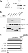
FIG. 1.
FIG. 1.
The acidic domain of IE1 is required for STAT2 binding. (A) Interaction of IE1 with STAT2 in HCMV-infected cells. HF cells were mock infected or infected with HCMV at an MOI of 2 (IFU/cell). (Left panel) At 24 h, immunoprecipitation was carried out with anti-STAT2 rabbit PAb or control IgG, followed by immunoblotting with anti-IE1 mouse MAb. (Right panels) The protein levels of STAT2 and IE1 in total cell lysates of infected and uninfected cells were measured by immunoblotting. (B) CoIP of IE1 with STAT2 in cotransfected cells. 293T cells were cotransfected with myc-STAT2 and wild-type or mutant HA-IE1 expression plasmids. (Upper panel) At 48 h, total cell lysates were immunoprecipitated with anti-Myc antibody, followed by immunoblotting with anti-HA antibody. (Lower panels) Input protein levels of STAT2 and IE1 proteins in total cell lysates as measured by immunoblotting. (C) Summary of the structure of IE1 proteins used in cotransfection assays. The amino-terminal region required for the NLS function (an open circle), the large central hydrophobic region, and the carboxyl-terminal acidic plus chromatin tethering domains (open boxes) within IE1 are indicated.
FIG. 2.
FIG. 2.
Generation of the recombinant T-BAC clone. (A) Genome structure of the T-BAC clone used in the present study. The F plasmid sequences containing the partition and replication functions (parA, parB, and repE), replication origin (ori), chloramphenicol resistance marker (Cmr), and the GFP eukaryotic expression cassette are indicated. (B) Genome structures of the T-BAC containing the wild-type or IE1(Δ421-475) gene. The region encompassing the MIE locus and the locations of the restriction enzyme sites used for mapping by Southern blot analysis are shown. The location for the 400-bp probe used for Southern blot analysis is shown. (C) Southern blot analysis. (Left) Ethidium bromide-stained total restriction DNA fragment patterns after EcoRV/BamHI digestion of three T-BAC DNAs (wild type [wt], Δ421-475, and revertant [R]) obtained by pulsed-field gel electrophoresis. (Right) Results of autoradiography after Southern blotting with 32P-labeled probe. Marker, λ-HindIII/EcoRI. (D) Infectivity of the transfected T-BAC DNAs in permissive HF cells. HF cells were electroporated with 2 μg of wild-type [wt], IE1(Δ421-475) mutant, or revertant [R] T-BAC DNAs. Each reaction also included 1 μg of plasmid pCMV71 encoding pp71 and 1 μg of plasmid pEGFP-C1. The cells were monitored for the spreading of the GFP signals. The GFP images and phase-contrast images were taken at 12 days after electroporation.
FIG. 3.
FIG. 3.
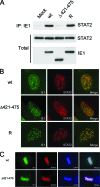
The acidic domain of IE1 is required for STAT2 binding in virus-infected cells. (A) HF cells were mock infected or infected with the wild-type, IE1(Δ421-475), or revertant viruses at an MOI of 2 IFU per cell. (Upper panel) At 24 h, total cell lysates were immunoprecipitated with anti-IE1 MAb CH443 (10 μg per 15 mg of total proteins), followed by immunoblotting with anti-STAT2 antibody. (Lower panels) The total protein levels of STAT2 and IE1 proteins in the cell extracts are also shown after immunoblotting. (B) HF cells were infected with recombinant viruses at an MOI of 1 IFU per cell. At 3 h postinfection, the cells were fixed with methanol, followed by double-label IFA with anti-IE1 and anti-STAT2 antibodies. (C) Metaphase association of wild-type or Δ421-475 mutant IE1. HF cells were infected with wild-type or IE1(Δ421-475) mutant at an MOI of 1 IFU per cell. At 72 h postinfection, the cells were fixed with methanol, followed by double-label IFA with anti-IE1 and anti-STAT2 antibodies. DAPI was used to stain DNA.
FIG. 4.
FIG. 4.
Analysis of the growth of recombinant viruses. (A and B) Analysis of growth curves in recombinant virus-infected cells. HF cells in 12-well plates were infected with wild-type, IE1(Δ421-475) mutant, or revertant virus at an MOI of 3 (A) or 0.01 (B) IFU/cell. The time course results shown represent the total infectious center units of infectious virus present in 2 ml of culture supernatant at the indicated sampling times. Note that the growth curves of wild-type and revertant viruses are indistinguishable, whereas those of mutant viruses (both 1 and 2) show different patterns (but almost identical) compared to those of wild-type and revertant viruses. (C) Comparison of viral growth between the IE1(Δ421-475) and CR208 viruses. HF cells were infected with the IE1(Δ421-475) and its parent viruses (reconstituted from the T-BAC clone), or the CR208 and its parent Towne viruses at an MOI of 0.5 or 0.1. At 5 days after infection, the total progeny virus titers were determined by measuring the numbers of IE2-positive infectious centers. (D) The images of GFP signals in infected cell cultures were taken at 10 days after infection at an MOI of 0.01 (IFU/cell). (E and F) Comparison of the accumulation of viral proteins in recombinant virus-infected cells. HF cells in six-well plates were infected with the recombinant viruses at an MOI of 3 (E) or 0.01 (F) IFU/cell. At the indicated time points, the total cell lysates were prepared and subjected to SDS-PAGE (8%), and immunoblotting was then performed with antibodies specific for IE1/IE2 and delayed-early p52(UL44) proteins. β-Actin was detected on the same blot as a loading control. wt, Wild type; R, revertant.
FIG. 5.
FIG. 5.
Comparison of the growth of recombinant viruses in cells pretreated with IFN-β. HF cells in 12-well plates were untreated or pretreated with 100 U of IFN-β for 24 h and then infected with wild-type, IE1(Δ421-475) mutant, or revertant virus at an MOI of 5 (IFU/cell). (A) The images of GFP signals in infected cells were taken at 7 days after infection. (B) The production of progeny virions at 10 days after infection from untreated and IFN-β-pretreated cells was measured by infectious center assays. The results shown are the mean values and standard errors of four independent experiments. (C) ChIP assays for the loading of STAT2 on the ISG54 promoter in cells infected with recombinant viruses were performed. HF cells were mock infected or infected with wild-type, IE1(Δ421-475), or revertant virus at an MOI of 2 IFU per cell for 12 h. As a positive control, HF cells were treated with IFN-β (1,000 U/ml) for 2 h. ChIP assays were conducted as described in Materials and Methods. The amounts of coprecipitated DNA were quantified by real-time PCR and normalized to input. The results shown are the mean values and standard errors of three independent experiments. (D) Expression of IFN-induced genes in cells infected with recombinant viruses. HF cells were infected with recombinant viruses or treated with IFN-β as in panel C. mRNAs for ISG54, CXCL10, MxA, and IFN-β were quantified by quantitative RT-PCR. The amounts of mRNAs in cells infected with viruses or treated with IFN-β over those in mock-infected cells are indicated as the fold induction. wt, wild type; R, revertant.
FIG. 6.
FIG. 6.
Effect of sumoylation of IE1 on STAT2 binding. (A) Structural map of the two plasmids used to produce the sumoylated IE1 protein in E. coli. pT-E1E2S1 expressed E1, E2, and the cleaved active form of SUMO-1. Note that pT-E1E2S1 and pGST-IE1 contain different antibiotic markers and replication origins. (B) CoIP assays using bacterially produced proteins. The GST-IE1 and SUMO-1-modified GST-IE1 fusion proteins were incubated with the control cell lysates or in vitro-translated myc-STAT2 or myc-UL44 proteins. After immunoprecipitation of the samples with anti-myc antibody, the bound proteins were fractionated by SDS-PAGE and visualized by immunoblotting with anti-IE1 antibody (right). One-sixth of the proteins used in the binding reaction were shown by immunoblotting (for myc-tagged proteins, left) and Coomassie blue staining (for GST fusion proteins, middle) as input controls. (C) CoIP assay in virus-infected cells. U373 astrocytoma/glioblastoma cell lines that constitutively overexpress flag-SUMO-1 were mock infected or infected with HCMV at an MOI of 10 (IFU/cell). At 72 h, the total cell lysates were prepared with CoIP buffer containing 0.5 mM N-ethylmaleimide. The expression levels of unmodified IE1, SUMO-modified IE1, and STAT2 in total lysates were shown by immunoblotting with anti-IE1 mouse MAb and anti-STAT2 rabbit PAb (left). Total cell lysates were immunoprecipitated with anti-STAT2 antibody, and immunoblot analysis was conducted with antibodies for IE1 and STAT2 (right). The amounts of unmodified IE1 and SUMO-modified IE1 were measured quantitatively using Scion Image software (Scion Corp., Maryland) and are indicated as a percentage.
FIG. 7.
FIG. 7.
Effect of the carboxyl-terminal SUMO-1 fusion of IE1 on the abilities of IE1 to bind to STAT2 and to repress the IFN-induced gene expression. (A) Interaction of the IE1-SUMO-1 fusion with HDAC3 in cotransfected cells. 293T cells were cotransfected with plasmids encoding myc-tagged HDAC3 and hemagglutinin (HA)-tagged wild-type IE1 or IE1-SUMO1 fusion. The total cell lysates were prepared at 48 h and immunoprecipitated with anti-myc antibody, and SDS-PAGE and immunoblotting with anti-HA antibody were performed (top panel). Immunoblots of the total cell extracts with anti-myc or anti-HA antibody to show the expression levels are shown (bottom panels). (B) Disruption of PML-NBs by IE1-SUMO-1 fusion. HF cells expressing wild-type IE1 or IE1-SUMO1 fusion were fixed in paraformaldehyde, followed by double-label IFA with anti-IE1 (6E1) and anti-PML (PML-C) antibodies. (C) GST pull-down assays. The bacterially purified GST, GST-IE1, and GST-IE1-SUMO-1 proteins immobilized to glutathione-Sepharose beads were incubated with in vitro-translated STAT2 protein. The bound proteins were fractioned by SDS-PAGE and visualized by immunoblotting with anti-STAT2 antibody. One-fifth of the GST or GST fusion proteins used in the binding reaction were shown by Coomassie blue staining, and one-tenth of those in STAT2 were shown by immunoblotting as input controls. (D) CoIP assays using cotransfected cells. 293T cells were cotransfected with myc-STAT2 and the indicated IE1 constructs. At 48 h, CoIP assays were performed as described for Fig. 1B. (E) The reporter assays using the ISG54 ISRE-luciferase construct. 293T cells were cotransfected with 0.5 μg of the ISG54 ISRE-luciferase reporter construct and 0.1 μg of empty vector or plasmid expressing the intact IE1 or IE1-SUMO-1 fusion protein. At 24 h, cells were left untreated or were treated with IFN-β (1,000 U/ml) for 8 h, and luciferase reporter assays were then conducted. The results shown are the mean values and standard errors of three independent experiments. The expression levels of IE1 proteins in the extracts were shown by immunoblotting.

Similar articles

Cited by

References

    1. Abate, D. A., S. Watanabe, and E. S. Mocarski. 2004. Major human cytomegalovirus structural protein pp65 (ppUL83) prevents interferon response factor 3 activation in the interferon response. J. Virol. 7810995-11006. - PMC - PubMed
    1. Ahn, J. H., E. J. Brignole III, and G. S. Hayward. 1998. Disruption of PML subnuclear domains by the acidic IE1 protein of human cytomegalovirus is mediated through interaction with PML and may modulate a RING finger-dependent cryptic transactivator function of PML. Mol. Cell. Biol. 184899-4913. - PMC - PubMed
    1. Ahn, J. H., and G. S. Hayward. 2000. Disruption of PML-associated nuclear bodies by IE1 correlates with efficient early stages of viral gene expression and DNA replication in human cytomegalovirus infection. Virology 27439-55. - PubMed
    1. Ahn, J. H., and G. S. Hayward. 1997. The major immediate-early proteins IE1 and IE2 of human cytomegalovirus colocalize with and disrupt PML-associated nuclear bodies at very early times in infected permissive cells. J. Virol. 714599-4613. - PMC - PubMed
    1. Baldick, C. J., Jr., A. Marchini, C. E. Patterson, and T. Shenk. 1997. Human cytomegalovirus tegument protein pp71 (ppUL82) enhances the infectivity of viral DNA and accelerates the infectious cycle. J. Virol. 714400-4408. - PMC - PubMed

Publication types

LinkOut - more resources