

doi: 10.1128/JVI.01204-08.
Epub 2008 Aug 6.
Fusion of enhanced green fluorescent protein to the pseudorabies virus axonal sorting protein Us9 blocks anterograde spread of infection in mammalian neurons
Affiliations
- PMID: 18684822
- PMCID: PMC2566268
- DOI: 10.1128/JVI.01204-08
Item in Clipboard
Fusion of enhanced green fluorescent protein to the pseudorabies virus axonal sorting protein Us9 blocks anterograde spread of infection in mammalian neurons
J Virol.
2008 Oct.
Abstract
Pseudorabies virus encodes a membrane protein (Us9) that is essential for the axonal sorting of virus particles within neurons and anterograde spread in the mammalian nervous system. Enhanced green fluorescent protein (GFP)-tagged Us9 mimicked the trafficking properties of the wild-type protein in nonneuronal cells. We constructed a pseudorabies virus strain that expressed Us9-GFP and tested its spread capabilities in the rat visual system and in primary neuronal cultures. We report that Us9-EGFP does not promote anterograde spread of infection and may disrupt packing of viral membrane proteins in lipid rafts, an essential step for Us9-mediated axonal sorting.
Figures

Analysis of Us9-EGFP mediated anterograde spread in the rat visual system. (A) Schematic representation of the genome of PRV 164. A CMV Us9-GFP expression cassette was inserted into the gG locus (striped box upstream of gD), and the endogenous Us9 open reading frame was deleted (black box between gE and Us2). IR, inverted repeat. (B) Approximately 2.5 × 10⁵ PFU of either PRV Becker or PRV 164 was injected into the vitreous humor of Sprague-Dawley male rats. At the time of imminent death, the animals were sacrificed and the brains were fixed and sliced into 35-μm-thick coronal sections with a freezing microtome. Infected tissue was stained for PRV antigen or Us9-EGFP (with Rb133 or Us9 rabbit polyvalent antiserum, respectively). Areas reached by anterograde, transneuronal spread are highlighted in red. D, dorsal; V, ventral.
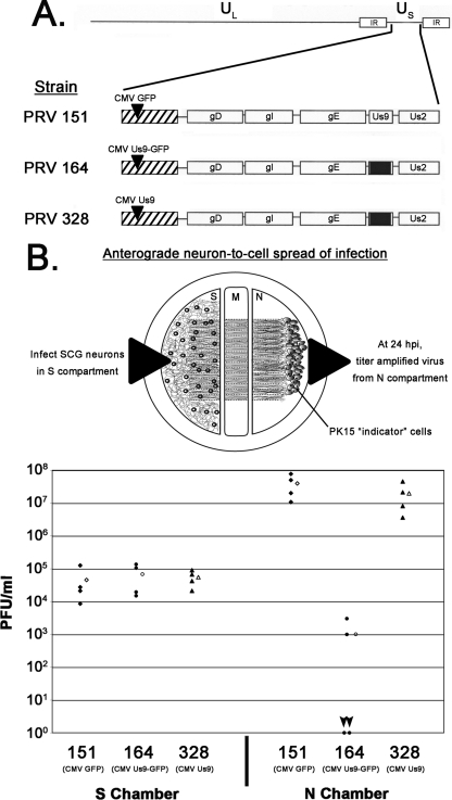
Dropping the EGFP tag from Us9 restores anterograde, neuron-to-cell spread in vitro. (A) Schematic of the PRV strains 151, 164, and 328. IR, inverted repeat. (B) SCG neurons were plated in the S compartment and allowed to extend neurites into the N compartment for 2 weeks. Axons were guided by a series of grooves scratched into the surface of the tissue culture dish. After 2 weeks, a monolayer of indicator PK15 cells was plated on top of the axon termini in the N compartment. Cell bodies in the S compartment were then infected at an MOI of 10 with PRV 151, 164, or 328. Four chambers were used for each type of infection (closed symbols). At 24 hpi, medium and infected cells were harvested together from either the S or the N compartment. Total PFU/ml were determined for each chamber. The mean value for the four samples is denoted by the offset open symbol. Black arrowheads denote the two plates infected with PRV 164 that showed no anterograde, neuron-to-cell spread. M, methocellulose compartment.

Targeting of Us9-EGFP to detergent-resistant microdomains. Differentiated PC12 cells were infected with PRV 328 (A) or PRV 164 (B) for 12 h and then lysed with cold 1% Triton X-100. Lysates were separated on a discontinuous Optiprep density gradient (5%, 30%, and 40%) by ultracentrifugation at 4°C for 20 h (13). Detergent-insoluble complexes (i.e., lipid rafts) floated to the 5%/30% interface, while detergent-soluble proteins remained at the bottom of the gradient. Fractions were collected from the top to the bottom of the tube (1 ml each). Samples were subjected to sodium dodecyl sulfate-polyacrylamide gel electrophoresis, and Western blotting analysis was performed using biotinylated cholera toxin B subunit (for GM1) and antiserum to PRV Us9 and transferrin receptor (TfR). Numbers at right of each panel are molecular masses in kilodaltons.
Similar articles
-
Visualization of an alphaherpesvirus membrane protein that is essential for anterograde axonal spread of infection in neurons.mBio. 2012 May 2;3(2):e00063-12. doi: 10.1128/mBio.00063-12. Print 2012. mBio. 2012. PMID: 22448044 Free PMC article.
-
Role of Us9 phosphorylation in axonal sorting and anterograde transport of pseudorabies virus.PLoS One. 2013;8(3):e58776. doi: 10.1371/journal.pone.0058776. Epub 2013 Mar 19. PLoS One. 2013. PMID: 23527020 Free PMC article.
-
Targeting of pseudorabies virus structural proteins to axons requires association of the viral Us9 protein with lipid rafts.PLoS Pathog. 2008 May 16;4(5):e1000065. doi: 10.1371/journal.ppat.1000065. PLoS Pathog. 2008. PMID: 18483549 Free PMC article.
-
The role of virion membrane protein endocytosis in the herpesvirus life cycle.J Clin Virol. 2000 Aug;17(2):69-82. doi: 10.1016/s1386-6532(00)00084-6. J Clin Virol. 2000. PMID: 10942087 Review.
-
Use of pseudorabies virus to delineate multisynaptic circuits in brain: opportunities and limitations.J Neurosci Methods. 2000 Nov 15;103(1):51-61. doi: 10.1016/s0165-0270(00)00295-8. J Neurosci Methods. 2000. PMID: 11074095 Review.
Cited by
-
Molecular features contributing to virus-independent intracellular localization and dynamic behavior of the herpesvirus transport protein US9.PLoS One. 2014 Aug 18;9(8):e104634. doi: 10.1371/journal.pone.0104634. eCollection 2014. PLoS One. 2014. PMID: 25133647 Free PMC article.
-
Proteomic Comparison of Three Wild-Type Pseudorabies Virus Strains and the Attenuated Bartha Strain Reveals Reduced Incorporation of Several Tegument Proteins in Bartha Virions.J Virol. 2022 Dec 21;96(24):e0115822. doi: 10.1128/jvi.01158-22. Epub 2022 Dec 1. J Virol. 2022. PMID: 36453884 Free PMC article.
-
Single-Particle Tracking of Virus Entry in Live Cells.Subcell Biochem. 2023;106:153-168. doi: 10.1007/978-3-031-40086-5_5. Subcell Biochem. 2023. PMID: 38159226 Review.
-
Comparison of the pseudorabies virus Us9 protein with homologs from other veterinary and human alphaherpesviruses.J Virol. 2009 Jul;83(14):6978-86. doi: 10.1128/JVI.00598-09. Epub 2009 May 6. J Virol. 2009. PMID: 19420087 Free PMC article.
-
The Attenuated Pseudorabies Virus Vaccine Strain Bartha Hyperactivates Plasmacytoid Dendritic Cells by Generating Large Amounts of Cell-Free Virus in Infected Epithelial Cells.J Virol. 2022 Jun 22;96(12):e0219921. doi: 10.1128/jvi.02199-21. Epub 2022 May 23. J Virol. 2022. PMID: 35604216 Free PMC article.
References
Publication types
MeSH terms
Substances
Grants and funding
LinkOut - more resources
Full Text Sources
