. 2008 Aug;20(8):2205-20.
doi: 10.1105/tpc.108.058339. Epub 2008 Aug 1.

Arabidopsis protein disulfide isomerase-5 inhibits cysteine proteases during trafficking to vacuoles before programmed cell death of the endothelium in developing seeds

Affiliations

Arabidopsis protein disulfide isomerase-5 inhibits cysteine proteases during trafficking to vacuoles before programmed cell death of the endothelium in developing seeds

Christine Andème Ondzighi et al. Plant Cell. 2008 Aug.

Abstract

Protein disulfide isomerase (PDI) oxidizes, reduces, and isomerizes disulfide bonds, modulates redox responses, and chaperones proteins. The Arabidopsis thaliana genome contains 12 PDI genes, but little is known about their subcellular locations and functions. We demonstrate that PDI5 is expressed in endothelial cells about to undergo programmed cell death (PCD) in developing seeds. PDI5 interacts with three different Cys proteases in yeast two-hybrid screens. One of these traffics together with PDI5 from the endoplasmic reticulum through the Golgi to vacuoles, and its recombinant form is functionally inhibited by recombinant PDI5 in vitro. Peak PDI5 expression in endothelial cells precedes PCD, whereas decreasing PDI5 levels coincide with the onset of PCD-related cellular changes, such as enlargement and subsequent collapse of protein storage vacuoles, lytic vacuole shrinkage and degradation, and nuclear condensation and fragmentation. Loss of PDI5 function leads to premature initiation of PCD during embryogenesis and to fewer, often nonviable, seeds. We propose that PDI5 is required for proper seed development and regulates the timing of PCD by chaperoning and inhibiting Cys proteases during their trafficking to vacuoles before PCD of the endothelial cells. During this transitional phase of endothelial cell development, the protein storage vacuoles become the de facto lytic vacuoles that mediate PCD.


Figures

Figure 1.
Figure 1.
Structure and Organization of the Arabidopsis PDI5 Gene and T-DNA Insertion Mutant Site. (A) Schematic representation of the PDI5 gene map (locus number AAD41430) and T-DNA insertion site location. The T-DNA (SALK_010645) insertion is located in the 300-nucleotide 5′ untranslated region (UTR) of the PDI5 gene at −248 bp upstream from the ATG codon. The gene contains nine exons (black boxes, numbered) separated by eight introns (thick black lines). (B) Schematic drawing of the fully spliced translated portion of PDI5 cDNA. The white arrowheads indicate the position of various domains, including the ER signal peptide (the bold AMRG at the N terminus in [C]), two thioredoxin catalytic domains (TRX and PWCghC in framed region in [C]), a unique peptide antigen (11 amino acids underlined, bold italics in [C]), and the ER retention motif Lys-Asp-Glu-Leu (KDEL) in the C terminus (bold, unframed in [C]). The numbers correspond to the position and to the numbers of amino acids. (C) Predicted protein sequence of PDI5 contains 501 amino acids and one stop codon.
Figure 2.
Figure 2.
Immunoblot Analysis of PDI5 in Protein Extracts from Wild-Type and pdi5Δ T-DNA Insertion Mutant Tissues. (A) SDS-PAGE and Coomassie blue staining of protein extracts from wild-type and pdi5Δ mutant tissues. Open arrowhead points to the 68-kD gel region where the PDI5 protein is expected to migrate. MW, molecular weight standard in kD; TP, total protein extract from whole plants; Fl, flowers; Lv, leaves; St, stems; Si, siliques; ImS, immature seeds; MS, mature seeds; Rt, roots. (B) Protein gel blot analysis of the gel shown in (A) using anti-PDI5 antibodies. The boxes highlight the lack of PDI5 protein in the T-DNA mutant.
Figure 3.
Figure 3.
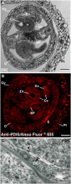
Localization of PDI5 in Immature Flowers. (A) to (C) Light, fluorescence, and electron microscopy images of transverse sections of immature florets of the wild-type plant. Gy, gynoecia; O, ovary; En, endothelial cells; Ov, ovule; Pt, petal; SP, septum; S, sepal. Bars = 100 μm in (A) and (B) and 0.2 μm in (C). (A) Differential interference contrast (DIC) micrograph illustrates the tissues and cellular organization of an early stage 12 Arabidopsis flower. The 12-stage floret contains the gynoecia organ, which encompasses the developing ovules, the septum, and the ovary. Petals surround all gynoecial tissues. (B) Immunofluorescence labeling of a cross-sectioned floret labeled with anti-PDI5 antibody-Alexa Fluor 555. Petals and the gynoecia organs, such as ovule, endothelium, septum, and ovary, are labeled. Hardly any labeling was seen over the sepals. (C) Electron micrograph of an ovule gynoecial cell immunolabeled with anti-PDI5 antibody. The gold label is seen over ER cisternae of the endothelial cells of the developing ovule.
Figure 4.
Figure 4.
Fluorescent Micrographs Showing the Confinement of PDI5 Expression to the Endothelium in Developing Seeds. (A) to (D) DIC micrographs of developing seeds. Pre-embryo stage ([A] to [A-3]), globular stage ([B] to [B3]), heart torpedo stage ([C] to [C3]), and early-bent cotyledon stage ([D] to [D3]). The endothelium (En) corresponds to the innermost layer of the integument and defines the interface between endosperm (E) and the future seed coat. In (B) to (D), black arrowheads point to the embryos (Em) at different stages of development. Bar = 100 μm for (A) to (D-3). (A-1) to (D-1) Autofluorescence micrographs of developing seeds viewed using a cyan fluorescent protein (CFP) filter set. (A-2) to (D-2) Immunofluorescence micrographs of developing seeds stained with Alexa Fluor 555 and labeled with anti-PDI5 antibodies. White arrowheads denote the endothelial cell layer. (A-3) to (D-3) Merged images of anti-PDI5 antibody-Alexa Fluor and cyan fluorescent protein.
Figure 5.
Figure 5.
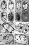
Structural Changes within Endothelial Cells and in the Endothelial Cell Layer during Seed Development. (A) to (H) Longitudinal sections through developing seeds of wild-type Arabidopsis, illustrating the stages of embryo (Em) and endosperm (E) development and the organization of the endothelial cell layer (En). (A) Pre-embryo stage. The endothelial cell layer surrounding the embryo sac is completed before embryo growth commences. (B) Two-celled embryo stage, with a prominent endothelial layer. (C) and (D) Globular embryo (C) and heart stages (D). The endothelial cell layer begins to thin as the embryo enlarges. (E) Torpedo embryo stage. The endothelial cell layer starts to break down (asterisks) as the cellular endosperm expands. (F) and (G) Early (F) and late (G) bent-cotyledon stages. Chalaza (CHZ) and micropilar (MZ) zones are shown. (H) Coated seed. (I) to (L) Electron micrographs of longitudinally sectioned endothelial cells at different stages of embryonic development. (I) Pre-embryo stage endothelial cell (see [A]). The cytoplasm exhibits a normal cellular organization with a large central LV, numerous small PSVs, and an amyloplast (AM) and is surrounded by a cell wall (CW) with a cuticular layer (Cu). (J) Heart stage endothelial cell (see [D]). Some PSVs are greatly enlarged and contain many electron-dense globoids (G). By contrast, the LVs are much smaller but more numerous than in (I). (K) Torpedo stage endothelial cell around the micropylar zone (see [E] and [F]). The large PSVs appear to be breaking down. (L) Bent-cotyledon stage (see [F] and [G]). The process of degradation encompasses the nucleus (N), organelles, and cell wall. Bars = 100 μm in (A) to (H), 3.00 μm in (I) and (K), 2.00 μm in (J), and 5.00 μm in (L).
Figure 6.
Figure 6.
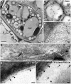
Characterization of Vacuole Types in Senescing Endothelial Cells by Means of Labeling with Anti-PDI5 and Anti-δ-TIP Antibodies. (A) Electron micrograph of a heart stage endothelial cell. The PSVs are larger than the LVs. Ch, chloroplast; Cu, cuticle; CW, cell wall. (B) and (C) Immunolocalization of PDI5 (open arrowheads) in an LV (B) and a PSV (C). (D) Immunolocalization of PDI5 in ER cisternae. (E) Immunolocalization of δ-TIP (filled arrowheads) in the tonoplast membrane of a PSV. (F) Double immunolabeling of a PSV with anti-PDI5 (15 nm gold) and anti-δ-TIP (10 nm gold) antibodies. Bars = 2.00 μm in (A), 0.4 μm in (B), 0.5 μm in (C), 0.2 μm in (D) and (F), and 0.1 μm in (E).
Figure 7.
Figure 7.
Validation of Protein Interaction with PDI5 Bait in the Yeast Two-Hybrid Assay and of in Vitro Inhibition of RD21 Protease Activity. (A) Standard β-gal activity measurements (OD570) were conducted according to (Serebriiskii and Golemis, 2000) and normalized to an equal culture density of yeast cells coexpressing PDI5 bait plus one of the interacting Cys proteases (43 [NM_123672], RD21A [NM_103612], or 19 [NM_112826]). Values of two negative controls (coexpression of Cys protease with empty bait vector; PDI5 bait with empty prey vector) were subtracted from the values of Cys proteases coexpressed with PDI5 bait and positive control. (B) Yeast cell culture densities (OD620) after 2 weeks of growth on His dropout medium with 1 mM 3-AT for each Cys protease coexpressed in the PDI5 bait cell line. The positive control (+cont) consisted of documented interacting partners (Epsin 1 is an interacting partner for the EH domain-containing region of Eps15) as described (Serebriiskii and Golemis, 2000). Values in (A) and (B) represent two experiments done in three replicates (mean ± sd). (C) Immunoblot of coimmunoprecipitation (Co-I.P.) products (Cys proteases RD21, top panel, or CP43, bottom panel). Open arrowhead indicates coimmunoprecipitated Cys protease (RD21 or CP43) using the anti-PDI5 antiserum. Recombinant RD21 and CP43 were detected with anti-HA tag antiserum. The left two lanes in each panel were controls for no coimmunoprecipiation (no Co-I.P.), in which 40 μg total E. coli protein containing recombinant PDI5, RD21, or CP43 was directly loaded on the gel. The heavy chain (hc) of the anti-PDI5 antisera used in the coimmunoprecipitation is shown in the Co-I.P. lanes. The band in the no Co-I.P. lane labeled RD21 refers to the recombinant RD21 protein that was a positive control for the antiserum used on the immunoblot. No HC protein was detected in this lane. Asterisk denotes a nonspecific background band present in all lanes. (D) In vitro RD21 Cys protease assay reveals that PDI5 inhibits RD21 activity. Recombinant RD21 activity was measured in the presence and absence of an equivalent amount of PDI5. Addition of 5 mM DTT or DTNB prior to adding PDI5 is noted. The y axis units are fluorescence emission at 530 ± 15 nm. The averages were derived from a sample size of three in six independent experiments (18 total averaged), and the error bars are sd. The a, b, and c refer to significantly different means (P < 0.05) as determined by analysis of variance.
Figure 8.
Figure 8.
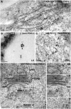
Coimmunolocalization of Cys Protease RD21 with PDI5 in Senescing Endothelial Cells. (A) Colocalization of anti-PDI5 (15 nm gold, open arrowheads) and anti-RD21 (10 nm gold, filled arrowheads) within ER-cisternae (ER). (B) Colocalization of anti-PDI5 (15 nm gold) and anti-RD21 (10 nm gold) antibodies in the LV. (C) Colocalization of anti-PDI5 (15 nm gold) and anti-RD21 (10 nm gold) antibodies in the PSV. (D) Colocalization of anti-PDI5 (15 nm gold) and anti-RD21 (10 nm gold) antibodies in the Golgi apparatus and TGN. Bars = 0.2 μm in (A), 0.1 μm in (B) and (C), 0.2 μm in (D), and 0.15 μm in the (ER), (G), (TGN), and (V) panels.
Figure 9.
Figure 9.
Quantitative Analysis of the Coimmunogold Labeling of Anti-PDI5 with Anti-RD21 Antibodies in LV and PSV of Senescing Endothelial Cells. Quantification of PDI5 and RD21 clusters (anti-PDI5C with anti-RD21C) and single (anti-PDI5 and anti-RD21) gold particles in LVs and PSVs of endothelial cells (n = 10 cells). Anti-PDI5- and anti-RD21-gold particles colocalize (a and a′) more frequently in LV than in PSV. The anti-RD21/anti-PDI5 labeling ratio is 1.2 over LVs. The number of gold-labeled clusters of anti-PDI5C with anti-RD21C (a and a′) in endothelial cell vacuoles (LVs and PSVs) was higher than the number of single gold particles in both vacuoles (b and b′).
Figure 10.
Figure 10.
The pdi5Δ Knockout Mutation Affects Seed Viability and Seed Set (A) and (B) Tetrazolium-based embryo viability test. Bar = 500 μm. (A) Top row: wild-type embryos show a uniform red color (viable seeds). Bottom row: wild-type embryos boiled for 30 min are light pink (negative control). (B) Top row: pdi5Δ embryos show variable degrees of red and pink coloring (white arrowheads). Bottom row: boiled mutant embryos are light pink. (C) and (D) Seed set in wild-type (C) and pdi5Δ mutant (D) siliques. Note the pdi5Δ mutant silique contains fewer seeds (open arrowheads) and gaps (filled arrowheads) (D) than the wild type (open arrowheads). Bars = 1 mm.

Similar articles

Cited by

References

    1. Andème-Onzighi, C., Sivaguru, M., Judy-March, J., Baskin, T.I., and Driouich, A. (2002). The reb1-1 mutation of Arabidopsis alters the morphology of trichoblasts, the expression of arabinogalactan-proteins and the organization of cortical microtubules. Planta 215 949–958. - PubMed
    1. Balmer, Y., Koller, A., del Val, G., Manieri, W., Schurmann, P., and Buchanan, B.B. (2003). Proteomics gives insight into the regulatory function of chloroplast thioredoxins. Proc. Natl. Acad. Sci. USA 100 370–375. - PMC - PubMed
    1. Beeckman, T., Rycke, R.D., Viane, R., and Inzé, D. (2000). Histological study of seed coat development in Arabidopsis thaliana. J. Plant Res. 113 139–148.
    1. Beers, E.P., Woffenden, B.J., and Zhao, C. (2000). Plant proteolytic enzymes: Possible roles during programmed cell death. Plant Mol. Biol. 44 399–415. - PubMed
    1. Beisson, F., Li, Y., Bonaventure, G., Pollard, M., and Ohlrogge, J.B. (2007). The acyltransferase GPAT5 is required for the synthesis of suberin in seed coat and root of Arabidopsis. Plant Cell 19: 351–368. - PMC - PubMed

Publication types

MeSH terms

Substances

LinkOut - more resources